Si deseas distinguir tus productos, servicios o ambos de los de otra empresa, es posible que necesites una marca o nombre comercial. Descubre qué son, en qué consiste su procedimiento de registro y qué implica.

Información sobre los plazos de presentación de solicitudes de transformación de marcas de la Unión Europea en marca nacional española. Más información


Si tienes un nuevo dispositivo, producto o procedimiento que resuelva un problema técnico o tenga una ventaja práctica, existen distintas formas de protegerlo en España y en otros países. Descubre cómo hacerlo.

¿Tu innovación reside en la estética, la ornamentación o la apariencia de tu producto? Protégela mediante un diseño industrial. Descubre qué derechos confiere el registro y cómo realizar la tramitación.

Las indicaciones geográficas protegen el nombre de un producto originario de una zona geográfica, a la cual le debe una determinada calidad, reputación u otra característica. Descubre qué son, en qué consiste su procedimiento de registro y qué beneficios conceden.

Las patentes publicadas en todo el mundo son una valiosa fuente de información científica, técnica y comercial.

Si eres emprendedor/a o una empresa y quieres potenciar y mejorar la rentabilidad de tu negocio protegiendo de forma adecuada los activos intangibles de tu organización, en este espacio encontrarás lo necesario.


 355
resultados
355
resultados
 Última actualización
01/05/2026 [08:11:00]
Última actualización
01/05/2026 [08:11:00]


 Resultados 25 a 50 de 355

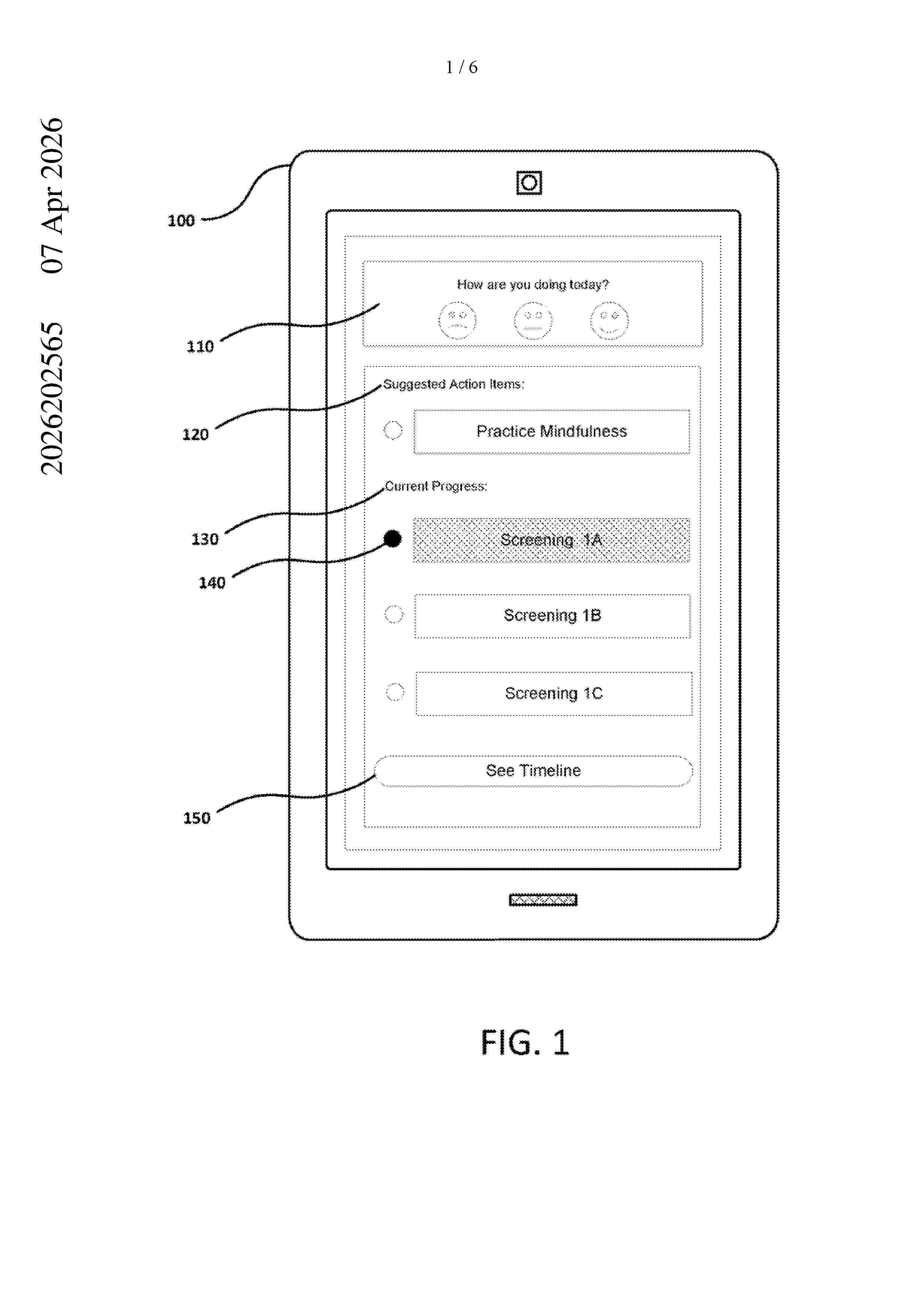
Resumen de: AU2026202565A1
Approaches for generating a set of biomarkers and generating a summary and one or more recommendations based, at least in part, upon the biomarkers are provided. Sensor data associated with a user may be received. The sensor data may be analyzed to generate a set of biomarkers. One or more biomarkers of the set may be combined to generate one or more additional biomarkers. A summary and one or more recommendations may be generated based, at least in part, upon the generated set of biomarkers and the additional biomarkers. The generated summary and the one or more recommendations may be provided for display on a client device. on a client device. pr p r

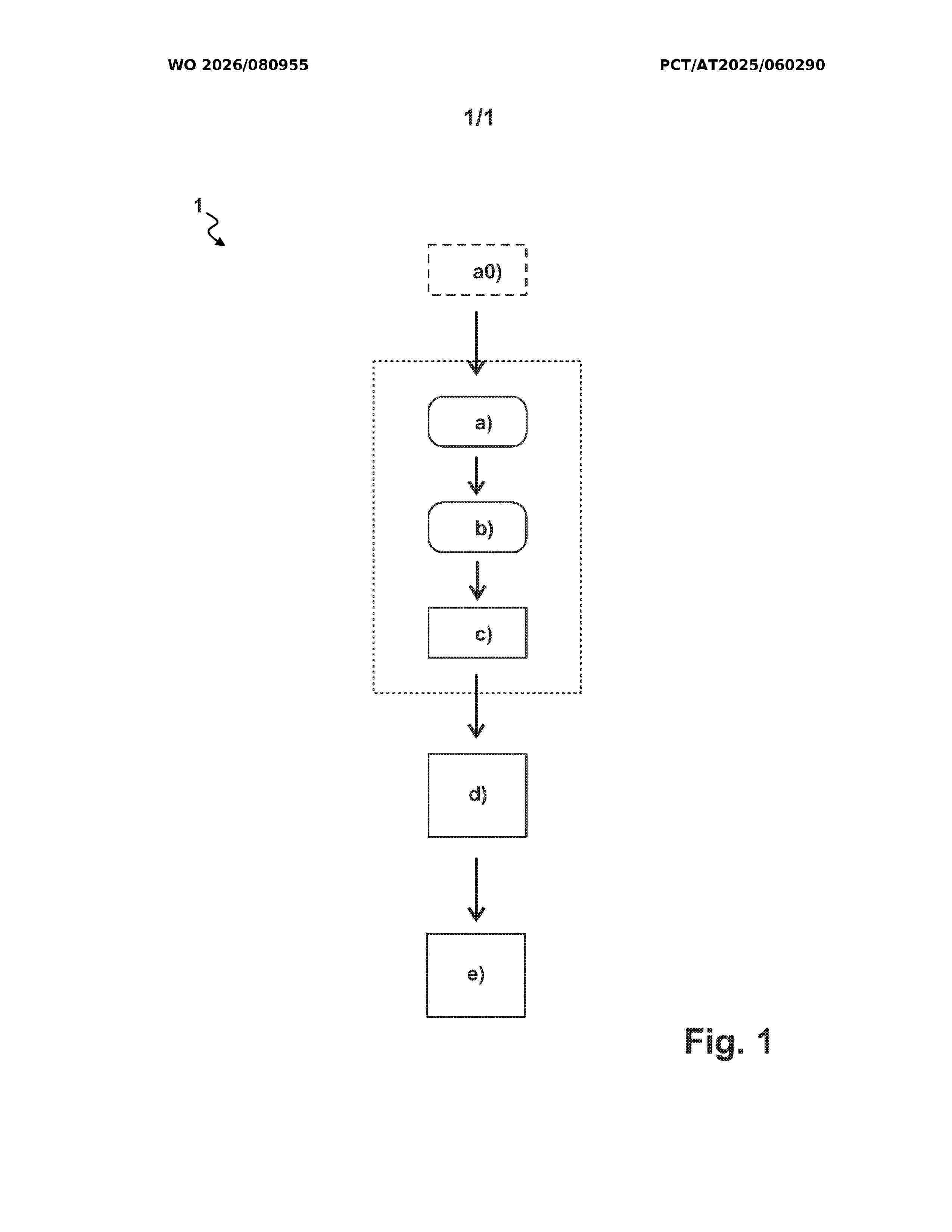
Resumen de: WO2026080955A1
The invention relates to a method for producing a template for a bone plate for treating a fracture of a bone of a human or animal, in particular for a bone plate for treating a pelvic fracture of a human. According to the invention, in order to produce the template in a patient-specific manner with high practical applicability, a digital data set of an image of bone fragments of the fracture is obtained using an imaging method, in particular an MRI or CT or X-ray method, wherein the method comprises the following steps: a) creating a digital, in particular virtual, three-dimensional bone model of the bone fracture fragments on the basis of the data set; b) determining a digital template design adapted to the bone model; c) producing, in particular 3D printing, the template on the basis of the determined template design. The invention also relates to a data processing device, to a system for producing the template, and to a computer program product.
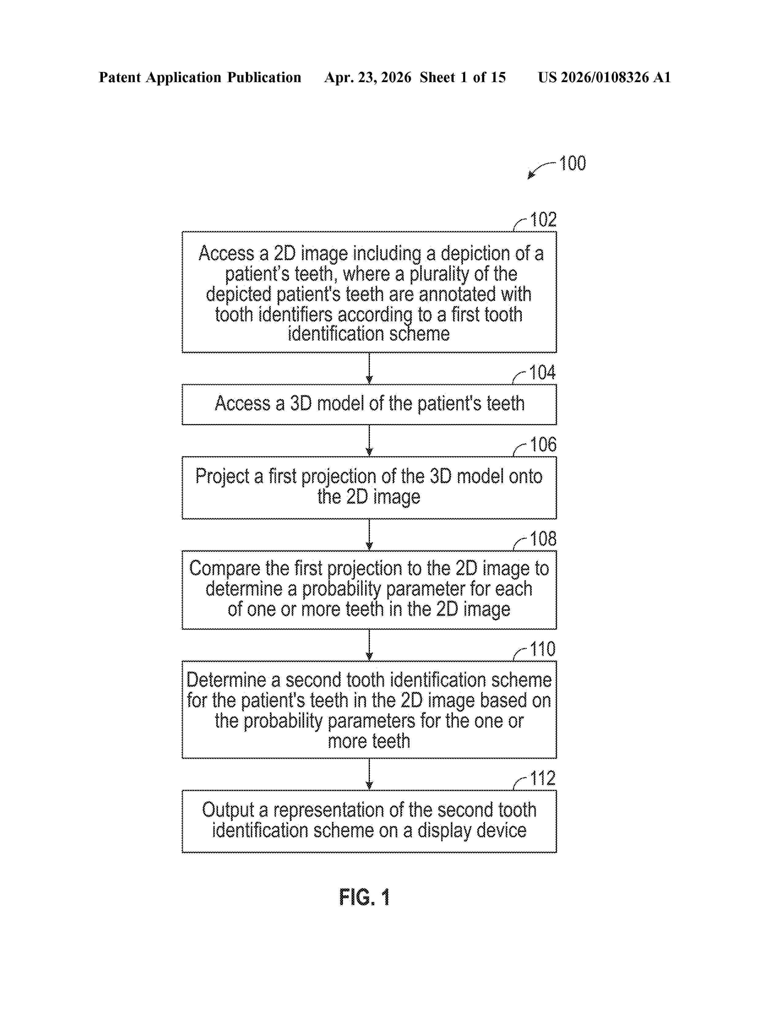
Resumen de: US20260108326A1
Systems and methods for identifying teeth in a patient image are provided. For example, a computer-implemented method can include, by one or more processors, accessing a 2D image including a depiction of a patient's teeth, where a plurality of the depicted patient's teeth are annotated with tooth identifiers according to a first tooth identification scheme. The computer-implemented method can further include transmitting, to a server computing device, the 2D image, and receiving from the server computing device, a second tooth identification scheme for the patient's teeth in the 2D image. The second tooth identification scheme can be generated by accessing a 3D model of the patient's teeth, projecting the 3D model onto the 2D image, comparing the projection to the 2D image to determine a probability parameter for each of one or more teeth in the 2D image, and determining, based on the probability parameters, the second tooth identification scheme.
Resumen de: US20260108182A1
0000 Systems and methods are directed to screening a subject for motor and cognitive impairment. A method for screening motor and cognitive impairment comprises administering a spiral tracing assessment to a subject, and receiving a response thereto; assessing the subject's performance based on the response; collecting performance parameters based on the subject's performance on the spiral tracing assessment; providing the performance parameters and the response to a pretrained machine learning model; predicting, using the pretrained machine learning model, a likelihood of the patient having a cognitive impairment; determining a motor and cognitive impairment status of the patient based on the prediction; and outputting the motor and cognitive impairment status.
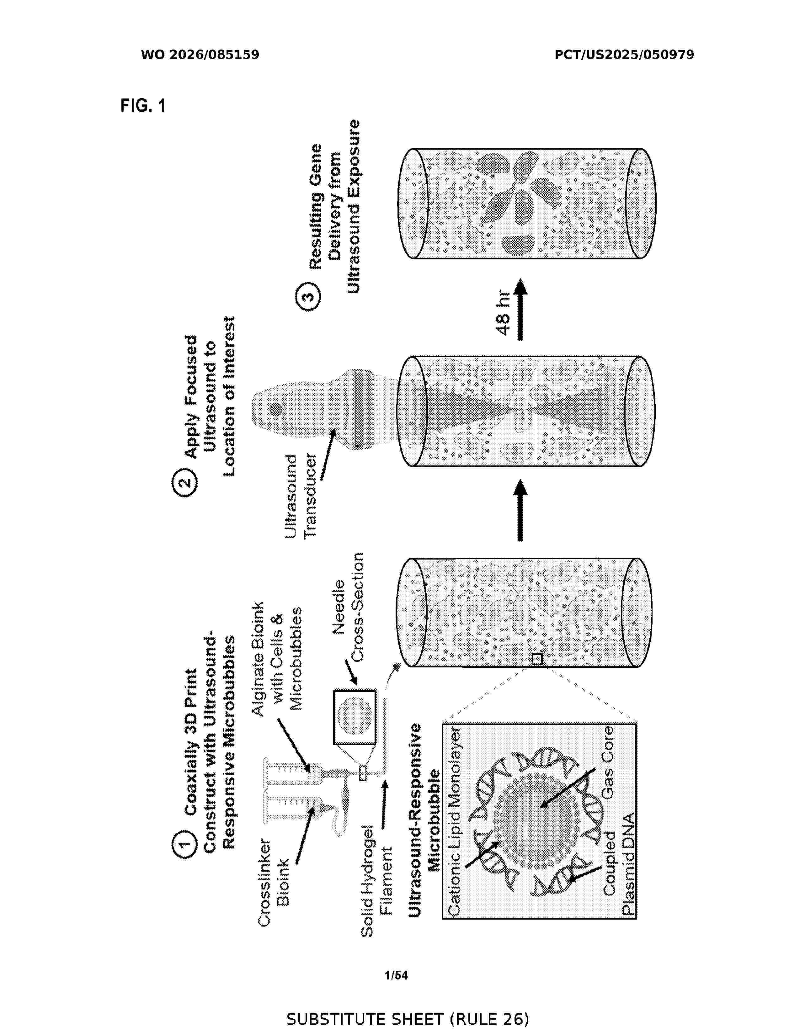
Resumen de: WO2026085159A1
Disclosed are methods and compositions to enable bioprinting of energy-responsive bioinks. The embodiments of the present disclosure relate to ultrasound-controlled gene delivery to a target cell in a three-dimensional bioprinted cell construct. The compositions include, for example, a hydrogel scaffold with a plurality of filament structures printed on a substrate; and a collagen cast gel that encases the hydrogel scaffold; wherein the compositions are configured to receive a population of microparticle-coupled cargo for controlled delivery to one or more target cells by application of ultrasound energy. Also disclosed are methods of creating transgenic target cells using the disclosed compositions.
Resumen de: US20260108304A1
A modified digital library and an algorithm able to make six functions for the improvement of most implant planning software used for the design of the custom surgical guide are provided. The prior art implant planning software available on the market, improved by the modified digital library and algorithm presented here, is essential to guide the digital design of both the custom surgical guide and the CAD-CAM cemented prosthesis onto straight or angled prefabricated abutments, through preliminary determination of abutment position with respect to an appropriate system of reference planes in correspondence with digital crown design. Furthermore, in a future perspective, the improved implant planning software presented here could be usefully employed in robot-assisted dental implant surgery.
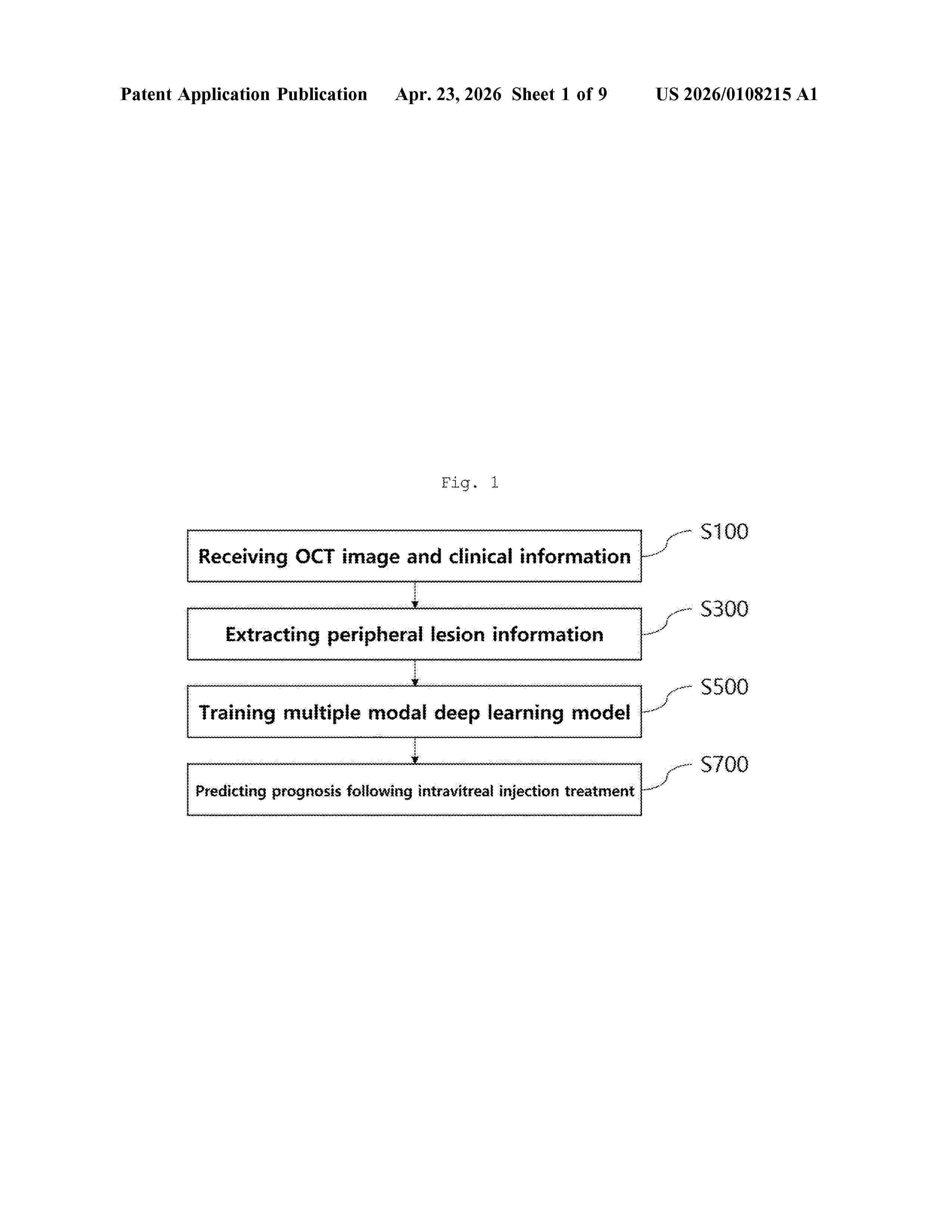
Resumen de: US20260108215A1
0000 The present invention relates to a method for predicting the prognosis of intravitreal injection treatment, comprising the steps of: receiving OCT images and clinical information of a plurality of diabetic macular edema patients; extracting one or more major lesion information from the OCT images; training a multi-modal deep learning model by inputting the OCT images, the clinical information, and the one or more major lesion features, and outputting the intravitreal injection treatment prognosis of the diabetic macular edema patients; and predicting the prognosis of intravitreal injection treatment by inputting the OCT images, the clinical information, and the major lesion features of a specific diabetic macular edema patient to the trained multi-modal deep learning model.
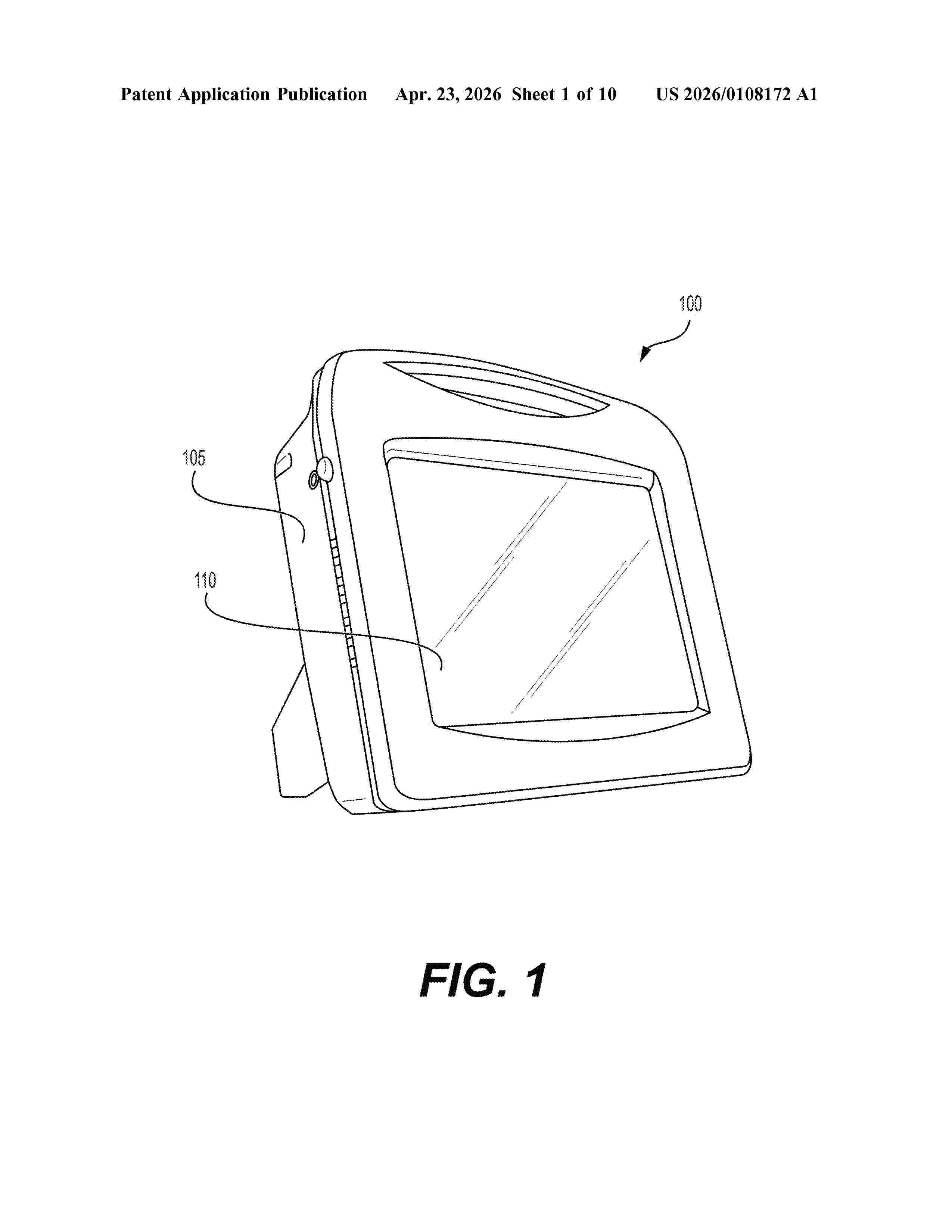
Resumen de: US20260108172A1
0000 Disclosed is a bioimpedance measurement system: A stabilized high frequency current generator is connected to padset electrodes via a patient cable. Electrodes are connected to an adaptive circuit that conditions the resulting voltage signal and converts it to digital form. Firmware performs signal acquisition and relays data to the device.
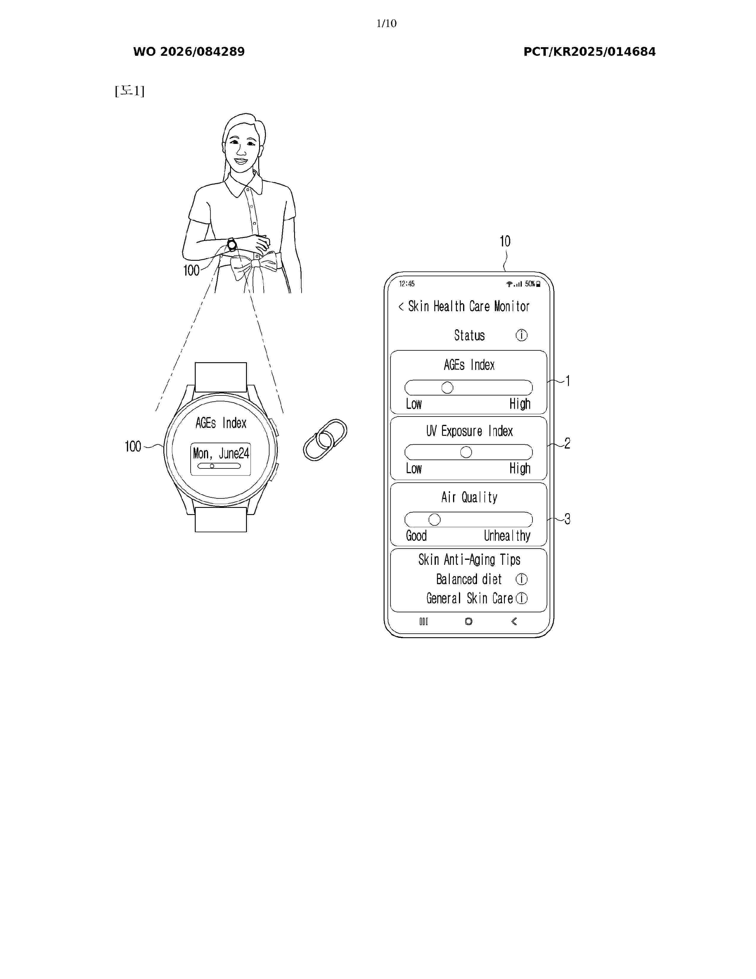
Resumen de: WO2026084289A1
An electronic device according to the present disclosure comprises: a communication interface; a memory, which stores instructions and includes one or more storage media; and at least one processor including processing circuitry, wherein, when executed individually or collectively by the at least one processor, the instructions instruct the electronic device to: receive advanced glycation end products (AGEs) data of a user from a wearable device; acquire weather data on the basis of position information of the user wearing the wearable device; input the AGEs data and the weather data into an artificial intelligence model; and allow the artificial intelligence model to provide a recommendation message generated, the artificial intelligence model being trained to: acquire state information of the user on the basis of the AGEs data, and generate the recommendation message for guiding a behavior of the user on the basis of an ultraviolet index and an air quality index included in the weather data and the state information.
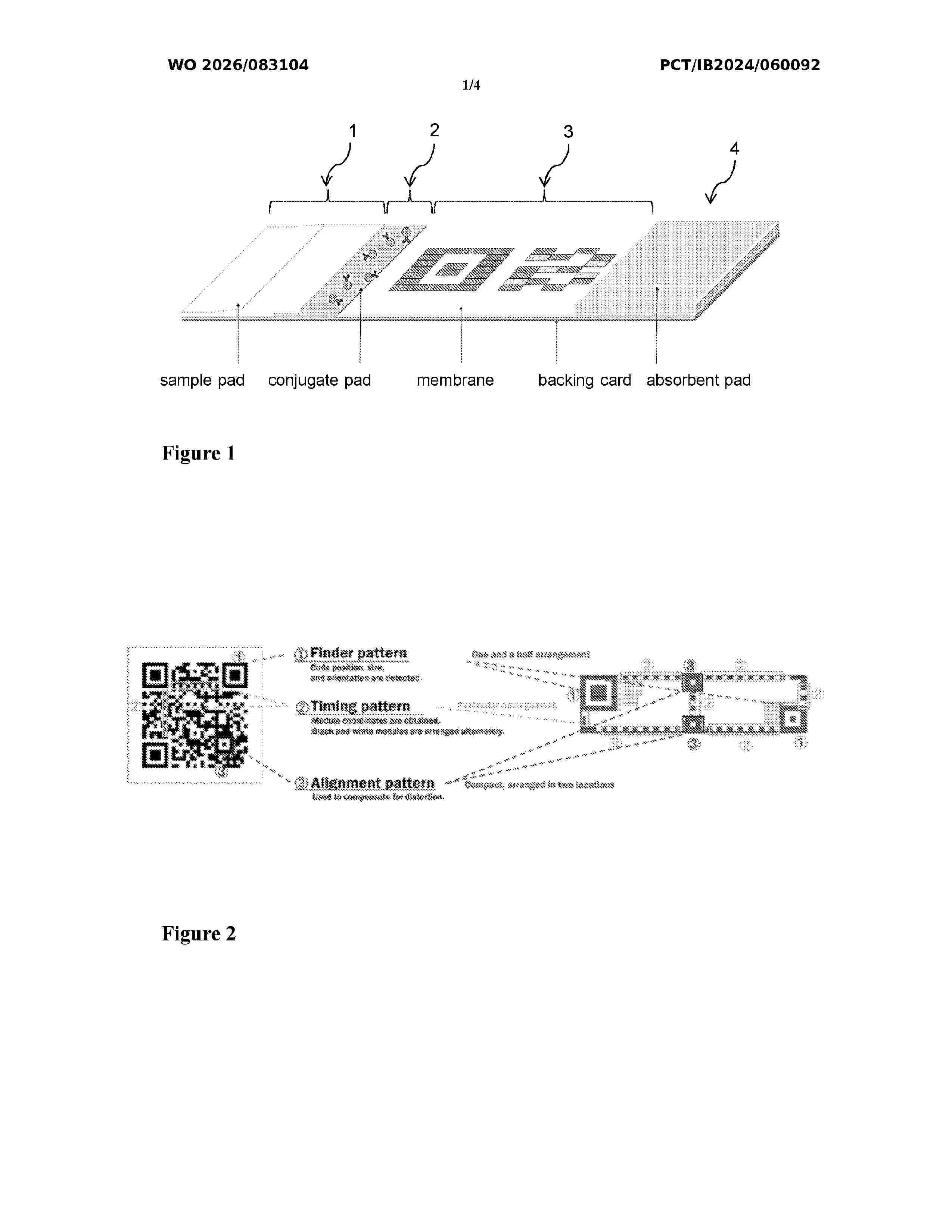
Resumen de: WO2026083104A1
This invention relates to a method of manufacturing a membrane for lateral flow assay or rapid diagnostic assay, a device comprising a membrane for detecting an analyte in a sample and the use of the device for detecting an analyte in a sample, wherein a two-dimensional barcode of a plurality of reagent dots comprising bioreceptors is patterned or printed on the membrane, the plurality of reagent dots being arranged in form of a two-dimensional barcode visible after the completion of the test and in which a certain information associated with the analyte is embedded, and to the use of said device.
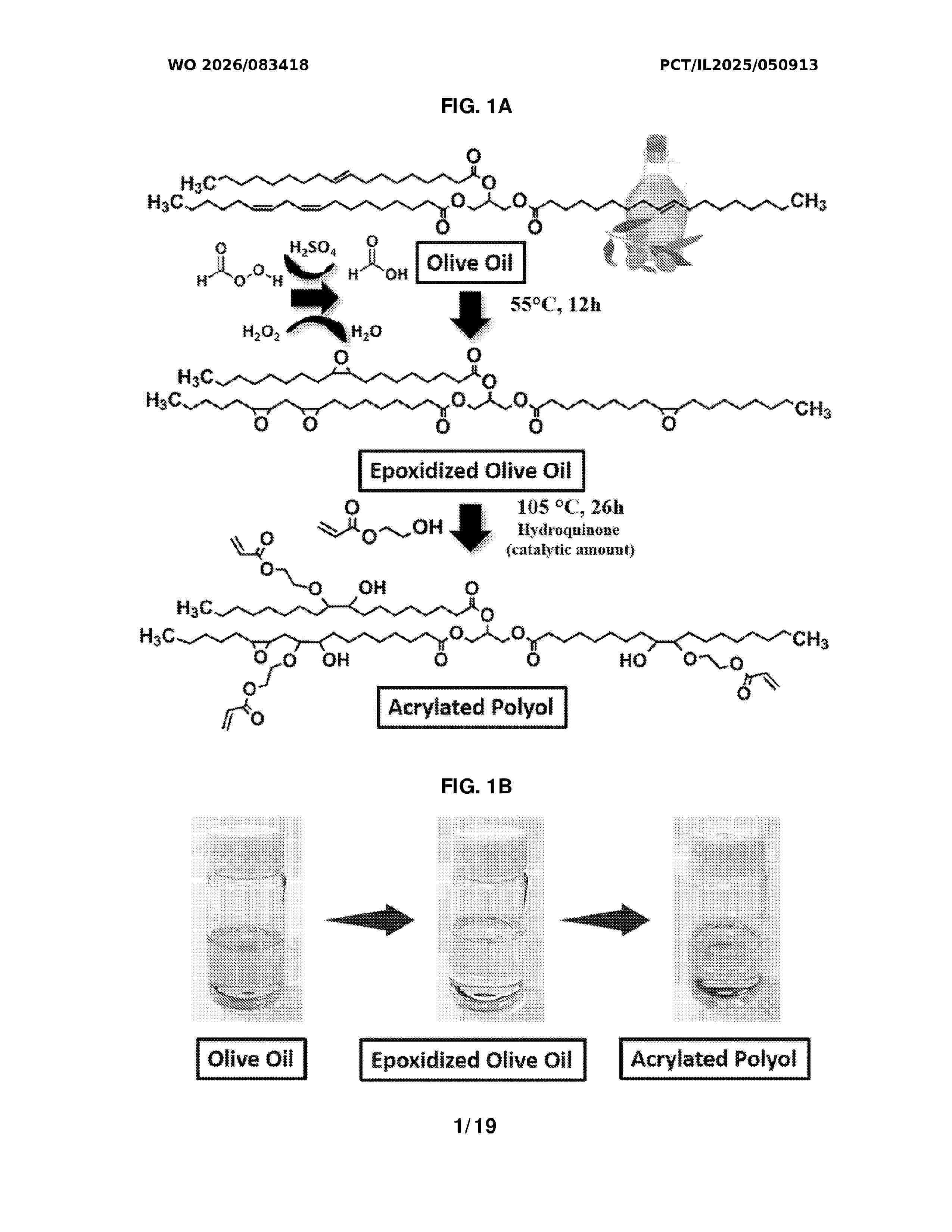
Resumen de: WO2026083418A1
The present invention provides a photopolymerisable composition for 4D printing, comprising an acrylated polyol (AOPO) derived from a natural oil, such as olive oil, and acrylic acid as a comonomer. The composition is used in a solvent-free digital light processing (DLR) method to fabricate high-resolution, multifunctional objects. The resulting 4D printed objects are biocompatible, hemocompatible, and cytocompatible. They exhibit excellent temperature- responsive shape memory properties activated near body temperature, inherent antimicrobial activity against both gram-negative and gram-positive bacteria, and tuneable mechanical properties suitable for soft tissue engineering. Furthermore, the composition can be loaded with therapeutic agents, such as progesterone, to form biodegradable objects capable of sustained, controlled drug release. These properties make the material ideal for medical implants, cardiovascular stents, and drug delivery devices.
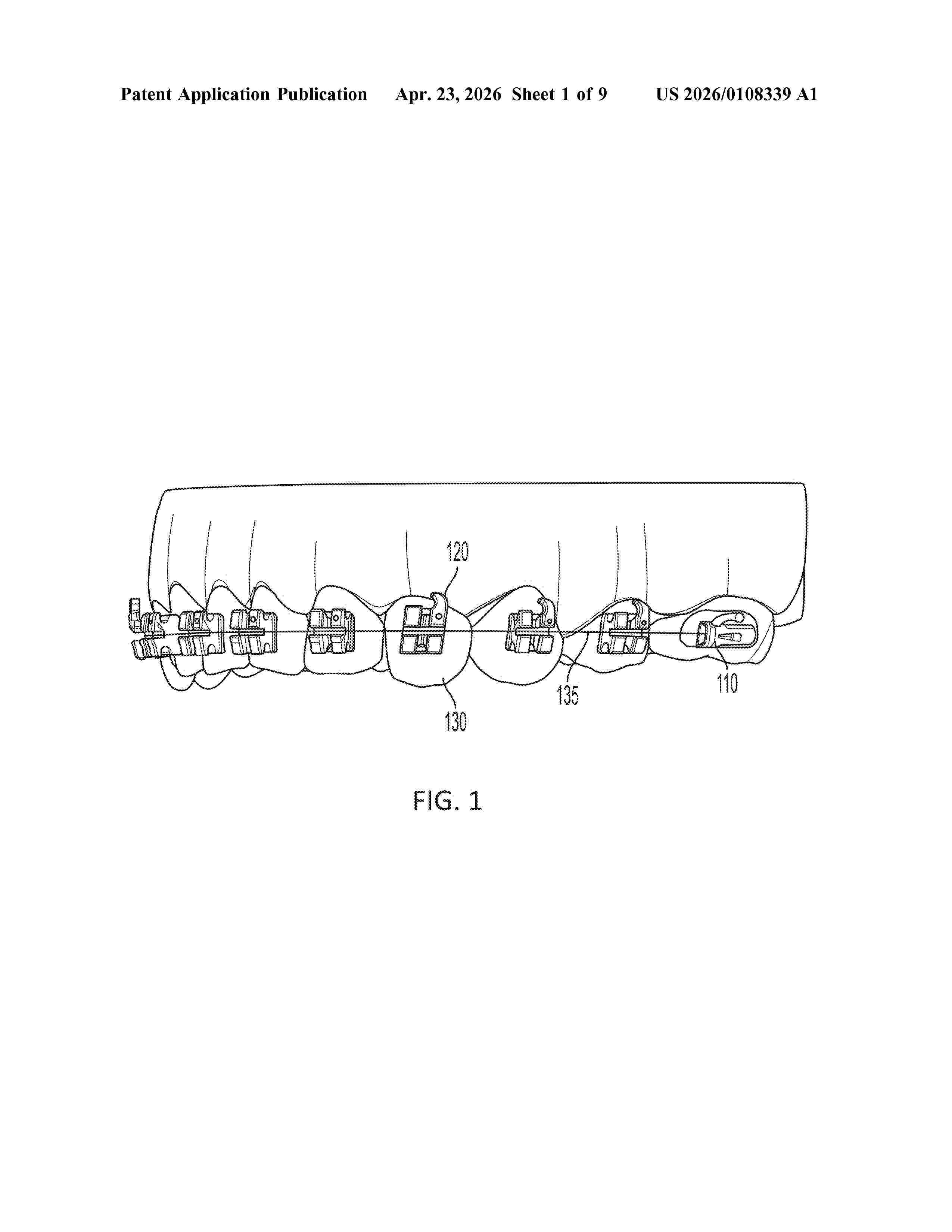
Resumen de: US20260108339A1
0000 A drug delivery device may include a component with a bonding location to be bonded to a surface of one or more teeth. A cavity may hold a formulation that includes a compound, and an orifice may release the compound outside the component.
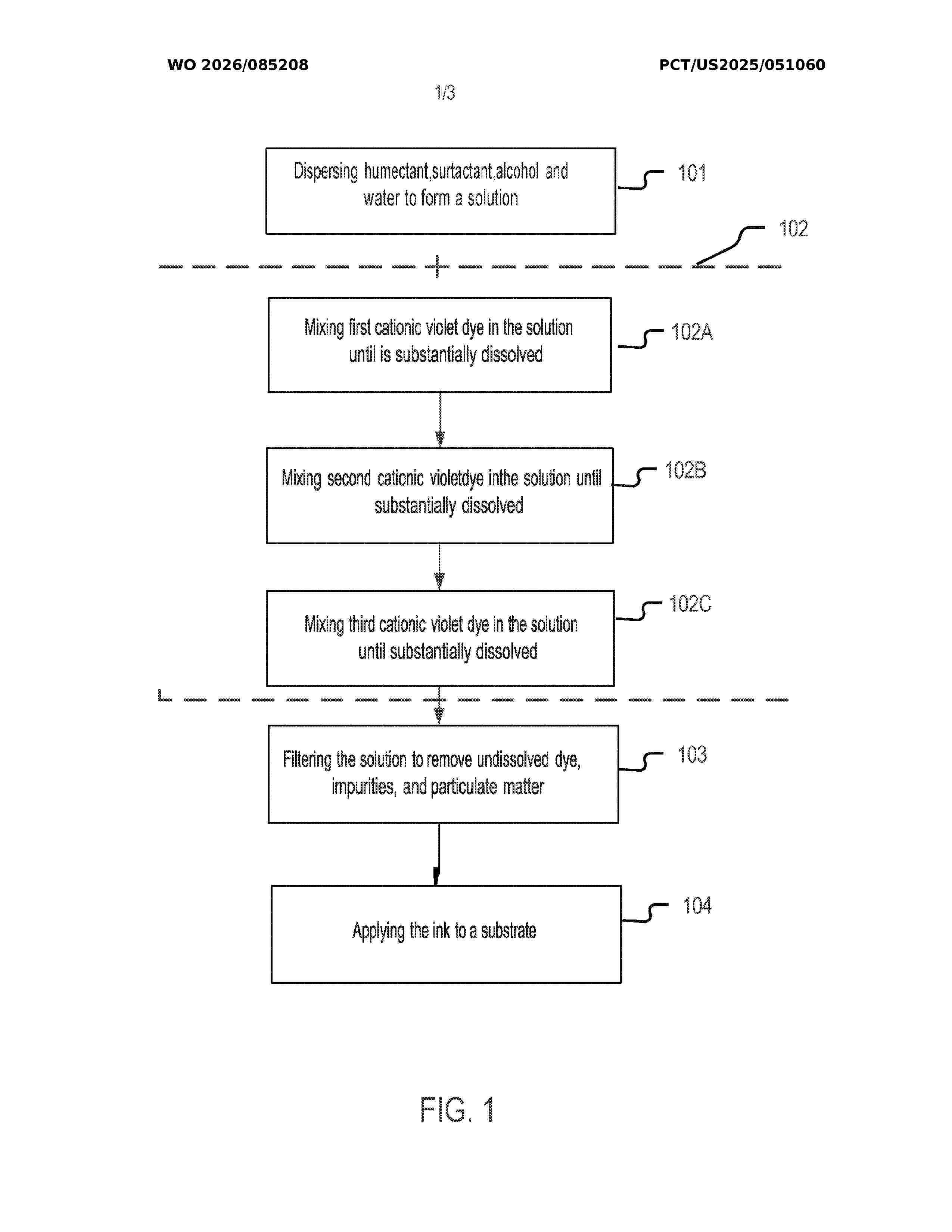
Resumen de: WO2026085208A1
An ink composition for a tattoo stencil and methods of making the same are disclosed herein. The ink composition comprises a plurality of cationic violet dyes in combination with a humectant, optionally along with surfactants and/or alcohol. It exhibits enhanced color vibrancy and color reproduction of the transferred tattoo design. The disclosed ink composition also exhibits excellent stability under thermal conditions and is suitable for long-term storage.
Resumen de: AU2026202539A1
A SYSTEM AND METHOD FOR EVALUATING THE LEVEL OF HARASSMENT OF INSECTS ON A PLURALITY OF ANIMALS The presently disclosed subject matter aims to a system and method for evaluating the level of harassment of flying insects on a plurality of animals located at a given area, 5 the system comprising a processing circuitry configured to: obtain one or more ear movement patterns associated with at least one ear of at least one animal of the plurality of animals, each ear movement pattern is associated with respective ear movement characteristics; for at least part of the one or more ear movement patterns, determine whether their respective ear movement characteristics meet a predefined rule; and, 10 determine whether a number of ear movement patterns whose ear movement characteristics met the predefined rule meets an action requirement rule. A SYSTEM AND METHOD FOR EVALUATING THE LEVEL OF pr p r
Resumen de: WO2024226130A1
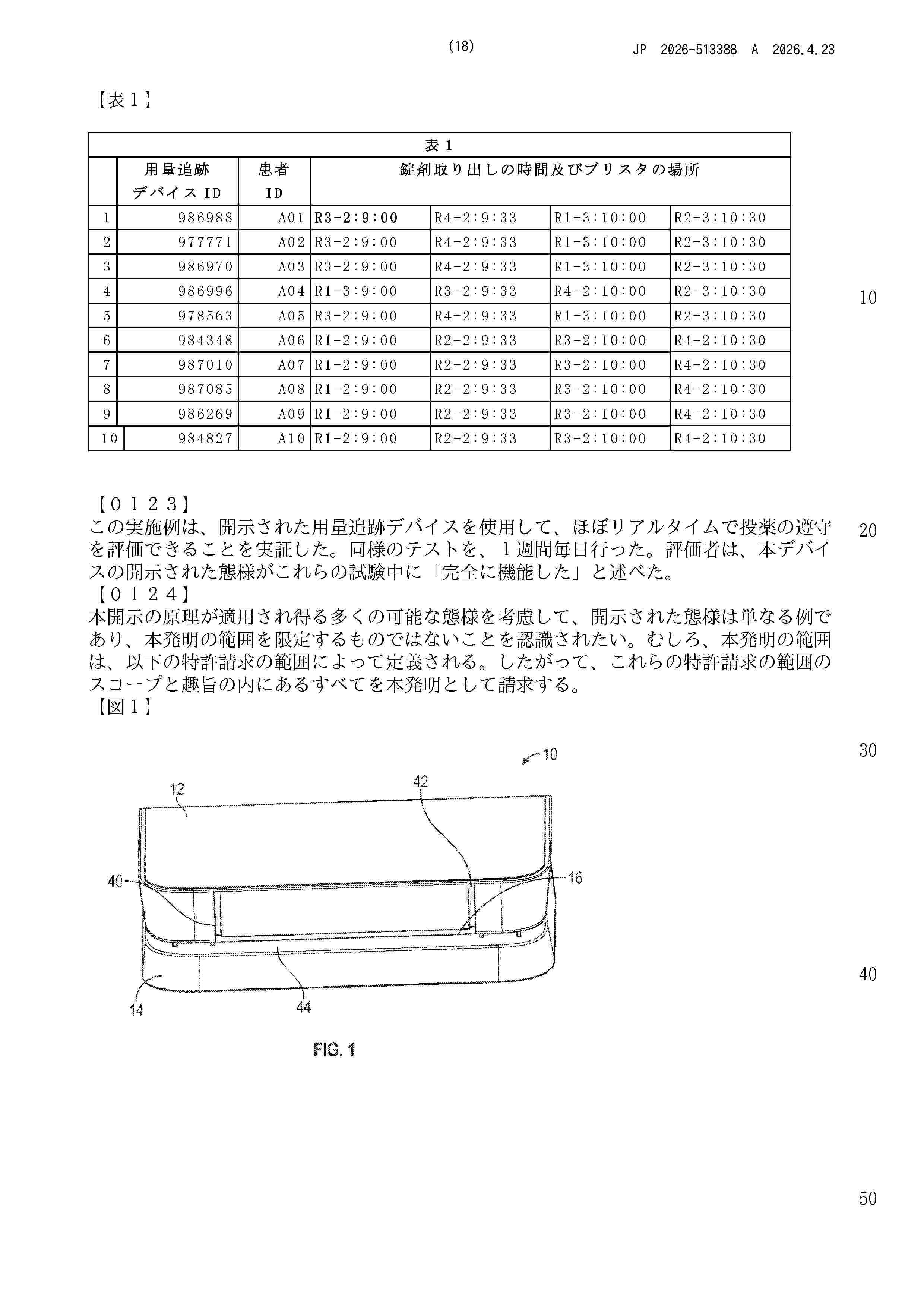
Aspects of a dose tracking device configured for use with commercial packages or "blister cards" of medicaments are disclosed. Disclosed tracking devices track the removal of medicament doses from the "blister cards" with which they are associated. Certain disclosed aspects concern an electronic tracking module comprising plural voltage sensors electrically coupled to a circuit trace that is applied to a medicament blister card obtained commercially or provided by a health care provider. The circuit trace comprises plural circuits that align with medicament blisters containing a medicament dose. When a dose is dispensed from a blister, a corresponding circuit of the circuit trace is broken, indicating that the dose has been dispensed. Patient dosing information can be conveyed to a remote server for monitoring and data storage.
Resumen de: WO2024201253A1
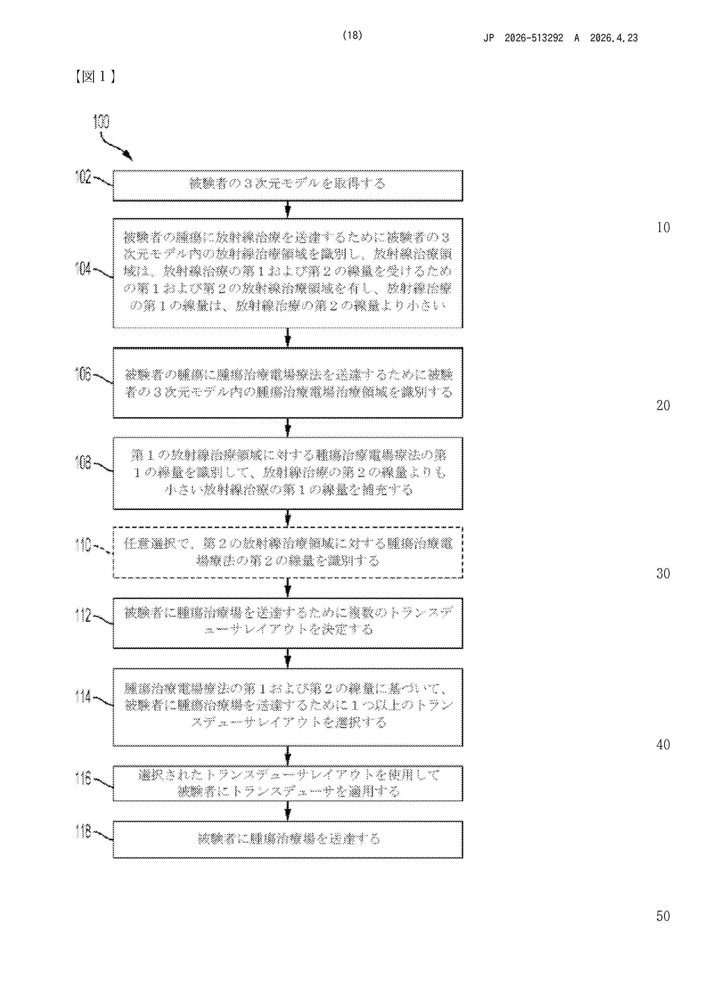
A computer-implemented method comprising: obtaining a three-dimensional model of a subject; identifying a radiation therapy region in the model for delivering radiation therapy to a tumor of the subject, the radiation therapy region comprising a first and second radiation therapy regions to respectively receive a first and second dosage of radiation therapy; identifying a tumor treating fields therapy region in the model for delivering tumor treating fields therapy to the tumor, the tumor treating fields therapy region comprising the first radiation therapy region; identifying a first dosage for the tumor treating fields therapy for the first radiation therapy region to compensate for the first dosage of radiation therapy being smaller than the second dosage of radiation therapy; and selecting one or more transducer layouts for delivering tumor treating fields to the subject based on the first dosage for the tumor treating fields therapy for the first radiation therapy region.
Resumen de: CN121311220A
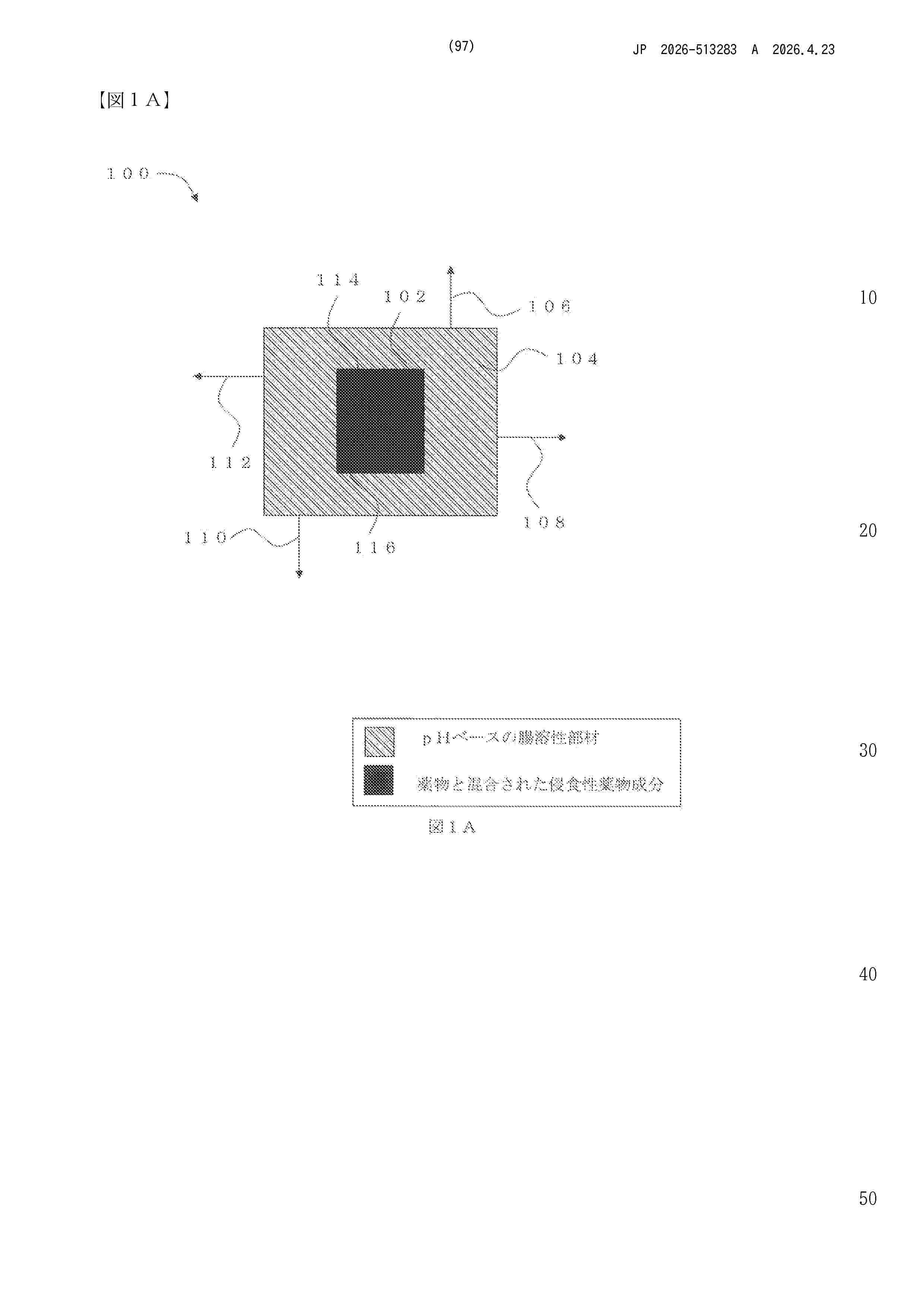
In some aspects, the present disclosure relates to oral pharmaceutical dosage forms configured to release a drug at a desired location in the colon of an individual. As provided in certain aspects, the oral pharmaceutical dosage forms include a pharmaceutical component containing an erodible material mixed with the drug and a delay component not mixed with the drug, where the delay component is configured to prevent release of the drug from the pharmaceutical component until the oral pharmaceutical dosage form reaches the colon of the individual after administration. In other aspects, the present disclosure relates to methods of design, methods of preparation, such as the use of three-dimensional printing, and methods of treatment related to the oral pharmaceutical dosage forms described herein.
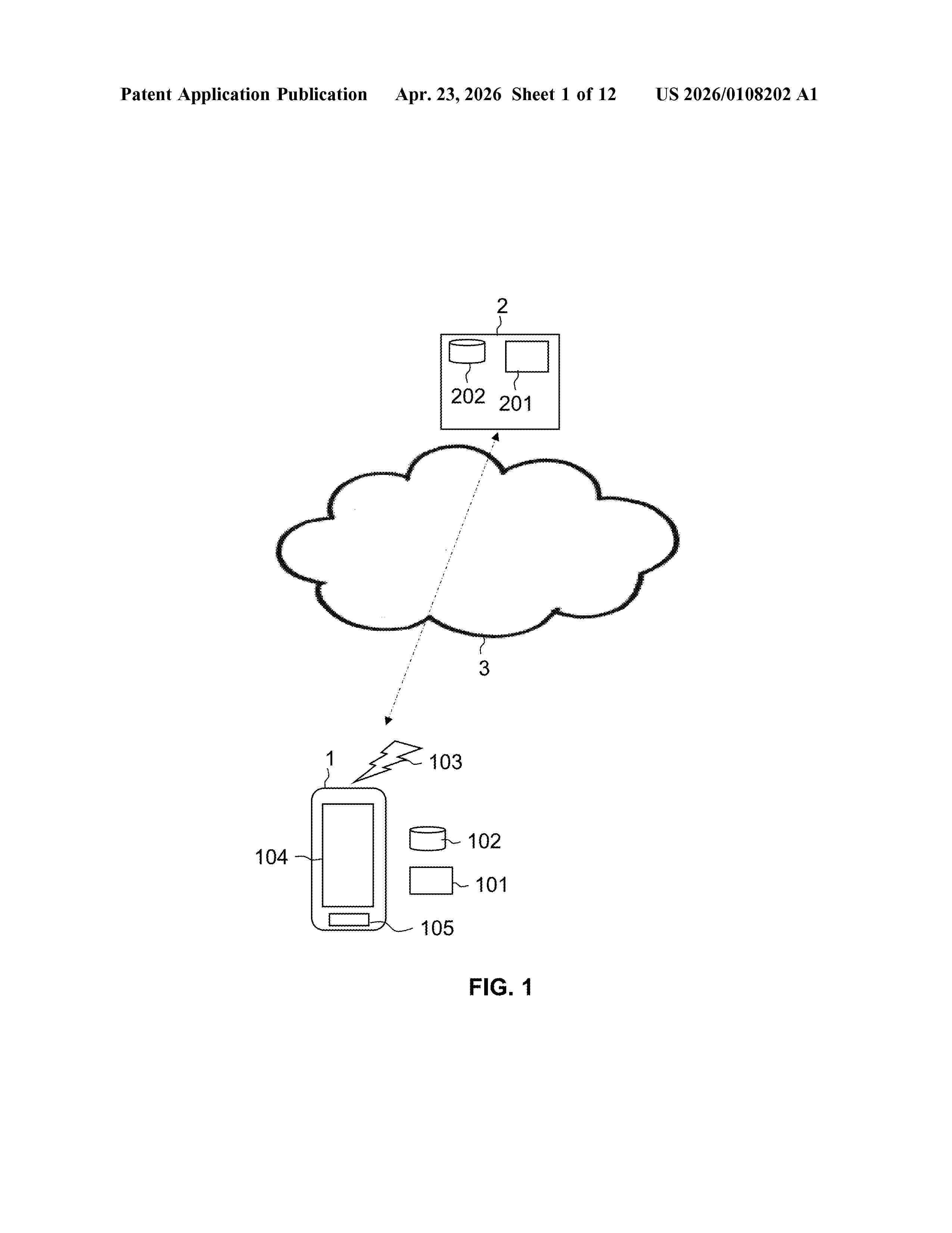
Resumen de: US20260108202A1
The application relates to assessing cognitive impairment and/or speech motor impairment. The method comprises analysing a voice recording from a word-reading test by identifying a plurality of segments of the voice recording that correspond to single words or syllables and determining the number of correctly read words in the voice recording and/or the speech rate. Determining the correct number of words in the recording may comprise computing one or more Mel-frequency cepstral coefficients for the segments, clustering the resulting vectors of values into n clusters, wherein each cluster has n possible labels, predicting a sequence of words in the voice recording using the labels associated with the clustered vectors of values, performing a sequence alignment between the predicted sequence of words and the sequence of words used in the word reading test, selecting the labels that result in the best alignment and counting the number of matches in the alignment.
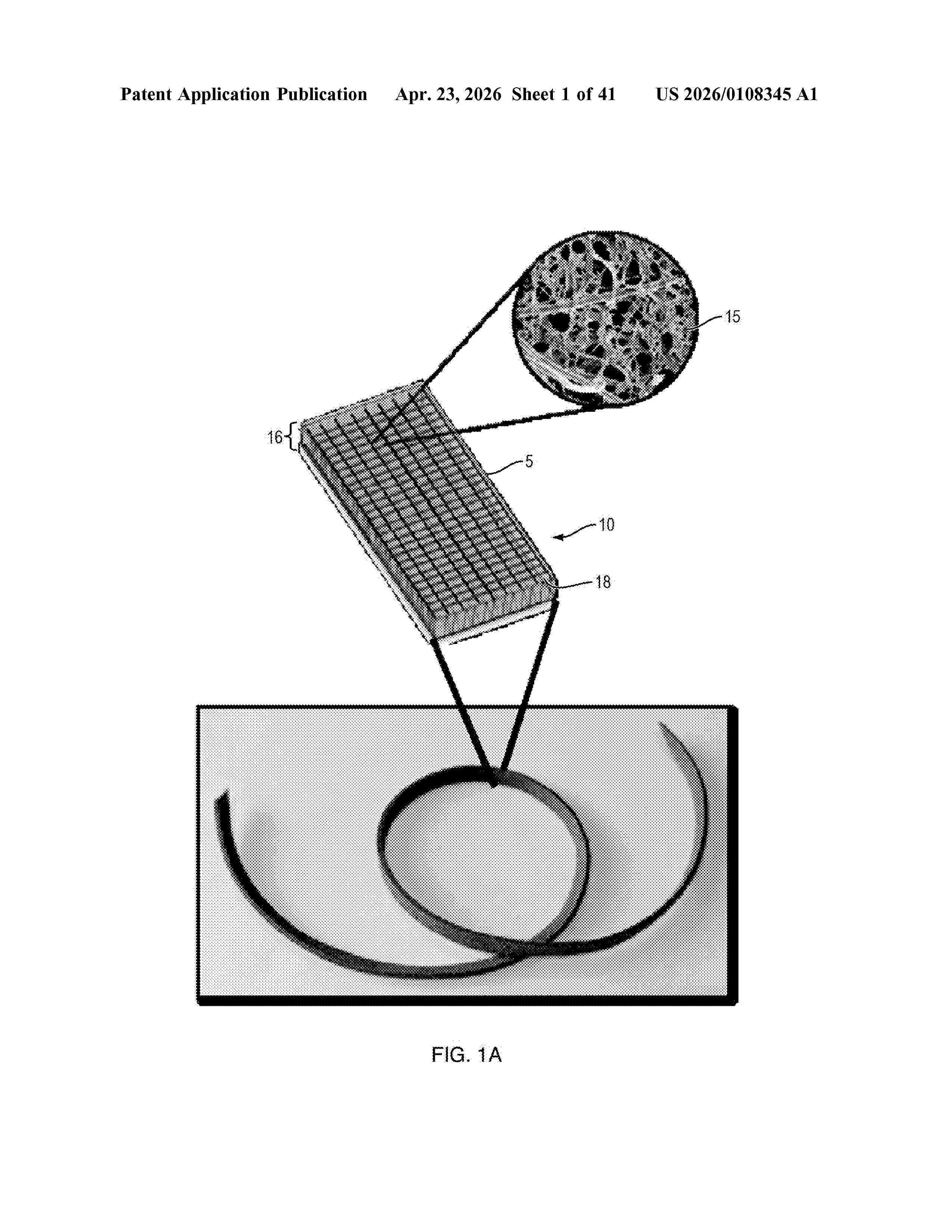
Resumen de: US20260108345A1
The disclosed composite scaffold provides a highly porous and flexible structure that substantially maintains its three-dimensional shape under tension and provides mechanical reinforcement of the repair or reconstruction-first via scaffold mechanical properties, and subsequently, through newly regenerated functional tissue as the scaffold is resorbed.
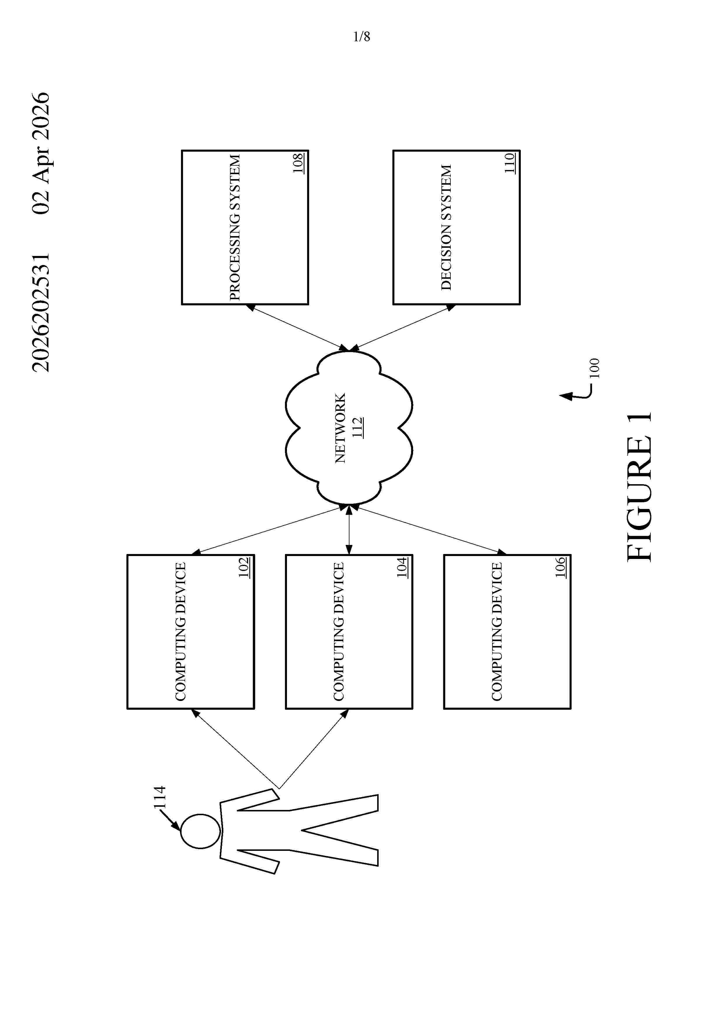
Resumen de: AU2026202531A1
Attorney Docket No. 04576.0100WOU1 / BSH.0124 Examples of the present disclosure describe systems and methods for automating clinical workflow decisions. In aspects, patient data may be collected from multiple data sources, such as patient records, imaging data, etc. The patient data may be processed using an artificial intelligence (AI) component. The output of the AI component may be used by healthcare professionals to inform healthcare decisions for patients. The output of the AI component and additional information relating to the healthcare decisions and healthcare paths may be provided as input to a decision analysis component. The decision analysis component may process the input and output an automated healthcare recommendation that may be used to further inform the healthcare decisions of the healthcare professionals. In some aspects, the output of the decision analysis component may be used to determine a priority or timeline for performing one or more actions relating to patient healthcare. pr p r
Resumen de: US20260108329A1
A method for creating a digital 3D model of a patient's dentition, according to the invention, provides that in a computing unit for a plurality of dentition variants, each comprising a combination of a digital 3D model of the upper jaw and a digital 3D model of the lower jaw of the patient, which differ from each other in the relative positioning of the upper and lower jaws, respectively: the digital 3D model of the upper jaw is combined with the digital 3D model of the patient's lower jaw in such a way that the upper and lower jaws rest on each other at a number of contact points without any spatial overlap of parts of the upper and lower jaws, andthe contact surface between the upper and lower jaw is determined, whereby an optimized denture variant is selected as a 3D model of the denture, taking into account the contact surface.
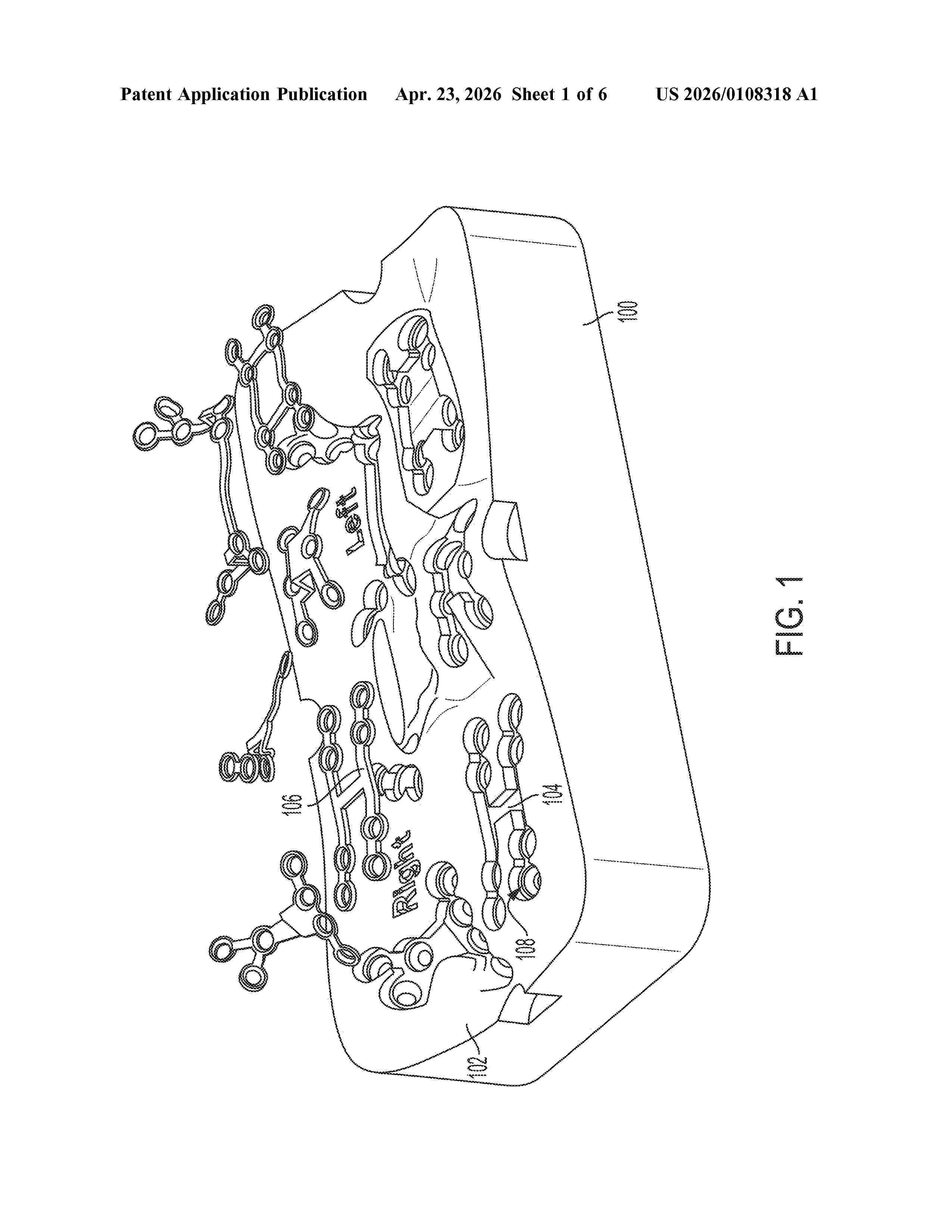
Resumen de: US20260108318A1
0000 In an aspect, a method of manufacturing a surgical kit includes providing one or more medical devices based on a three-dimensional (3D) image of an anatomical structure. The method additionally includes providing packaging based on the 3D image, and providing the surgical kit utilizing the packaging and the one or more medical devices. In another aspect, a surgical kit has one or more contoured packaging surface having a contour that matches a contour of an anatomical structure. The contoured packaging surface has one or more features for receiving one or more medical devices provided based on the contour in the 3D image. The surgical kit additionally has a lid connected under pressure with the one or more contoured packaging surface. The lid has apertures for sterilization of components of the one or more medical devices situated between the lid and the contoured packaging surface.
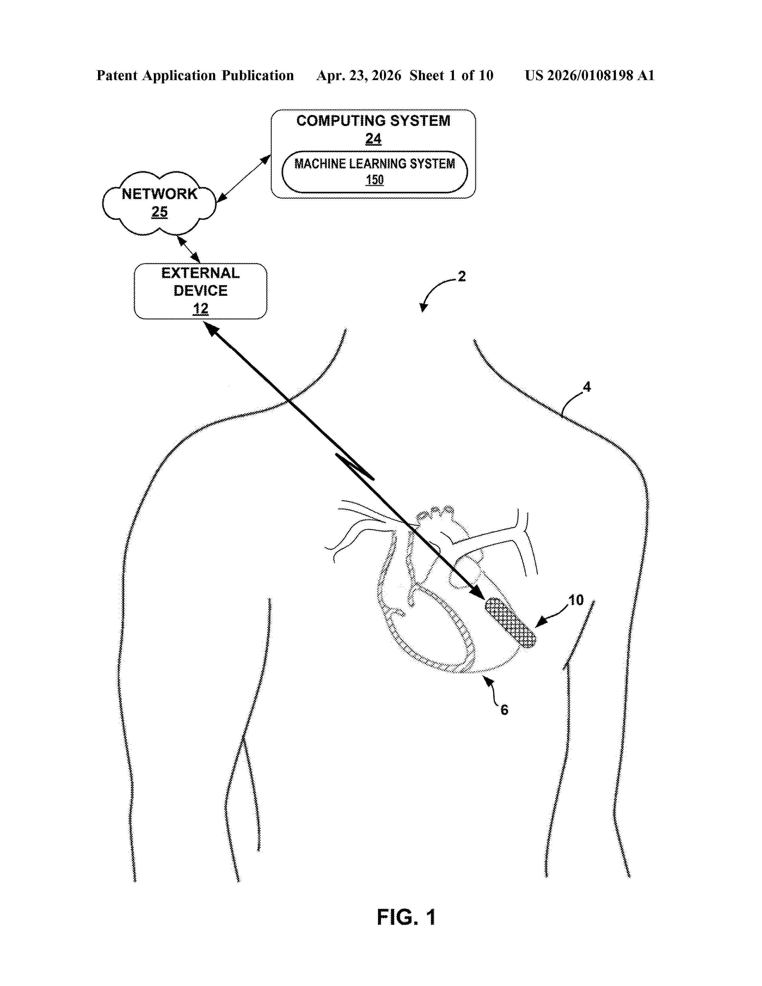
Resumen de: US20260108198A1
Techniques are disclosed for using both feature delineation and machine learning to detect cardiac arrhythmia. A computing device receives cardiac electrogram data of a patient sensed by a medical device. The computing device obtains, via feature-based delineation of the cardiac electrogram data, a first classification of arrhythmia in the patient. The computing device applies a machine learning model to the received cardiac electrogram data to obtain a second classification of arrhythmia in the patient. As one example, the computing device uses the first and second classifications to determine whether an episode of arrhythmia has occurred in the patient. As another example, the computing device uses the second classification to verify the first classification of arrhythmia in the patient. The computing device outputs a report indicating that the episode of arrhythmia has occurred and one or more cardiac features that coincide with the episode of arrhythmia.
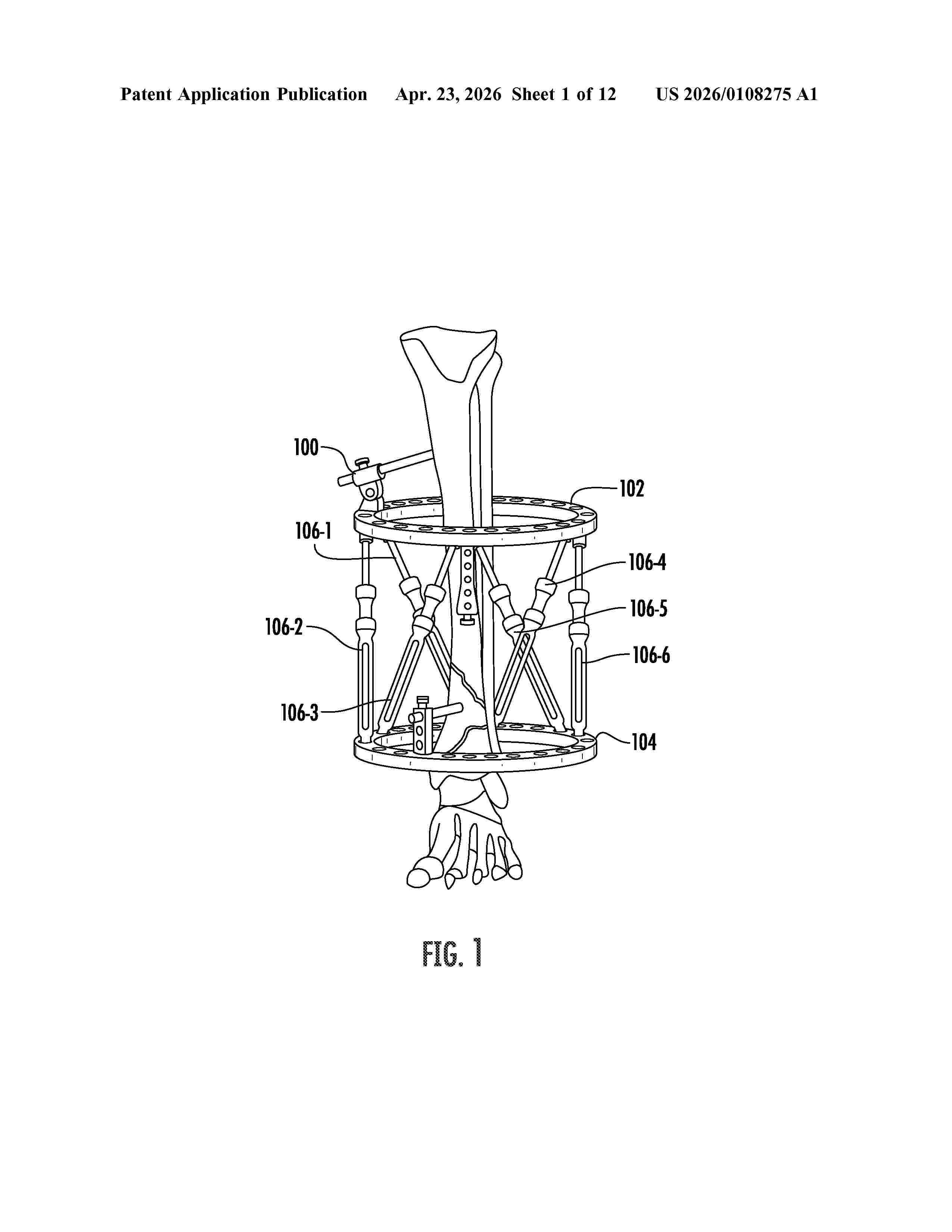
Resumen de: US20260108275A1
A spatial frame apparatus for positioning on a patient to treat an injury. The apparatus includes a frame having one or more motorized struts. One or more motorized struts include one or more motors configured to cause movement of one or more motorized struts in one or more directions in accordance with one or more treatment plans. The apparatus includes one or more processing 2024/102351 components communicatively coupled to one or more motorized struts and/or one or more motors. One or more processing components determine one or more loading parameters on the one or more motorized struts. The loading parameters define loading on the one or more motorized struts resulting from the frame being positioned and/or adjusted on the patient. The processing components execute at least one adjustment of one or more motorized struts based on the determined loading parameters.
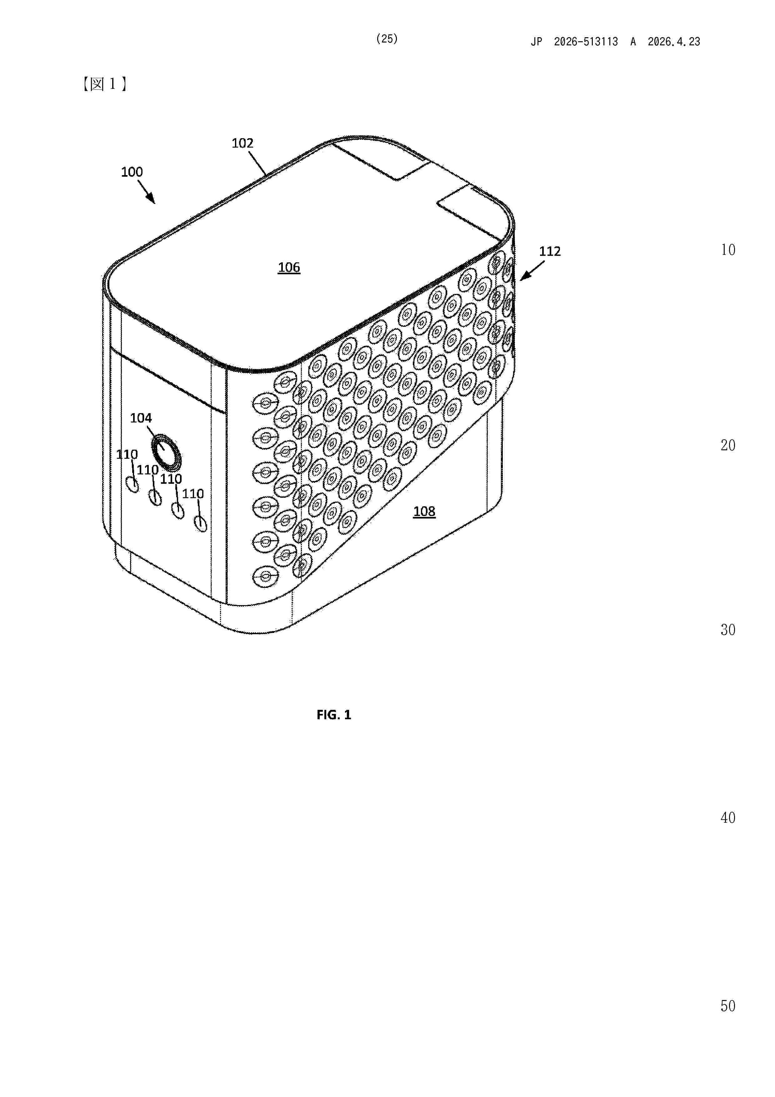
Nº publicación: JP2026513113A 23/04/2026
Solicitante:
ハークメディカルリサーチプロプライアタリーリミティド
Resumen de: WO2024086896A1
An apparatus including: a housing for containing/protecting/storing a plurality of containers/packages containing pills/medicaments; a power source in the housing; an actuator arrangement powered by the power source and configured to administer/deliver the pills/medicaments for a pre-selected patient; and a sensor powered by the power source to activate the actuator arrangement to allow the at least one package to be opened/accessed manually when the sensor detects a pre-defined identifier of the pre-selected patient.
 BOPI
BOPI
 Sede Electrónica