Si deseas distinguir tus productos, servicios o ambos de los de otra empresa, es posible que necesites una marca o nombre comercial. Descubre qué son, en qué consiste su procedimiento de registro y qué implica.

Información sobre los plazos de presentación de solicitudes de transformación de marcas de la Unión Europea en marca nacional española. Más información


Si tienes un nuevo dispositivo, producto o procedimiento que resuelva un problema técnico o tenga una ventaja práctica, existen distintas formas de protegerlo en España y en otros países. Descubre cómo hacerlo.

¿Tu innovación reside en la estética, la ornamentación o la apariencia de tu producto? Protégela mediante un diseño industrial. Descubre qué derechos confiere el registro y cómo realizar la tramitación.

Las indicaciones geográficas protegen el nombre de un producto originario de una zona geográfica, a la cual le debe una determinada calidad, reputación u otra característica. Descubre qué son, en qué consiste su procedimiento de registro y qué beneficios conceden.

Las patentes publicadas en todo el mundo son una valiosa fuente de información científica, técnica y comercial.

Si eres emprendedor/a o una empresa y quieres potenciar y mejorar la rentabilidad de tu negocio protegiendo de forma adecuada los activos intangibles de tu organización, en este espacio encontrarás lo necesario.


 444
resultados
444
resultados
 Última actualización
29/07/2026 [07:08:00]
Última actualización
29/07/2026 [07:08:00]


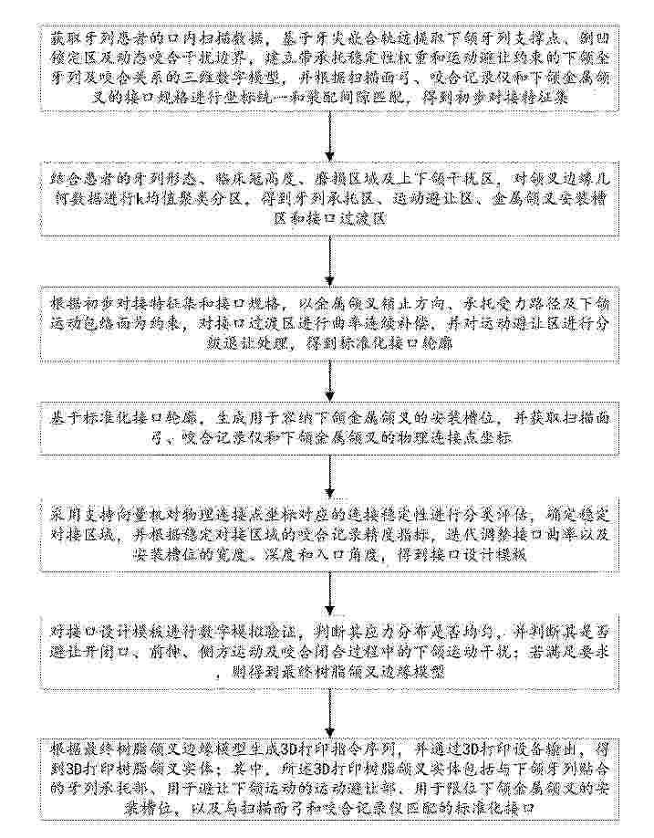
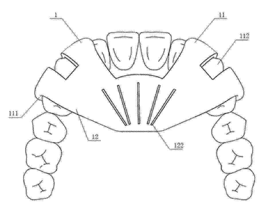
Resumen de: CN122442951A
本发明公开了用于复杂牙列患者的3D打印树脂颌叉固定方法及系统,涉及口腔数字化修复技术领域,包括获取牙列患者的口内扫描数据,基于牙尖嵌合轨迹提取下颌牙列支撑点、倒凹锁定区及动态咬合干扰边界,建立带承托稳定性权重和运动避让约束的下颌全牙列及咬合关系的三维数字模型,并根据扫描面弓、咬合记录仪和下颌金属颌叉的接口规格进行坐标统一和装配间隙匹配,得到初步对接特征集;该用于复杂牙列患者的3D打印树脂颌叉固定方法及系统,提高复杂牙列患者咬合记录和下颌运动分析的准确性、重复性及临床操作效率。

Resumen de: CN122440340A
本发明公开了一种CAD/CAM舌侧牙槽骨增量联合骨皮质切开导板,包括用于辅助上颌牙正畸的上颌导板和用于辅助下颌牙正畸的下颌导板;上颌导板包括上导板定位部和上导板体部,在上导板体部前侧一体化设置有上导板定位部,上导板定位部贴合扣紧在上颌牙上、使上导板体部贴合设置在上颌骨腭部;下颌导板包括下导板体部、下导板定位部和导板舌挡部,在下导板体部前侧一体化设置有下导板定位部、后侧一体化设置有导板舌挡部。制作方法包括:步骤一、获取数字化三维模型;步骤二、数字化导板设计。该发明保证了手术中植骨范围的准确性和植骨量的充分性,填补了舌侧术式的空白,推动了舌侧骨增量联合骨皮质切开术的广泛推广与规范化应用。
Resumen de: WO2025102118A1
The present disclosure relates to implants for teeth and bone. The implants are prepared from a polymer of the poly (aryl ether ketone) family, preferably poly ether ketone (PEK), and are activated with plasma to improve integration with teeth and bone. Methods and kits related to use of the implants are also disclosed.
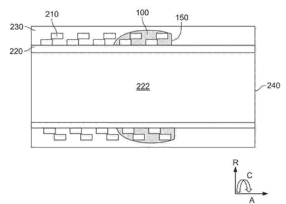
Resumen de: CN122440333A
本文中讨论了一种医疗设备,其包括用于标记所述医疗设备的标记物位置的标记物(100),所述医疗设备包括:所述医疗设备的管;不透射线的标记材料(100),其用于标记所述医疗设备的标记物位置;并且其中所述不透射线的标记材料(100)通过增材制造在流体中从所述医疗设备的标记物位置处添加到所述管上,从而形成固体不透射线的标记物(100),以用于增加所述医疗设备在径向方向(R)上的X射线不透性。特别地,管可以包括编织和/或盘绕的丝网(210)的纤维,并且该方法包括用标记材料固定编织和/或盘绕的丝网(210)的纤维的步骤。
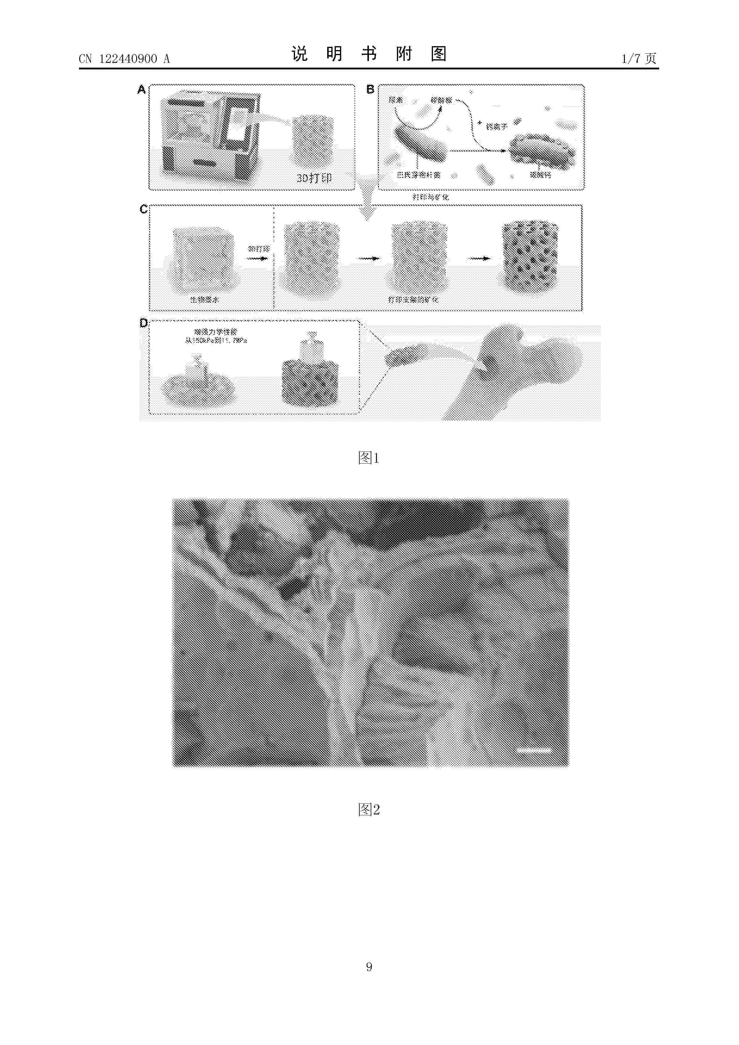
Resumen de: CN122440900A
本发明提供了一种高强度的细菌矿化仿生骨支架及其制备方法,该制备方法通过在水凝胶前体溶液中加入矿化细菌,利用3D打印制备具有精细宏观结构的水凝胶支架,后将该支架浸泡于含有尿素和矿物盐的矿化细菌培养液中,使细菌进行原位矿化,在微观尺度上形成由微米级矿化颗粒组成的三维网络,实现对天然骨组织的精确仿生。该制备方法具有能耗低、适用于工业化生产和低经济成本的优点。本发明的方法不仅显著提升了支架的力学性能,还优化了骨修复过程中所需的生理微环境,为骨组织再生提供了更加理想的生理基础。制得的仿生骨支架表现出优异的力学性能,压缩模量高达12.05MPa,并具有优异的良好的生物相容性和成骨活性。
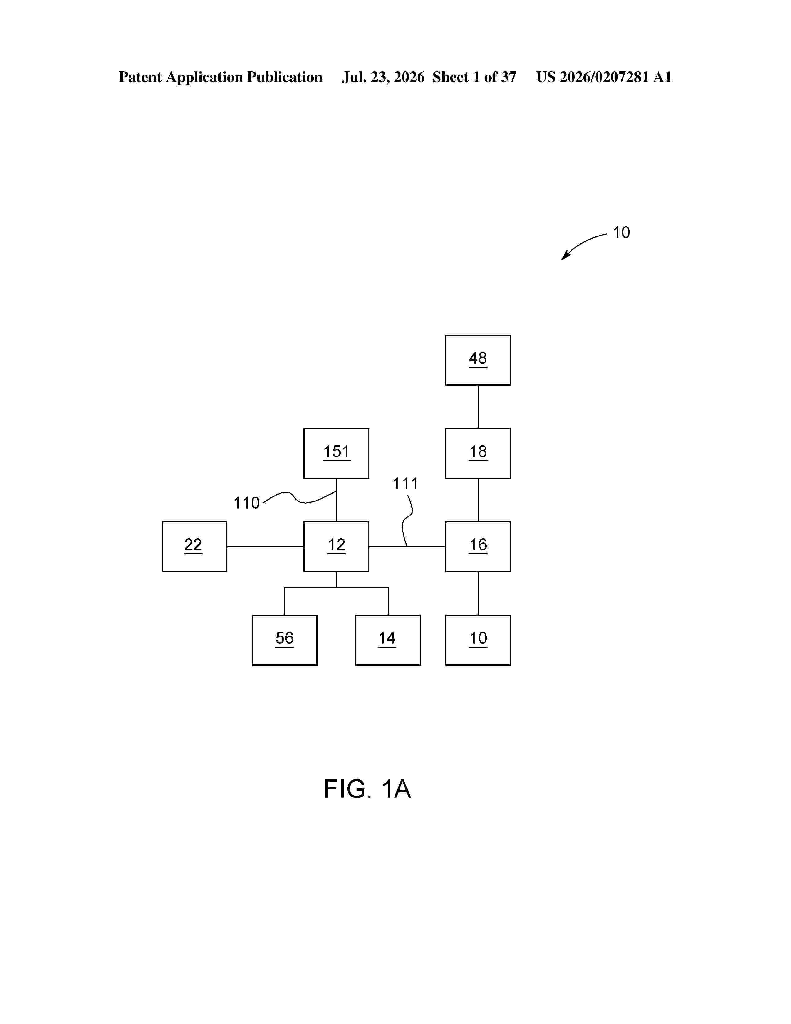
Resumen de: US20260207281A1
A robotic surgical system is disclosed that integrates a simulation module, artificial intelligence (AI), and a multimodal sensor array to enhance intraoperative decision-making and execution. The system generates a dynamic, patient-specific digital twin representing anatomical, physiological, and thermal data, continuously updated during surgery. Real-time sensor inputs, including force feedback, imaging, and biomarkers, are analyzed by an AI module to characterize tissue conditions, predict responses, and guide tool trajectories. The system adapts tool movement based on AI confidence scoring, margin proximity, and predictive modeling. A visualization interface overlays augmented insights onto live surgical views. The platform enables surgeon-specific and population-based learning, real-time feedback, and intraoperative handoff based on biometric or performance indicators. Embedded modules provide simulation, planning, deviation tracking, and control adjustment. This system supports autonomous, semi-autonomous, or surgeon-guided operation, promoting improved precision, safety, and outcomes across diverse surgical specialties.
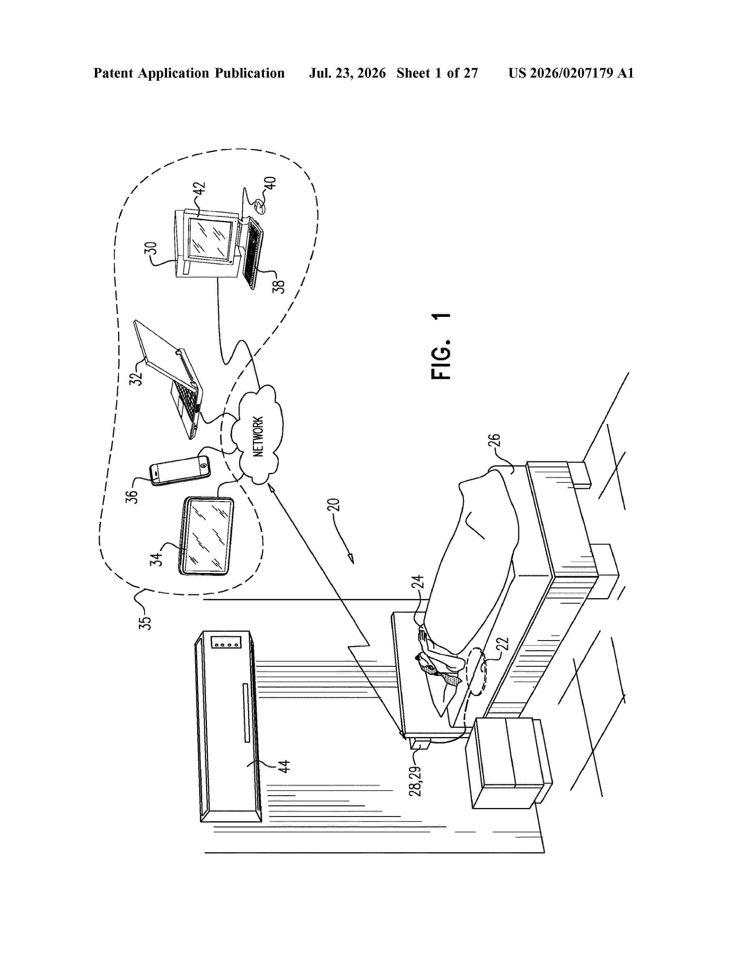
Resumen de: US20260207179A1
Apparatus and methods are described including using a sensor to monitor a sleeping subject and generate a signal in response to the monitoring. A noise control device is controlled in response to the signal.
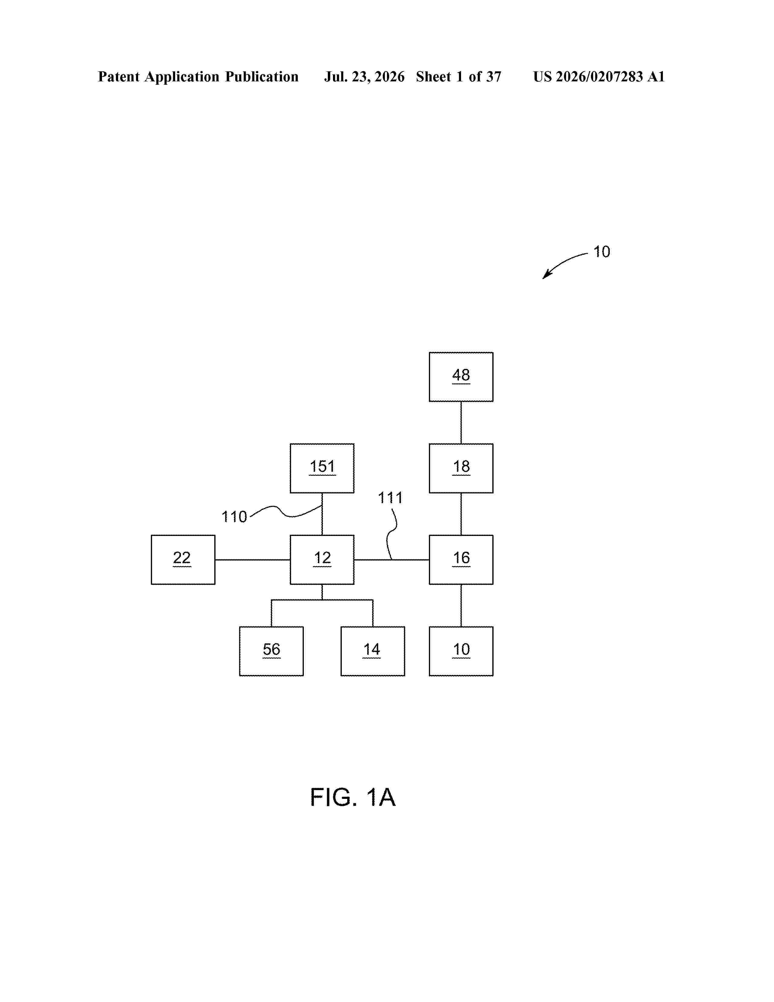
Resumen de: US20260207283A1
0000 A robotic surgical system is disclosed that integrates real-time surgeon behavior monitoring, biometric feedback, and adaptive AI-based decision support to enhance intraoperative performance and post-procedural analysis. The system includes a user behavior monitoring module to track surgeon interaction patterns, a biometric data aggregator collecting inputs such as heart rate variability, electrodermal activity, and eye tracking, and a feedback modulation engine operatively coupled to an AI inference engine. A phase classifier evaluates surgical criticality based on tool telemetry and contextual signals, enabling the system to dynamically escalate, suppress, or defer feedback. Surgeon-specific profiles guide system behavior across procedures, while a summary dashboard provides insights into feedback efficacy and surgeon responsiveness. The AI system continuously refines surgical guidance based on multimodal sensor data, surgeon preferences, and procedural outcomes, supporting personalized intraoperative adaptation and real-time risk mitigation. The invention enhances surgical safety, precision, and outcomes by leveraging intelligent automation and contextual surgeon state modeling.
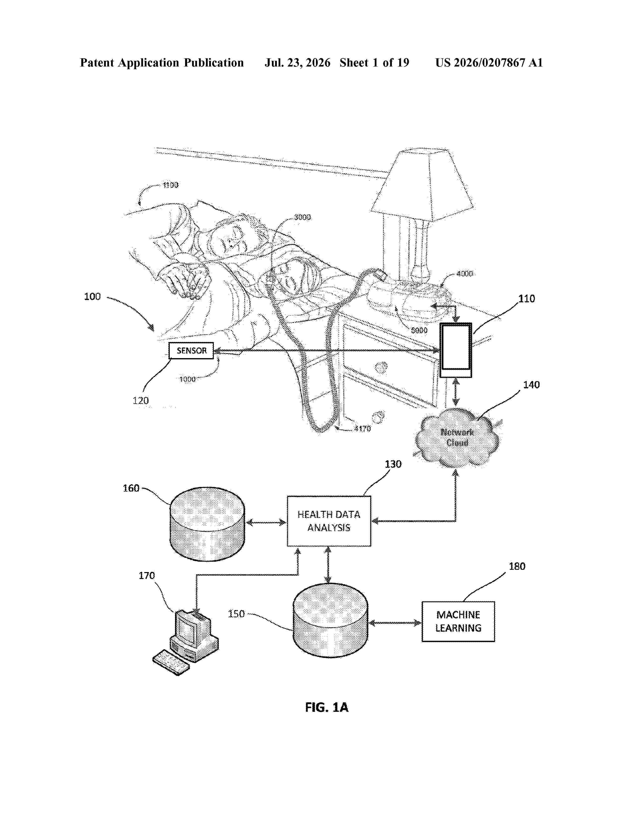
Resumen de: US20260207867A1
A system and method for applying treatment and efficiently collecting monitoring data from a patient is disclosed. The system includes a respiratory therapy device having a transmitter and an air control device to provide respiratory therapy to a patient. The respiratory therapy device sends either low or high resolution data to the health analysis engine. The high resolution data is transmitted based on the occurrence of an event detected based on the low resolution data. The respiratory therapy device collects operational data and transmits the collected operational data. A health analysis engine is in communication with the respiratory therapy device. The health analysis engine receives the collected data to determine a health condition of the patient based on the collected data.
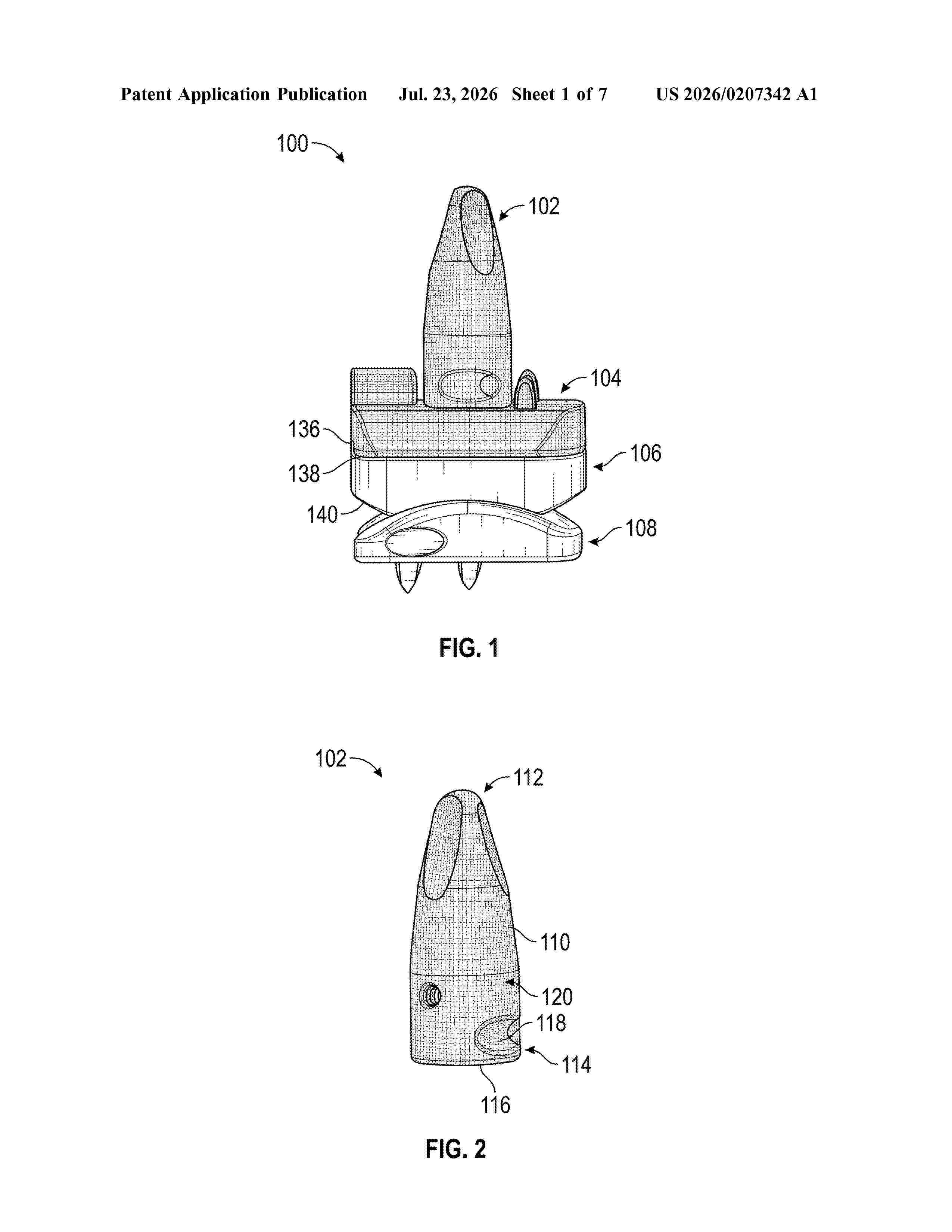
Resumen de: US20260207342A1
A gradient porous structure for one or more bone contacting surfaces of a total ankle replacement implant is provided. The gradient porosity of the porous structure may be optimized to match the natural variation of a patient's bone density, while the pattern of the porous structure may be optimized to maximize mechanical strength so that the implant is structurally stable within the ankle joint.
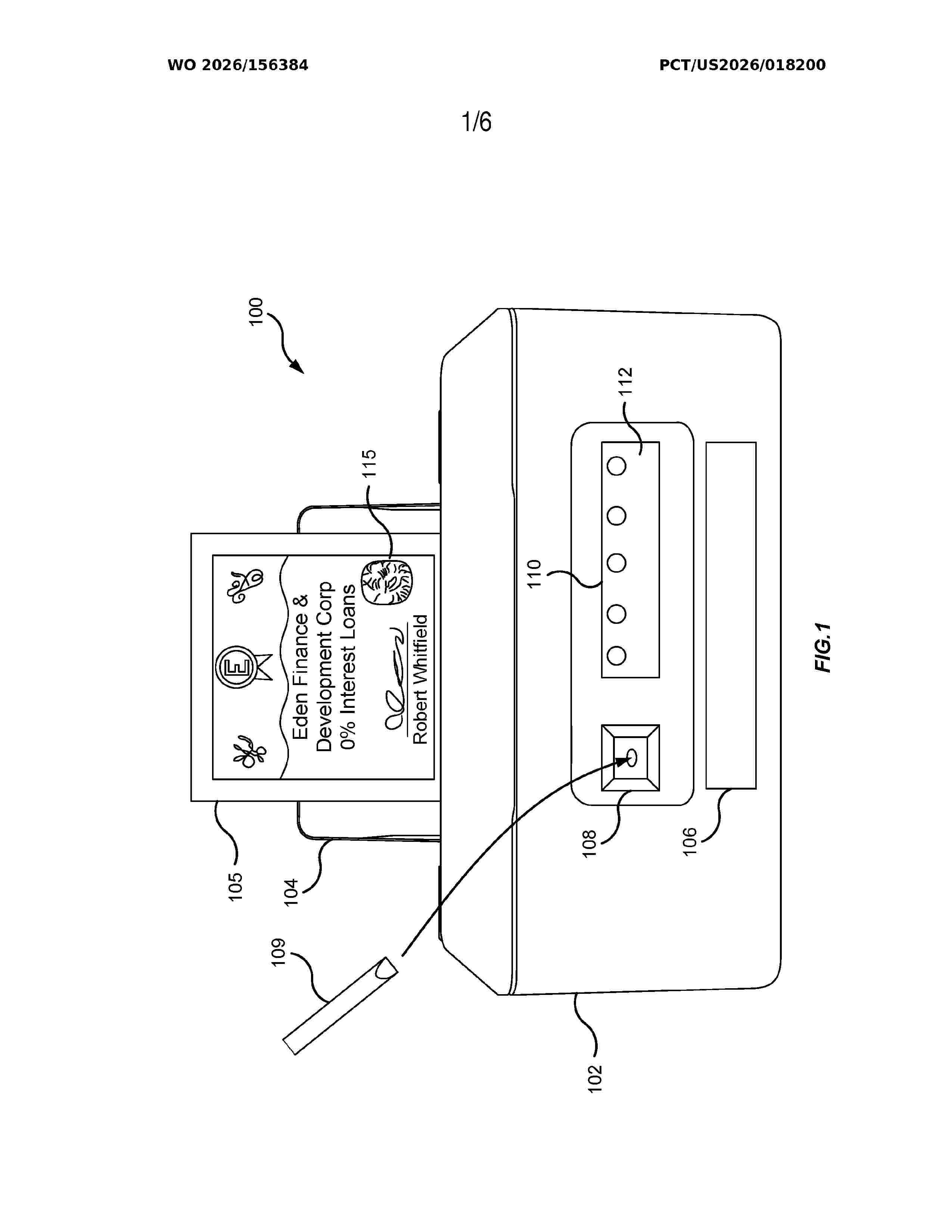
Resumen de: WO2026156384A1
Systems, apparatuses, and methods provide for processing for DNA fingerprint scanner systems. Such a DNA fingerprint scanner includes a body fluid DNA sampler, a printed DNA sampler, and a DNA analyzer. The body fluid DNA sampler is to receive body fluid. The printed DNA sampler is to sample a printed DNA infused fingerprint certificate. The DNA analyzer is coupled to the body fluid DNA sampler and the printed DNA sampler. The DNA analyzer is to determine DNA sequencing of the body fluid and the printed DNA infused fingerprint certificate.
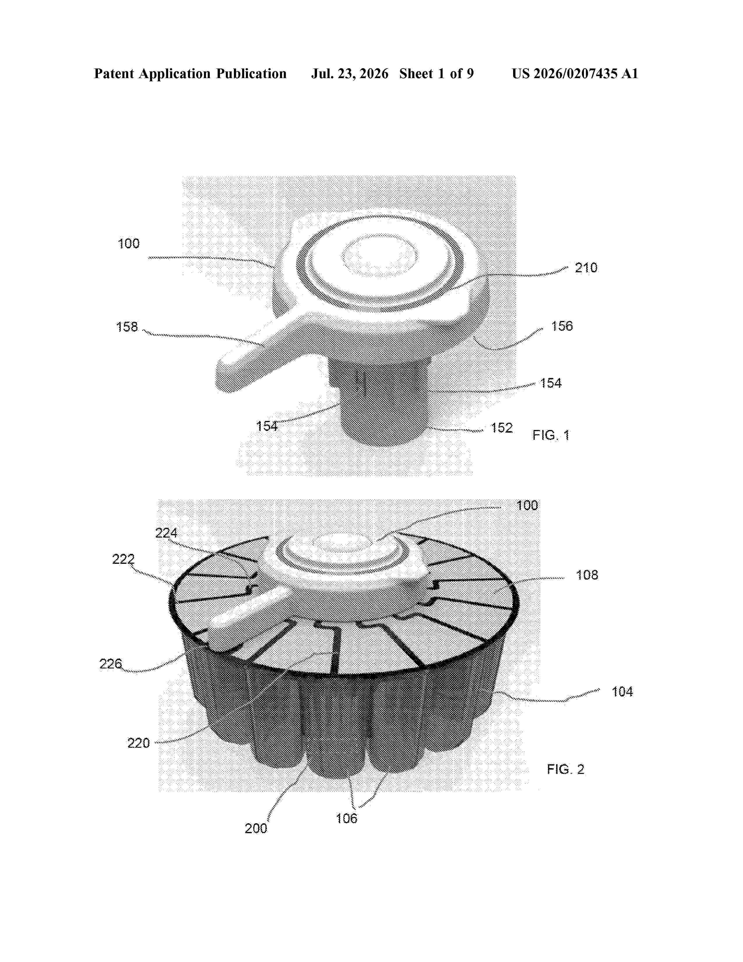
Resumen de: US20260207435A1
A system has a base member defining at least one chamber, and a cover cooperating with the base member to enclose the chamber. The cover includes at least one path associated with a respective chamber and positioned to be broken when the cover over the associated chamber is opened. A reader device has a housing sized to mate with a container enclosed by a cover, with the cover supporting a plurality of paths, with each path associated with and extending over each chamber. The reader device has first and second leads associated with a path of a respective chamber, and a controller configured to determine if a chamber is opened or unopened. A membrane for covering at least one chamber of a container is provided with a layer formed from an electrically non-conductive material, and a path formed from an electrically conductive material supported by the layer.
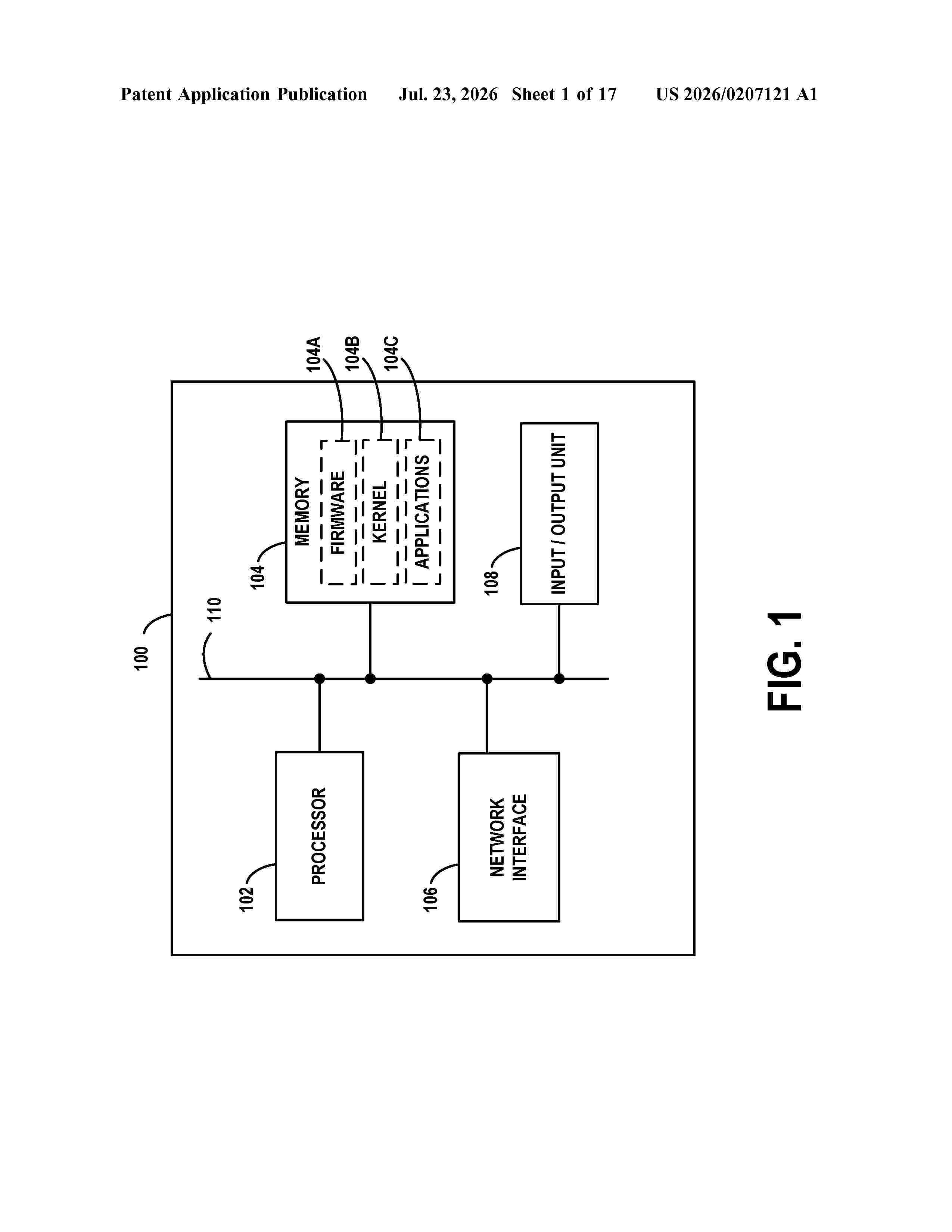
Resumen de: US20260207121A1
Example embodiments may include procedures for modeling segmentation and deformation of breast tissue, useable for surgical planning and possibly other procedures. These embodiments involve applying a tumor localization, tumor segmentation, and multi-tissue segmentation procedure to 3D image data of breast tissue, then combining the results of these procedures as well as anatomical feasibilities, in determining most likely tissue types for regions of the 3D image data. Additionally or alternatively, a finite element model can be used to represent the different tissue types in the 3D image data as elements with distinct physical characteristics. Such a model can be used to simulate gravity in a posterior direction to translate the elements from the prone position to a gravity-unloaded position and then from the gravity-unloaded position to a supine position. The translated elements can be interpolated into further 3D image data with identified locations of the tissue types.
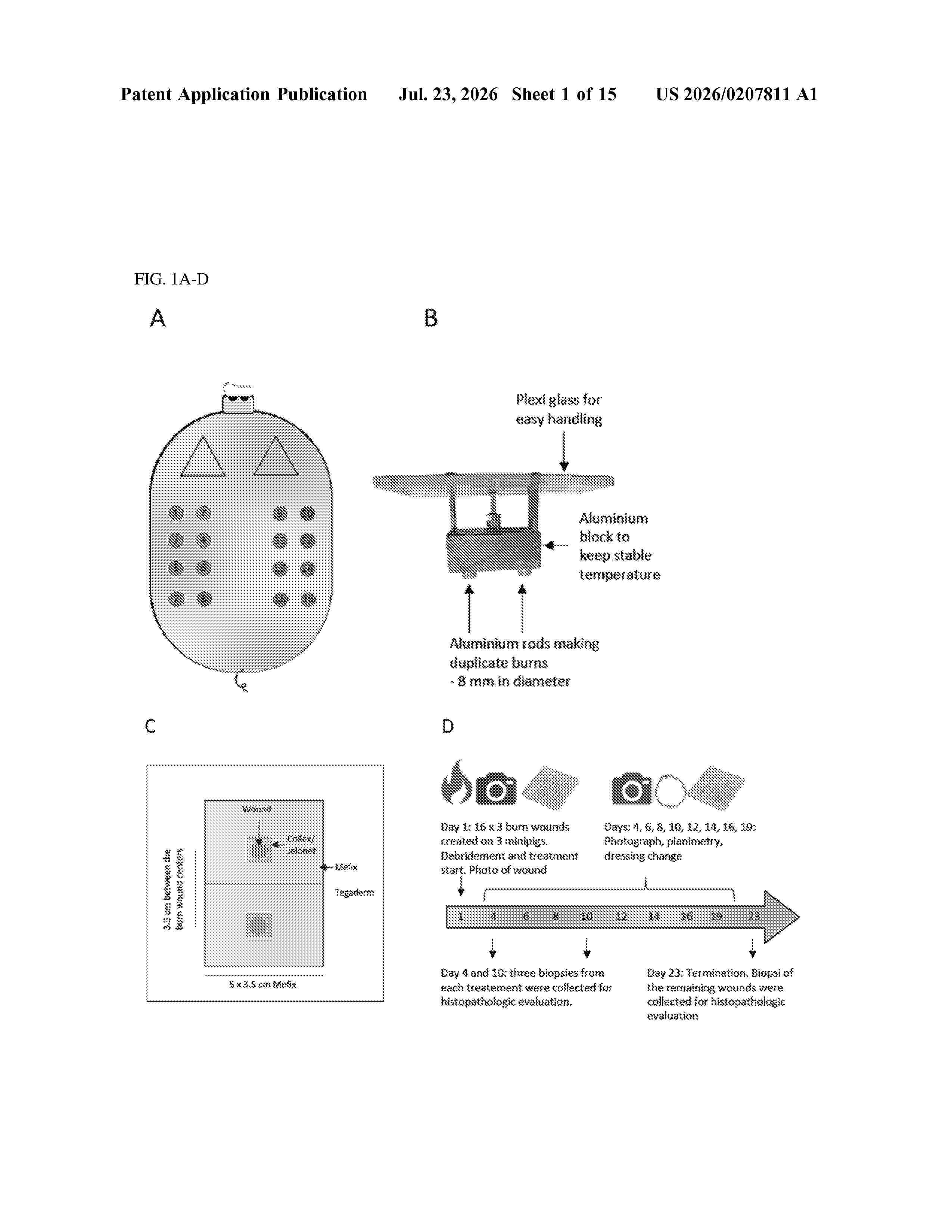
Resumen de: US20260207811A1
The present invention provides articles for the improved healing of wounds, preferably burn wounds. In some preferred embodiments, the article is a matrix formed from a mixture of two or more polysaccharides, the matrix comprising an active ingredient which is an extract from differentiable cells.
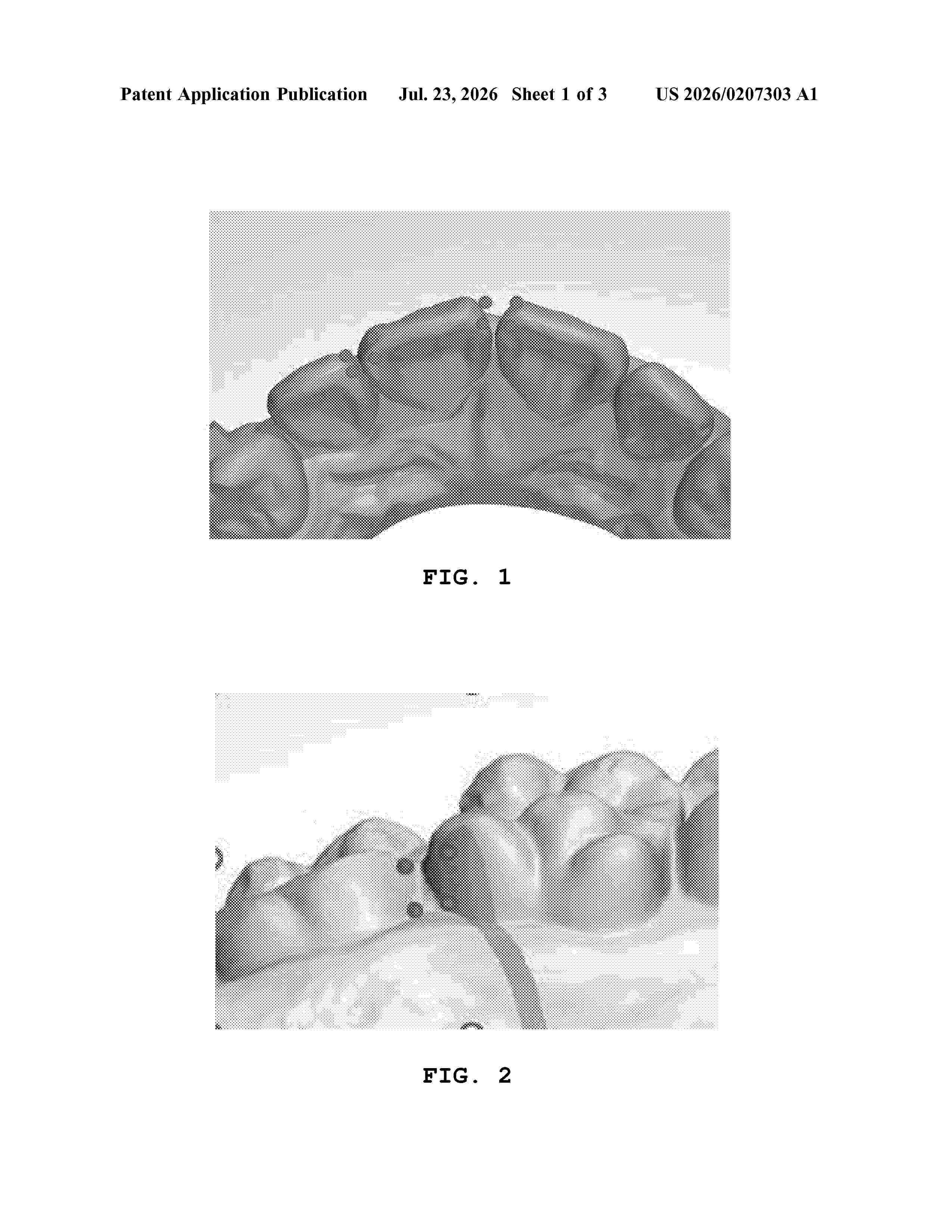
Resumen de: US20260207303A1
0000 A method for designing an orthodontic appliance using a computer comprises displaying a tooth arrangement image (S01), correcting an arrangement state of teeth in the tooth arrangement image (S02), correcting shapes of teeth in the tooth arrangement image (S03), displaying an orthodontic appliance on a screen according to the corrected tooth arrangement image (S04), and outputting the orthodontic appliance using a 3D printer (S05).
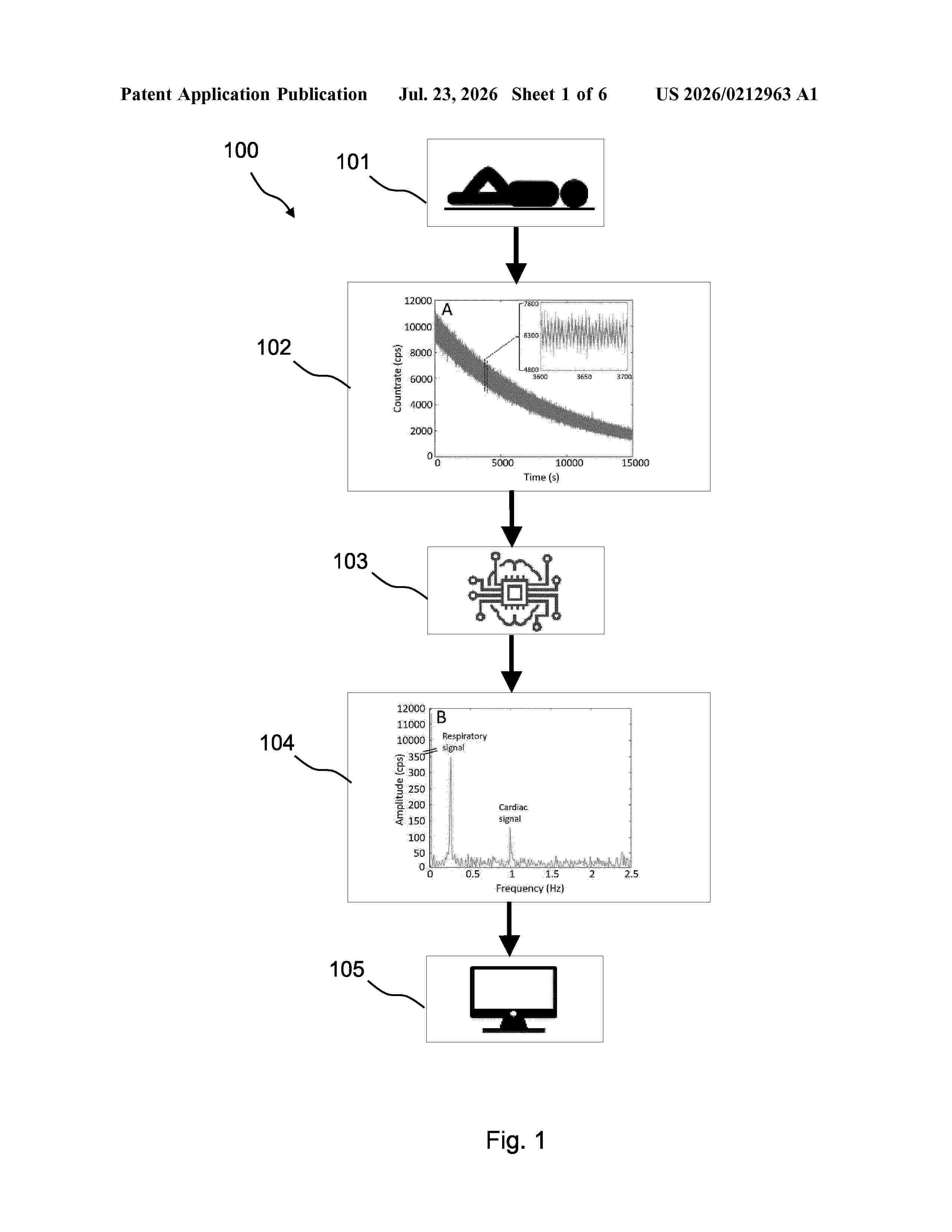
Resumen de: US20260212963A1
0000 The present disclosure relates to a technique to non-invasively and extra-corporeally measure the arterial concentration of a radiotracer administered to animal and/or human bodies or subjects with a radiation detector. In particular a computer implemented method for non-invasive and extra-corporally determining the arterial concentration of a radiopharmaceutical, for example a radiotracer, in an organ of an animal and/or human body is proposed, the computer implemented method comprising the steps of: i) receiving, from the moment of administration of a certain amount of the radiopharmaceutical in the animal and/or human body, by one or more radiation sensors positioned near or on a measurement location of an organ electromagnetic radiation emitted by the radio-pharmaceutical over time; ii) converting the received electromagnetic radiation in a time-sequence of radiation signals; iii) generating one or more time-frequency representations of the radiation signals by time-frequency transformation, and iv) analyzing the time-frequency representations for determining the arterial concentration of the radiopharmaceutical.
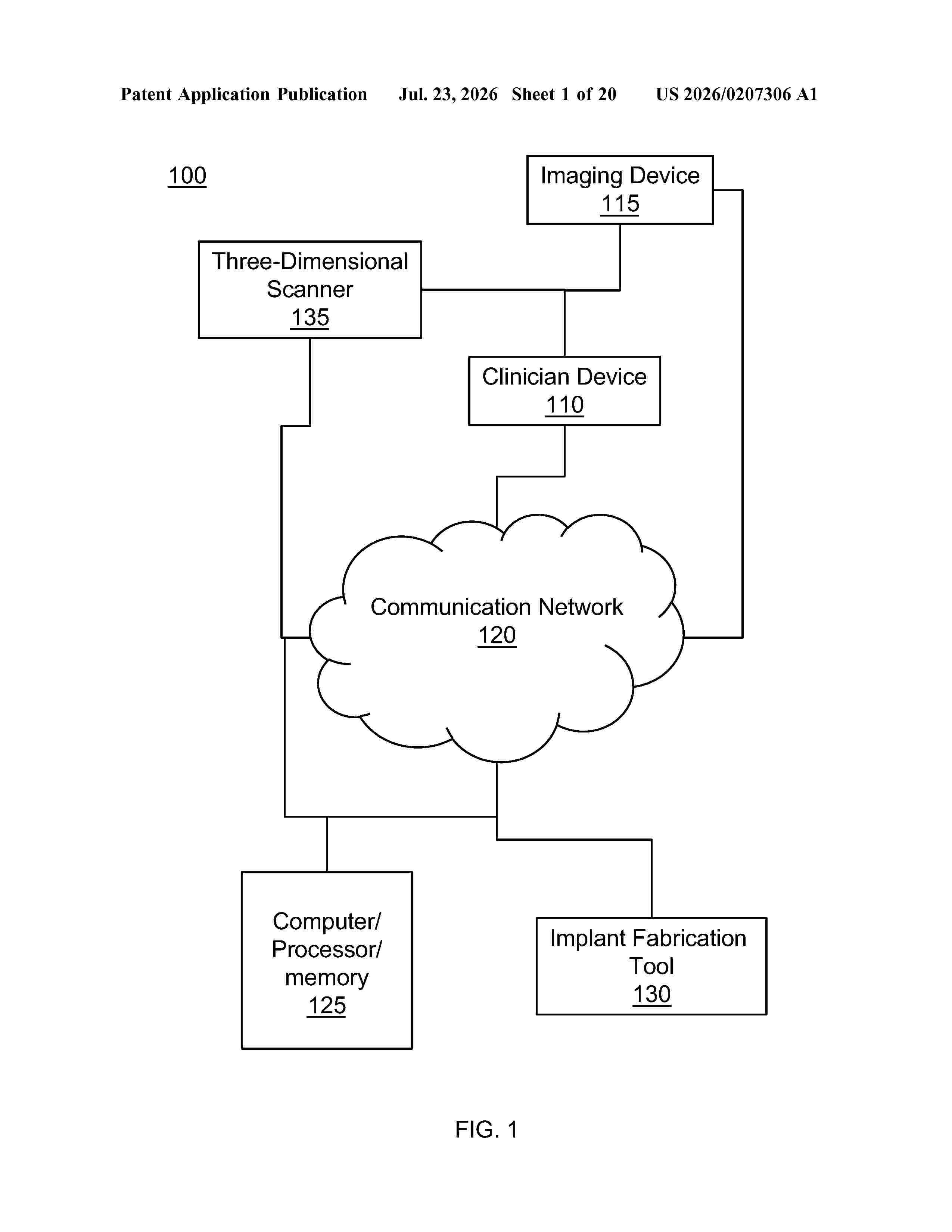
Resumen de: US20260207306A1
Root-analog dental implants include one or more coronal grooves that are sized, shaped, and/or configured to correspond to a size, shape, and/or position of a bone crest for an extracted tooth a root-analog dental implant is designed and manufactured to replace. The coronal grooves may be configured to engage with bone of an alveolar socket to enhance engagement between the root-analog dental implant and the alveolar socket particularly for the time period following seating the root-analog dental implant within the socket site and prior to osseointegration of the root-analog dental implant with the alveolar socket. This promotes stability of the root-analog dental implant within the socket site immediately following insertion and thereafter, which may decrease movement of the root-analog dental implant within the socket site, decrease a likelihood of foreign material such as bacteria or food entering the socket site, and/or reduce a risk of implant failure and jawbone injury.
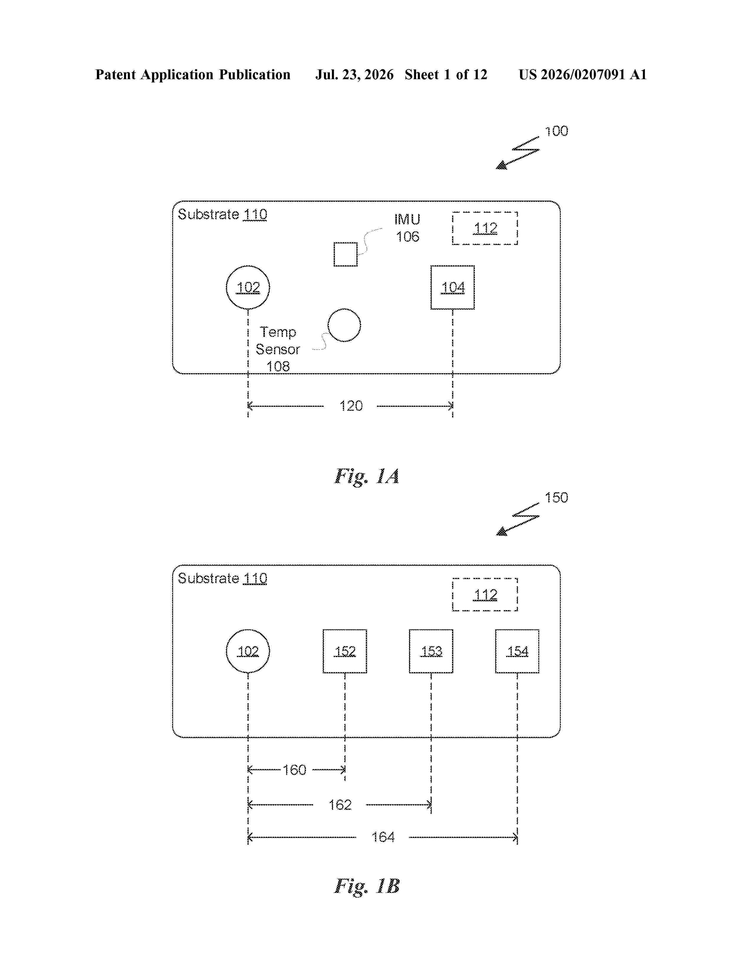
Resumen de: US20260207091A1
0000 A tissue oxygen saturation (StO<2>) parameter is calculated using a single light source and single detector of an optical device, where the light source and detector are separated by a separation distance. A single source-detector separation (SSDS) approach to measure StO<2 >in the tissue of a subject utilizes only intensity measurements of three different wavelengths of light emitted from the same light source (or one or more wavelengths from adjacent one or more light sources) at the detector. In addition to the three intensity measurements, the other inputs to the calculation are a differential pathlength factor (DPF) and extinction coefficients for both oxy-hemoglobin and deoxy-hemoglobin for the given wavelengths. The StO<2 >parameter can be calculated for each distinct source-detector pair of an optical device.
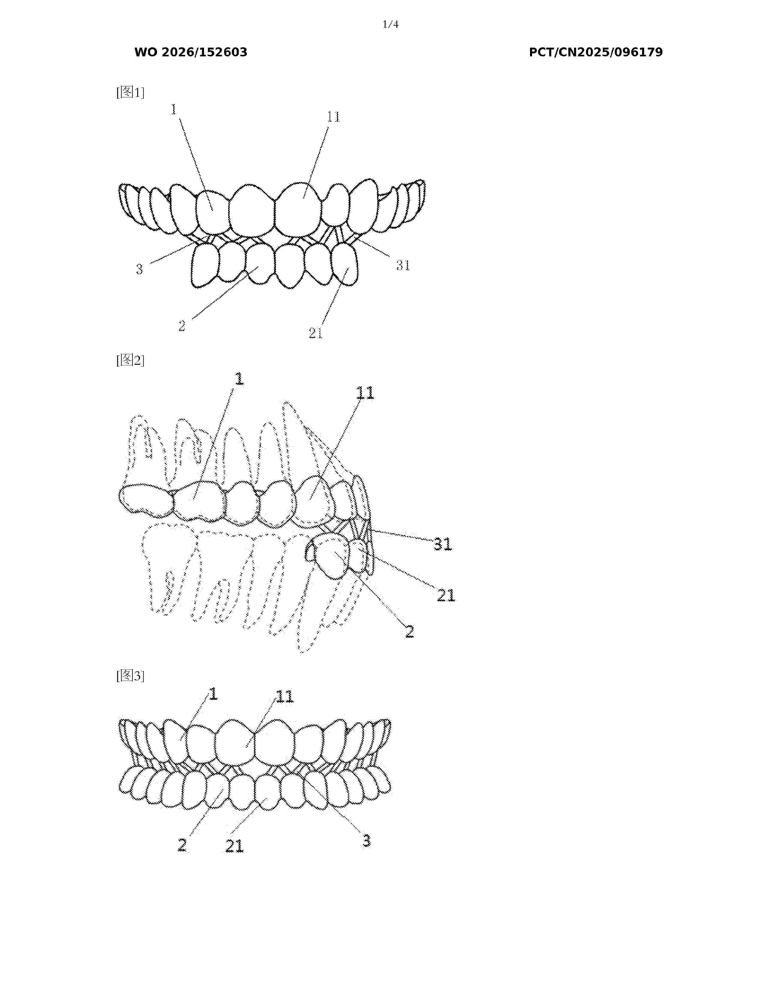
Resumen de: WO2026152603A1
An activator, comprising an upper jaw portion (1) and a lower jaw portion (2). The upper jaw portion (1) and the lower jaw portion (2) are connected by means of a connecting portion (3). The upper jaw portion (1), the lower jaw portion (2), and the connecting portion (3) are integrally manufactured. The upper jaw portion (1) at least covers maxillary anterior teeth, and the lower jaw portion (2) at least covers mandibular anterior teeth. The manufacturing method for the activator is a 3D printing method.
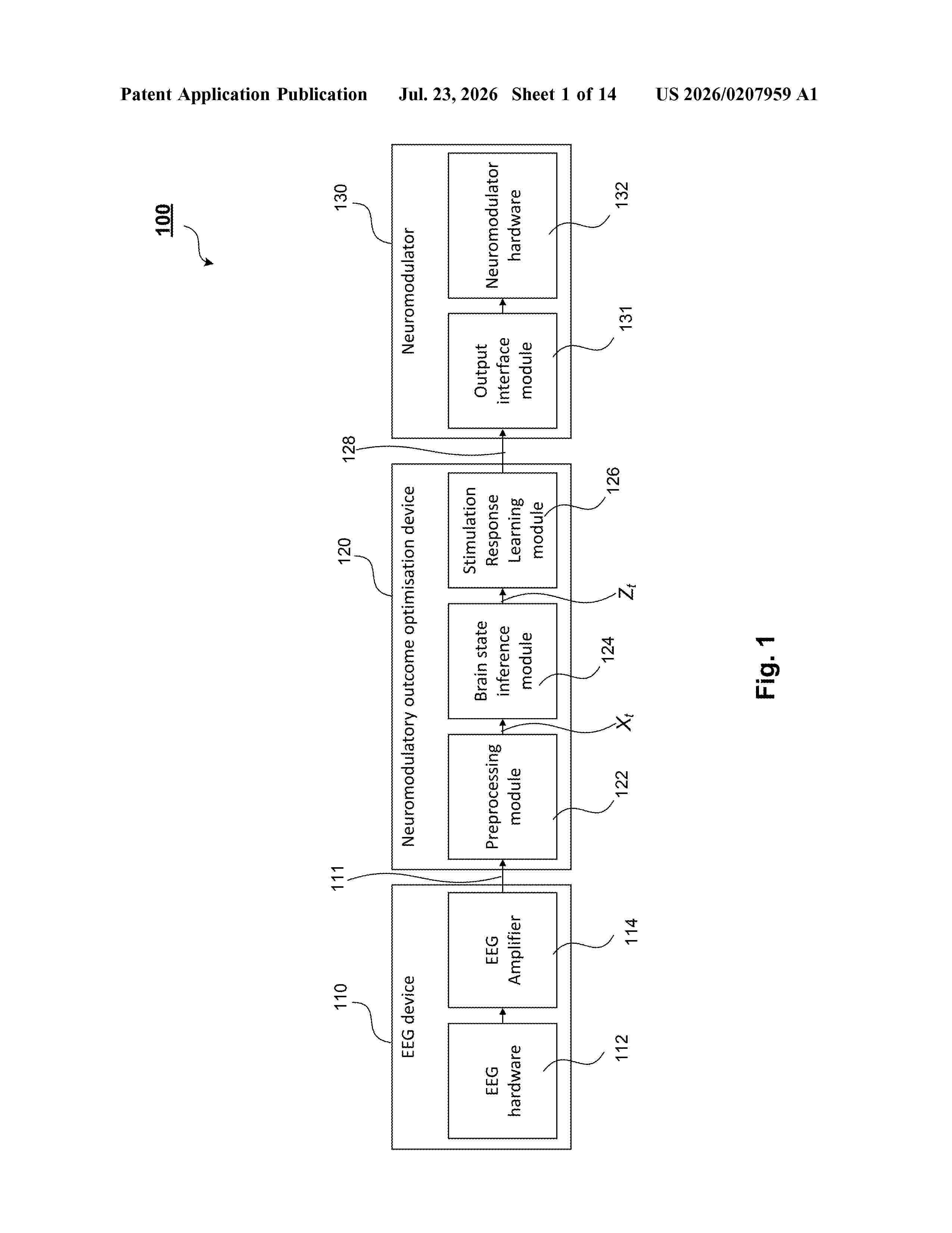
Resumen de: US20260207959A1
0000 Disclosed herein are a brain stimulation method and system for use on a mammal. The system includes: an electroencephalogram (EEG) headset (110) having sensors for detecting an electrical signal of a patient, and an EEG processing device (120) for determining a stimulation protocol based on that electrical signal. The EEG processing device (120) includes: a brain state inference module (124) for determining an inferred brain state of the patient; and a learning module (126) for learning the stimulation protocol based on the detected electrical signal and the inferred brain state. The system also includes a neuromodulator (130) for delivering stimuli to the patient based on the stimulation protocol.
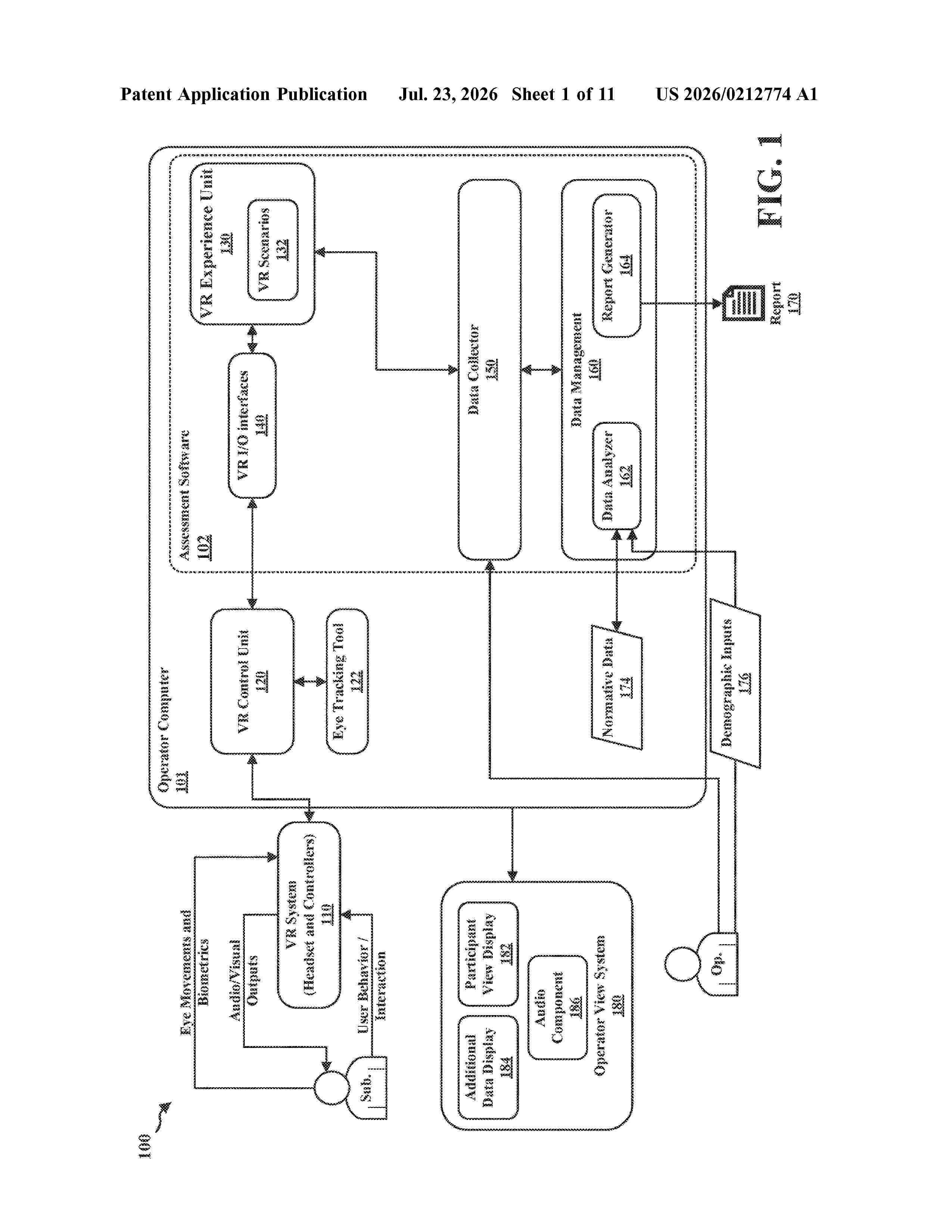
Resumen de: US20260212774A1
0000 Systems and methods provided in this disclosure explain the use of interactive narratives in VR to derive performance metrics for the assessment of individuals'socio-emotional skills. It also provides an explanation of how the change of narrative flow can be configured according to user responses, and how this changing nature of narratives can be used for more valid socio-emotional skills assessment for better prediction of real-life performance in targeted domains.
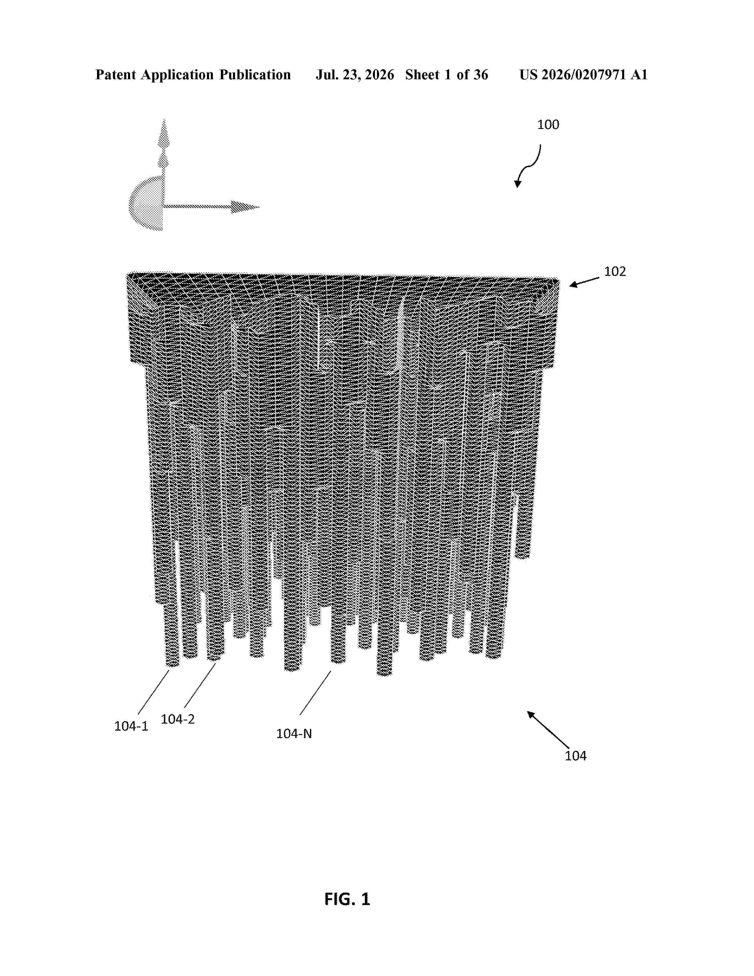
Resumen de: US20260207971A1
0000 A filter for radiation therapy, and other applications, is disclosed. The filter comprises a base and columns of varying heights extending from one face of the base. The column heights are determined by simulating the change in energy distribution of radiation passing through the filter and updating the column heights so as to more accurately match the desired output beam energy distribution. The method of filter design employs an inner loop of faster, but less accurate calculations and an outer loop of slower but more accurate calculations. The column heights for the filter are the last calculated values or the ones which produce the simulated output beam energy distribution which most closely matches the desired output beam energy distribution. The filter can be used in a radiation beam apparatus to treat a patient more quickly.
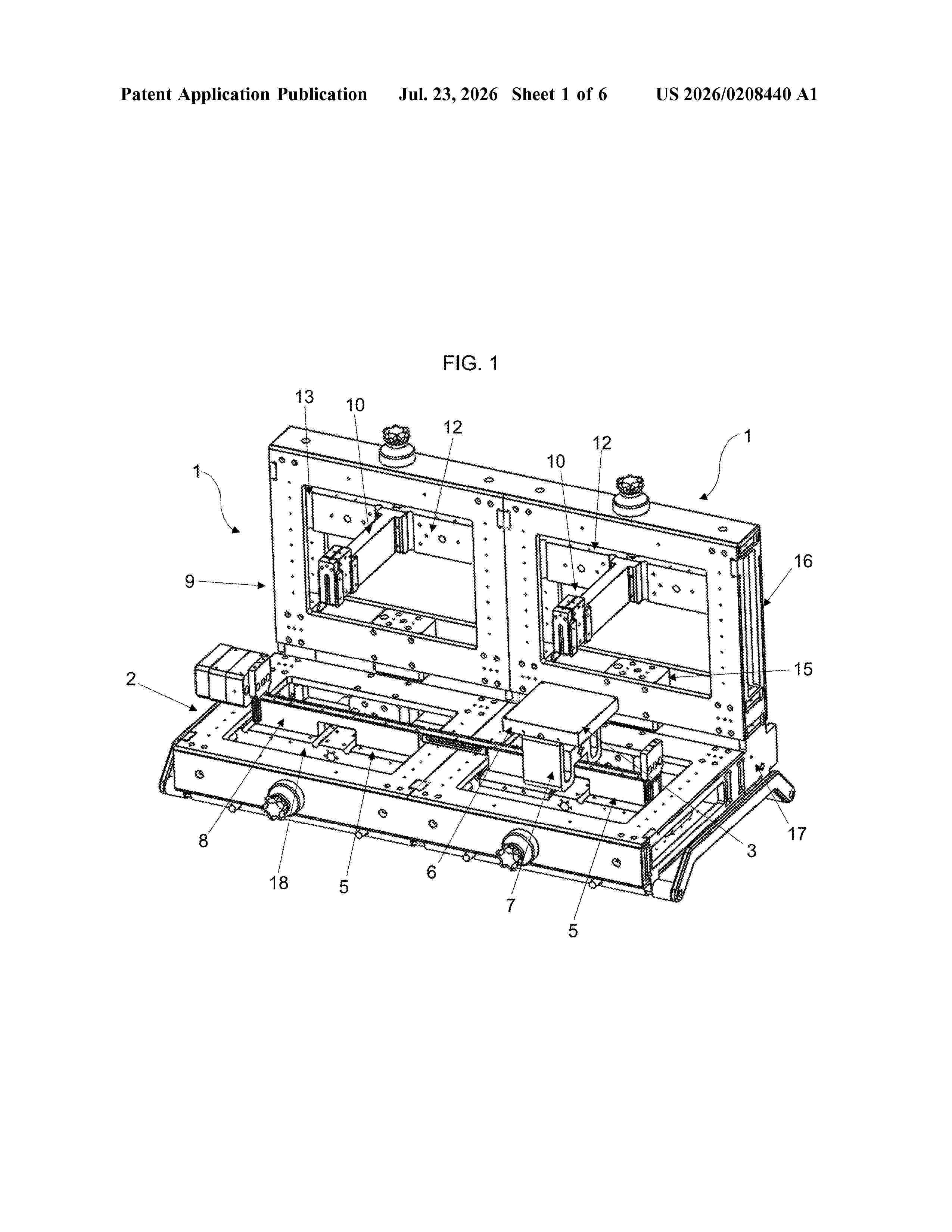
Resumen de: US20260208440A1
0000 A modular system (1) insertable into a confined environment (20), such as, for example, a tank containing a fluid, including: a base unit (2) comprising a work surface (3) that is movable along a working plane; an upright structure (9) constrained to one side of the base unit (2) and extending away therefrom; a support head (10) for supporting a tool or an accessory; said upright structure (9) comprising in turn: a guide means (11) for guiding the support head (10) along a direction (Z) transversal to the working plane; a sliding means (14) for sliding the support head (10) along the guide means (11); at least one motor (M) for the sliding means (14); a box-like casing (15) provided with sealing gaskets for housing said motor (M), said modular system (1) further comprising at least one tubular body (21) for cables of the motor (M).
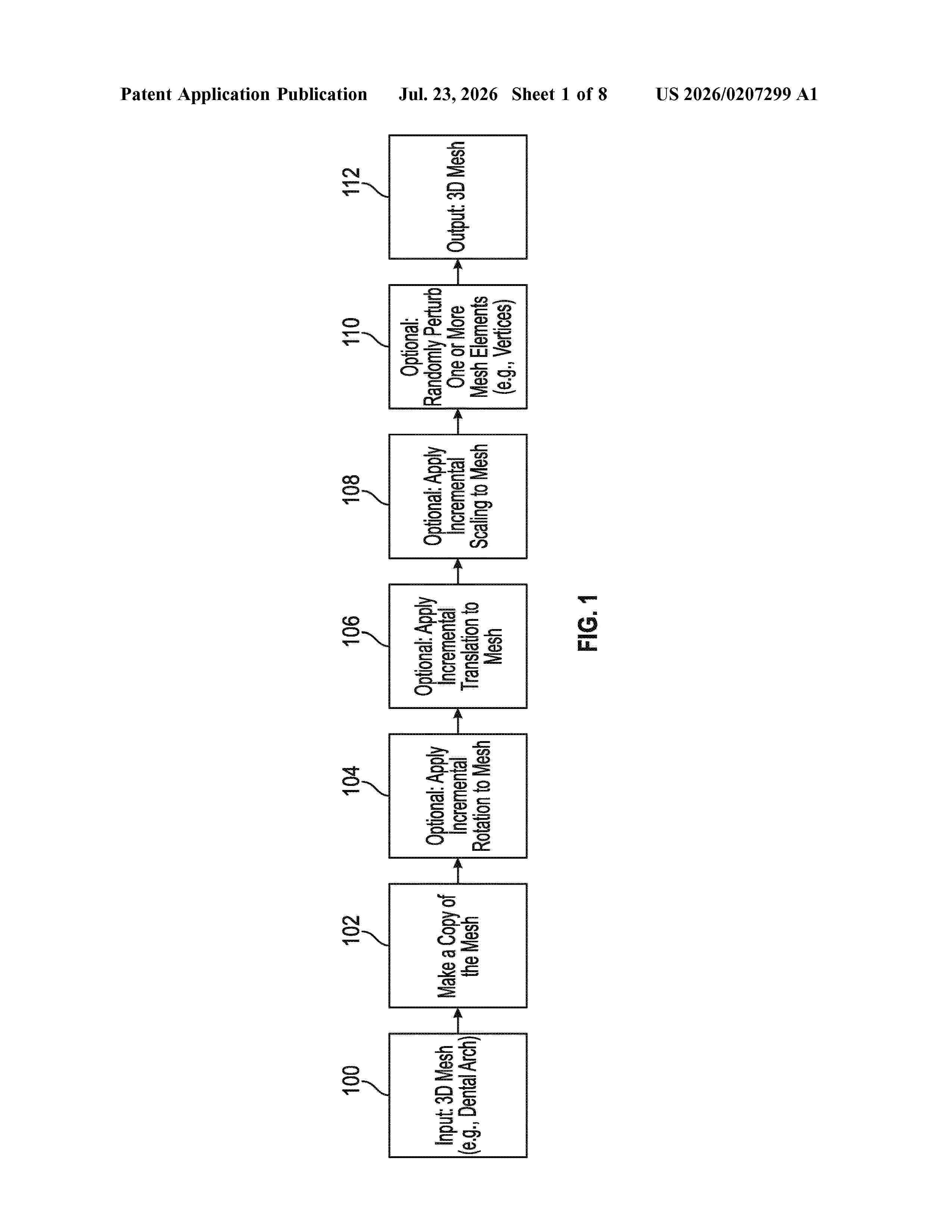
Resumen de: US20260207299A1
Systems and techniques are disclosed for generating and transferring pose information in three-dimensional (3D) representations of oral care data. The method involves receiving reference 3D representations and trial 3D representations of oral care data using processing circuitry of a computing device. Pose transfer neural networks are executed to assign pose information from the reference 3D representations onto the trial 3D representations. By incorporating the assigned pose information, one or more resulting 3D representations of oral care data are generated, wherein aspects of the trial 3D representations are modified to generate the resulting 3D representations. These resulting 3D representations, enriched with pose information, are then provided to one or more automated processes. These systems and techniques enable accurate and efficient analysis of oral care data, facilitating improved treatment planning and decision-making in the field of oral healthcare.
Nº publicación: US20260211979A1 23/07/2026
Solicitante:
SOLVENTUM INTELLECTUAL PROPERTIES CO [US]
Solventum Intellectual Properties Company
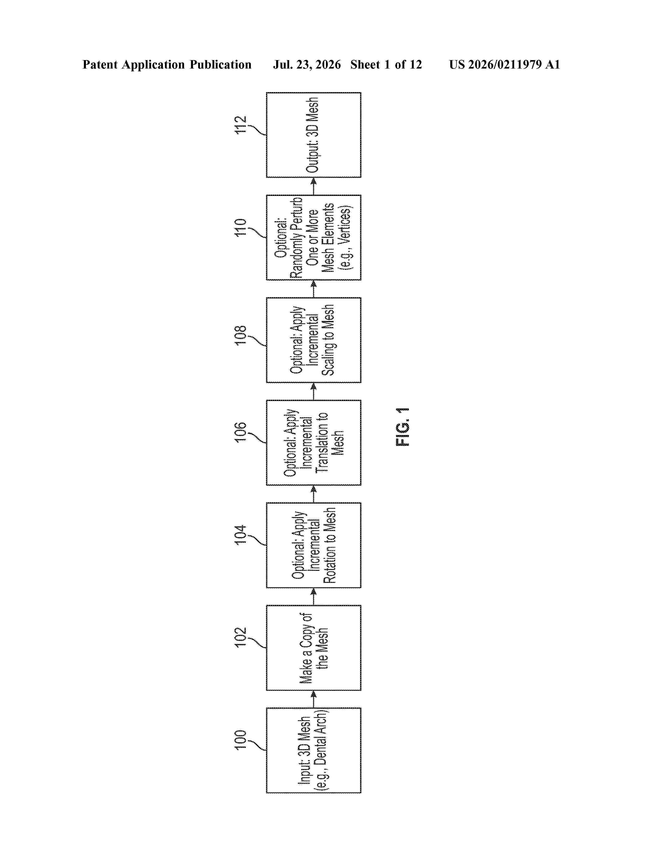
Resumen de: US20260211979A1
0000 Systems and techniques for classifying a 3D representation of oral care data are disclosed. The method involves receiving a first 3D representation comprising one or more mesh elements and providing it as input to a trained autoencoder network. The processing circuitry computes one or more mesh element features for the mesh elements and provides them to the trained autoencoder network. By executing the trained autoencoder network, the first 3D representation of oral care data is encoded into one or more latent space representations. These latent space representations are specifically designed for utilization by a machine learning model for the classification of the first 3D representation of oral care data. These systems and techniques enable accurate and efficient classification of 3D representations, enhancing the analysis and understanding of oral care data for improved diagnosis and treatment planning.
 BOPI
BOPI
 Sede Electrónica