Si deseas distinguir tus productos, servicios o ambos de los de otra empresa, es posible que necesites una marca o nombre comercial. Descubre qué son, en qué consiste su procedimiento de registro y qué implica.

Información sobre los plazos de presentación de solicitudes de transformación de marcas de la Unión Europea en marca nacional española. Más información


Si tienes un nuevo dispositivo, producto o procedimiento que resuelva un problema técnico o tenga una ventaja práctica, existen distintas formas de protegerlo en España y en otros países. Descubre cómo hacerlo.

¿Tu innovación reside en la estética, la ornamentación o la apariencia de tu producto? Protégela mediante un diseño industrial. Descubre qué derechos confiere el registro y cómo realizar la tramitación.

Las indicaciones geográficas protegen el nombre de un producto originario de una zona geográfica, a la cual le debe una determinada calidad, reputación u otra característica. Descubre qué son, en qué consiste su procedimiento de registro y qué beneficios conceden.

Las patentes publicadas en todo el mundo son una valiosa fuente de información científica, técnica y comercial.

Si eres emprendedor/a o una empresa y quieres potenciar y mejorar la rentabilidad de tu negocio protegiendo de forma adecuada los activos intangibles de tu organización, en este espacio encontrarás lo necesario.


 568
resultados
568
resultados
 Última actualización
17/10/2026 [07:24:00]
Última actualización
17/10/2026 [07:24:00]


 Resultados 250 a 275 de 568

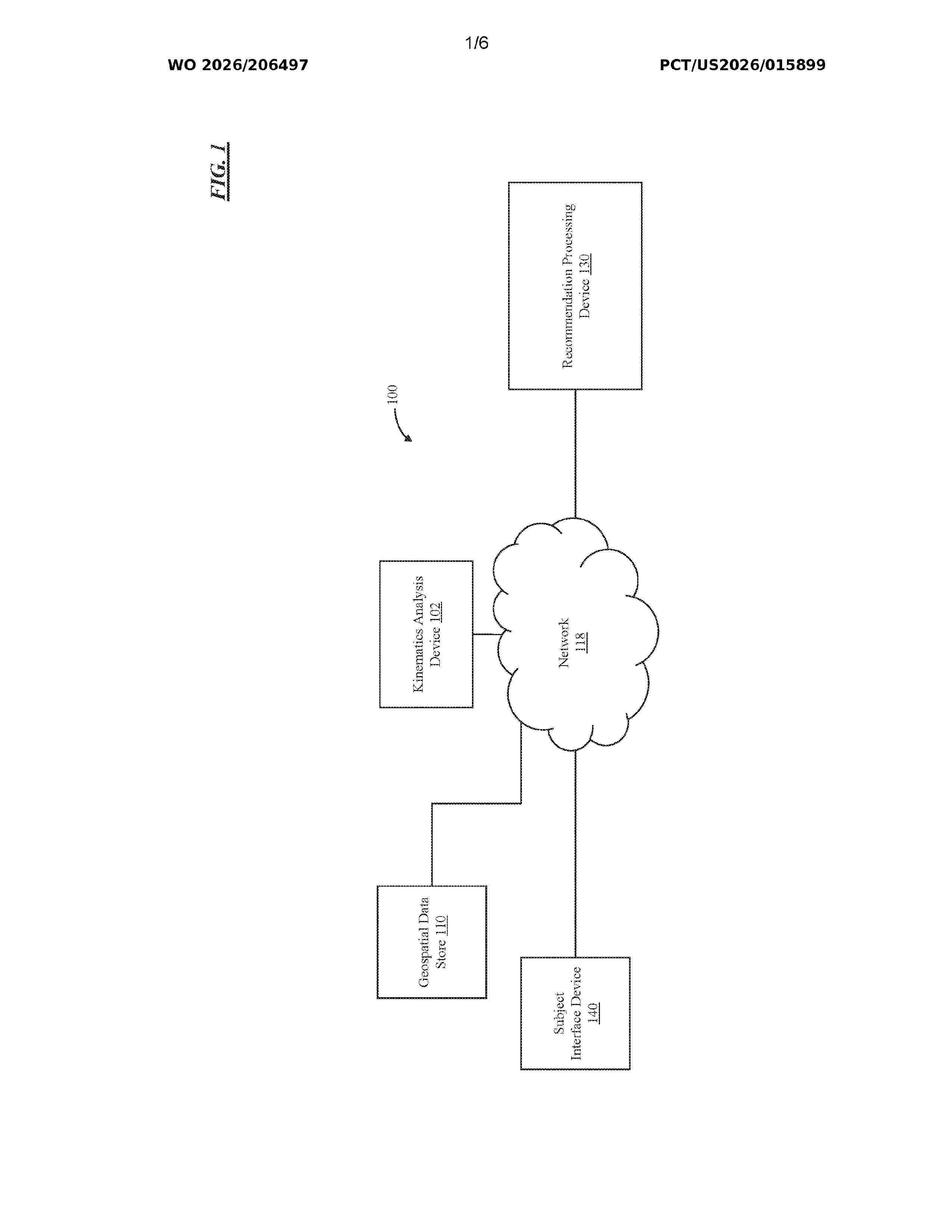
Resumen de: WO2026206497A1
A system preparing kinematic recommendations for athletes using artificial intelligence is disclosed. The system is programmed to sense personal physical data regarding a subject athlete, and optionally review environmental data, and historical performance and physical data related to the subject athlete. The system produces performance recommendations for that subject athlete, determines a best medium for providing the performance recommendations to the subject athlete based on their personal physical data, and provides the performance recommendations to the subject athlete via that determined best medium. The best medium can include an augmented reality interface worn by the athlete. The performance recommendations can be prepared by machine learning or artificial intelligence models designed to analyze athletic performances, and to communicate conversationally with the athlete.

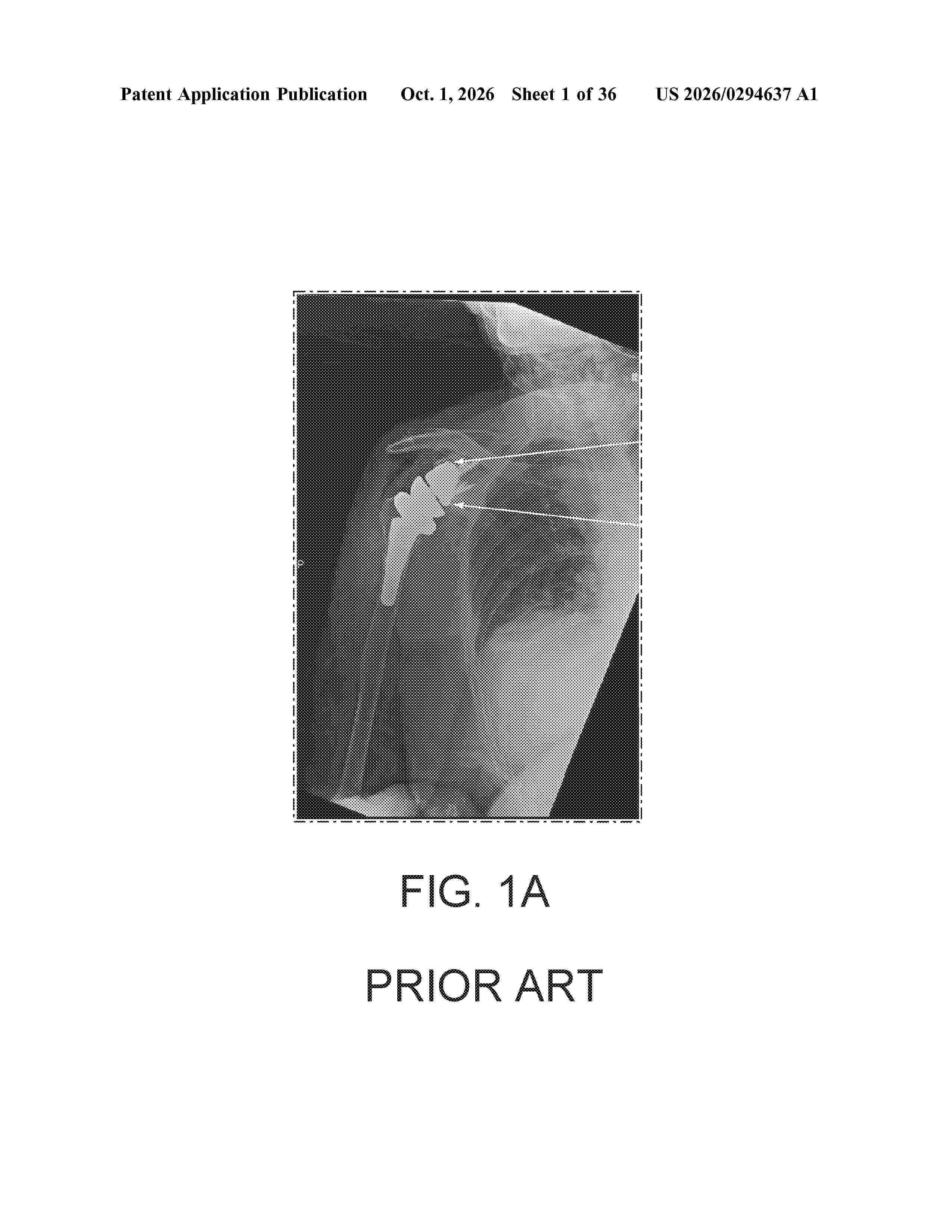
Resumen de: US20260294637A1
Systems and methods include a component of a bone implant that includes a patient specific bone contacting surface and, optionally, at least one protrusion adjacent to or extending from the patient specific bone contacting surface. The at least one protrusion is configured for engaging a bone preparation in the bone. To prepare the bone for receiving the component, one or more cutting tool templates may be used. The one or more cutting tool templates may be fabricated to have a patient specific bone contacting surface that engages the bone that is to receive the implant component. A cutting tool is engaged in one or more of one or more slots defined by the cutting tool template(s) to remove surface portions of the bone to create the bone preparation for engaging the protrusion. The patient specific bone contacting surface of the component engages the bone.
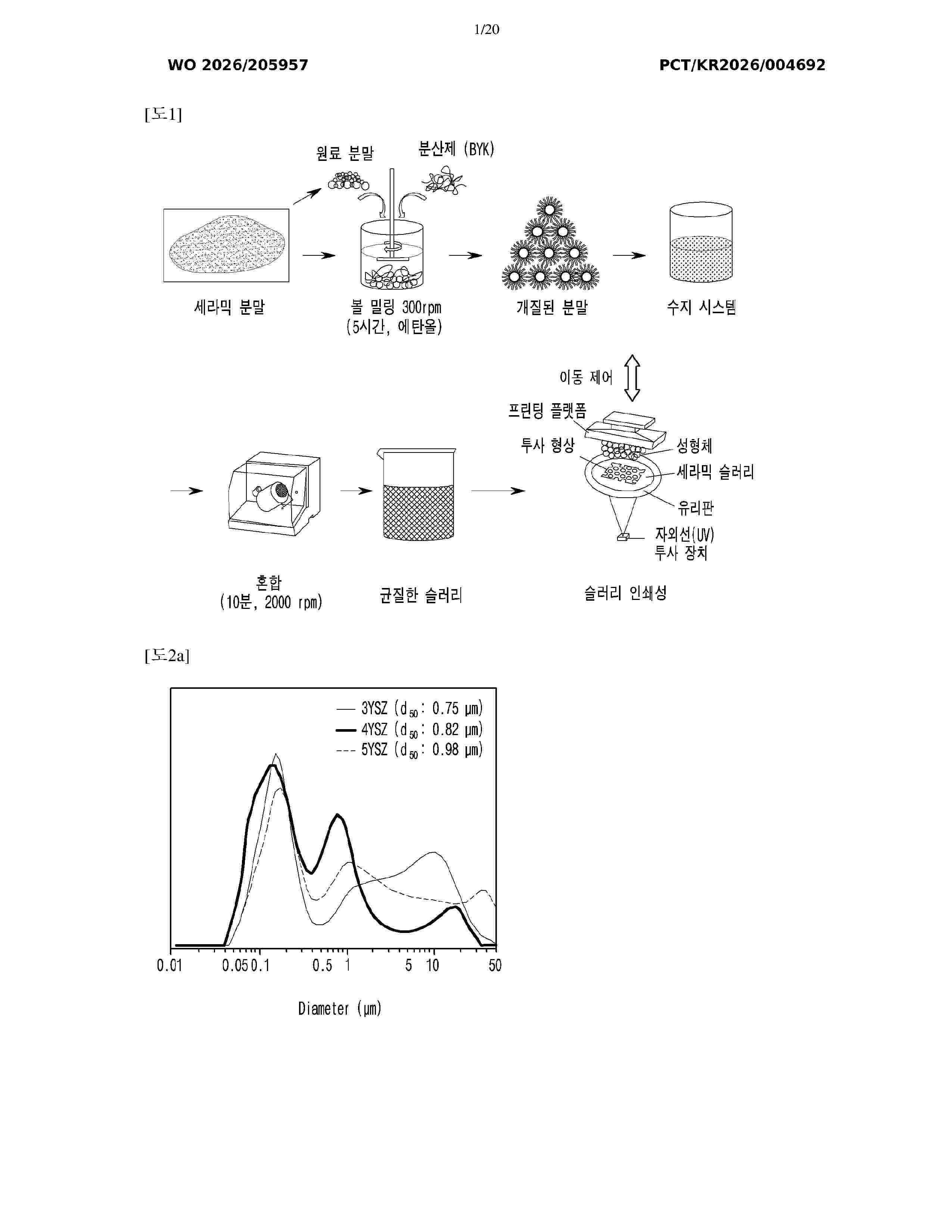
Resumen de: WO2026205957A1
The present invention relates to a dental zirconia 3D-printed article having color and translucency gradients and a manufacturing method therefor and, specifically, to a dental zirconia 3D-printed article and a manufacturing method therefor, the dental zirconia 3D-printed article comprising at least two types of yttria-stabilized zirconia (YSZ) doped with different contents of yttria (Y2O3), wherein the at least two types of yttria-stabilized zirconia are selectively doped with a colorant to have gradients in color and translucency.
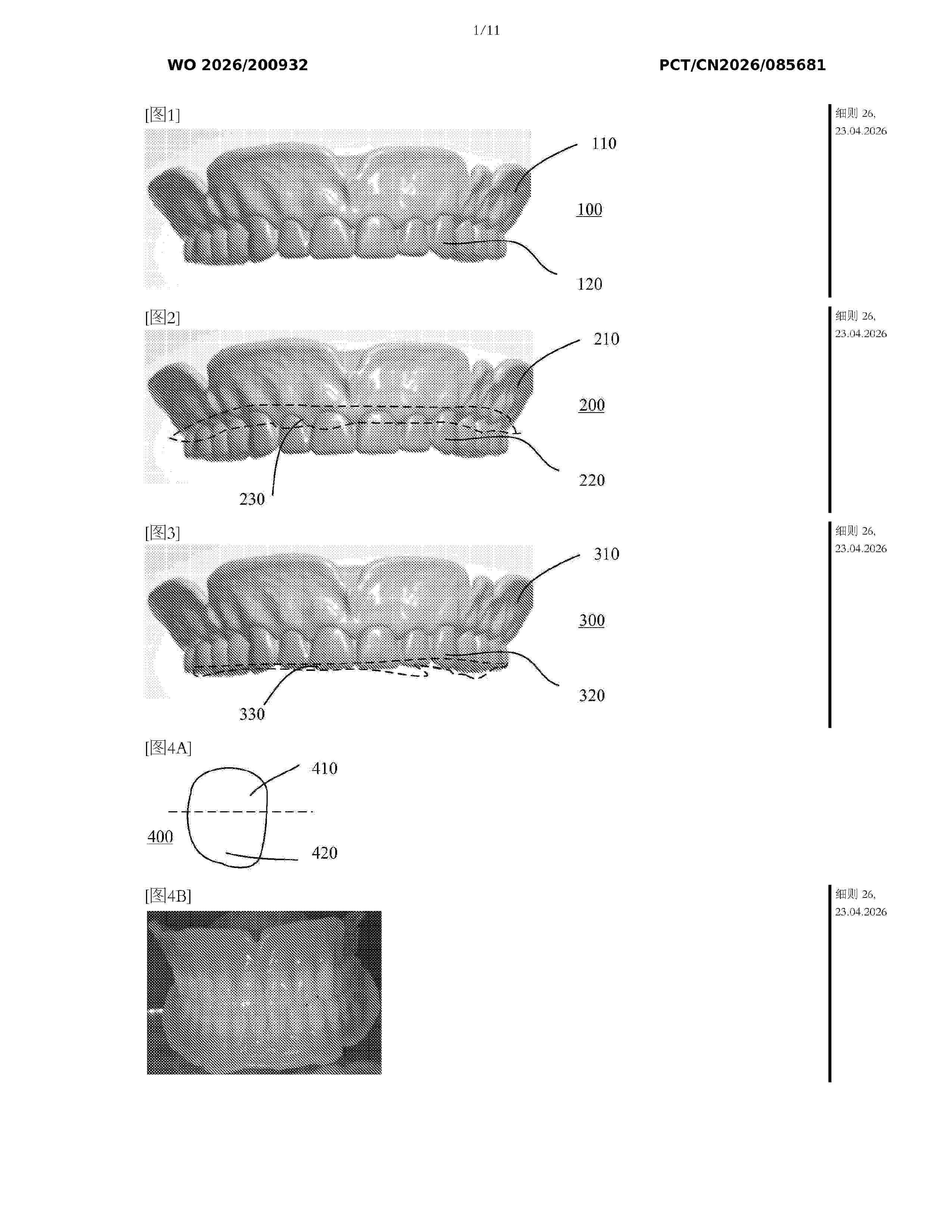
Resumen de: WO2026200932A1
The present application relates to a dental product formed by 3D printing, comprising: a first body, comprising a portion made of a first material; and a second body, connected to the first body and comprising a portion made of a second material, wherein the first material and the second material have different colors or transparencies. The dental product provided in the present application meets at least one application requirement.
Resumen de: WO2026201517A1
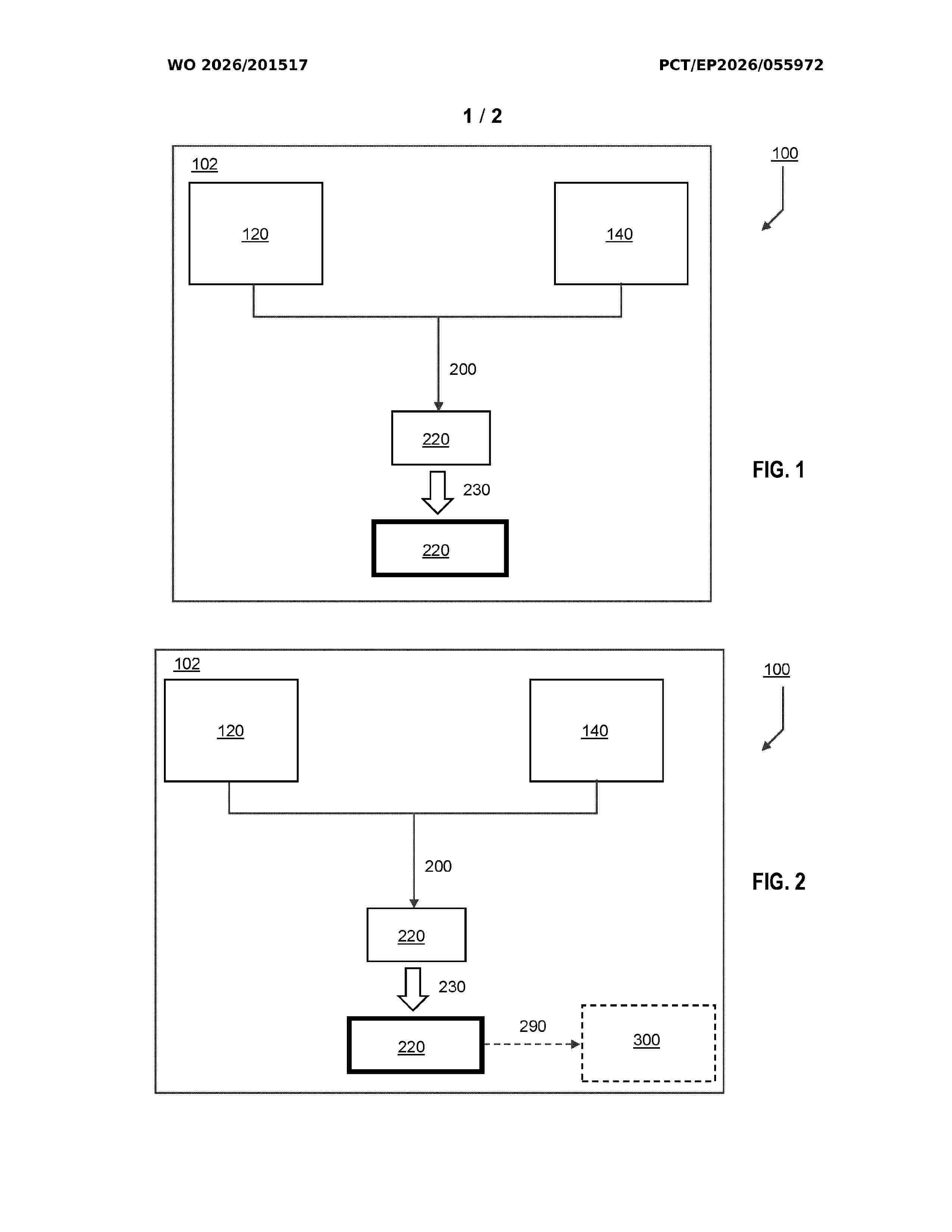
Provided herein is a method (100) for preparing a prosthetic crown for a subject or assisting a dental restoration programme comprising one or more dental sessions for fitting a prosthetic crown into a subject, the method comprising: receiving a planning dataset, 3D- PLAN (120), wherein the 3D-PLAN comprises three-dimensional dental measurement data of the subject as presented prior to an intervention, wherein: the intervention includes implanting an implant in a defined pose for later addition of the prosthetic crown; and the 3D-PLAN: is used for a planning and execution of the intervention; and is used for a preparation of a preliminary design of the prosthetic crown; receiving a local 3D post- intervention measurement dataset, L3D-POI (140), wherein the L3D-POI has been obtained by a three-dimensional contactless measurement of the intervention site, wherein the intervention site comprises the implant, wherein the implant has been fitted to a jawbone of the subject to support the prosthetic crown, and the L3D-POI includes a three- dimensional measurement data of at least a coronal portion of the in situ implant; determining (200), from the 3D-PLAN (120) and L3D-POI (140), a 3D patient specific dental component, 3D PSDC, dataset (220), from which the 3D PSDC is able to be prepared; and outputting (230) the 3D PSDC dataset (220) for transmission (290) to a device (300) for additive manufacturing, thereby allowing an additive manufacture of a 3D printed PSDC, wherein the 3D p
Resumen de: WO2026207093A1
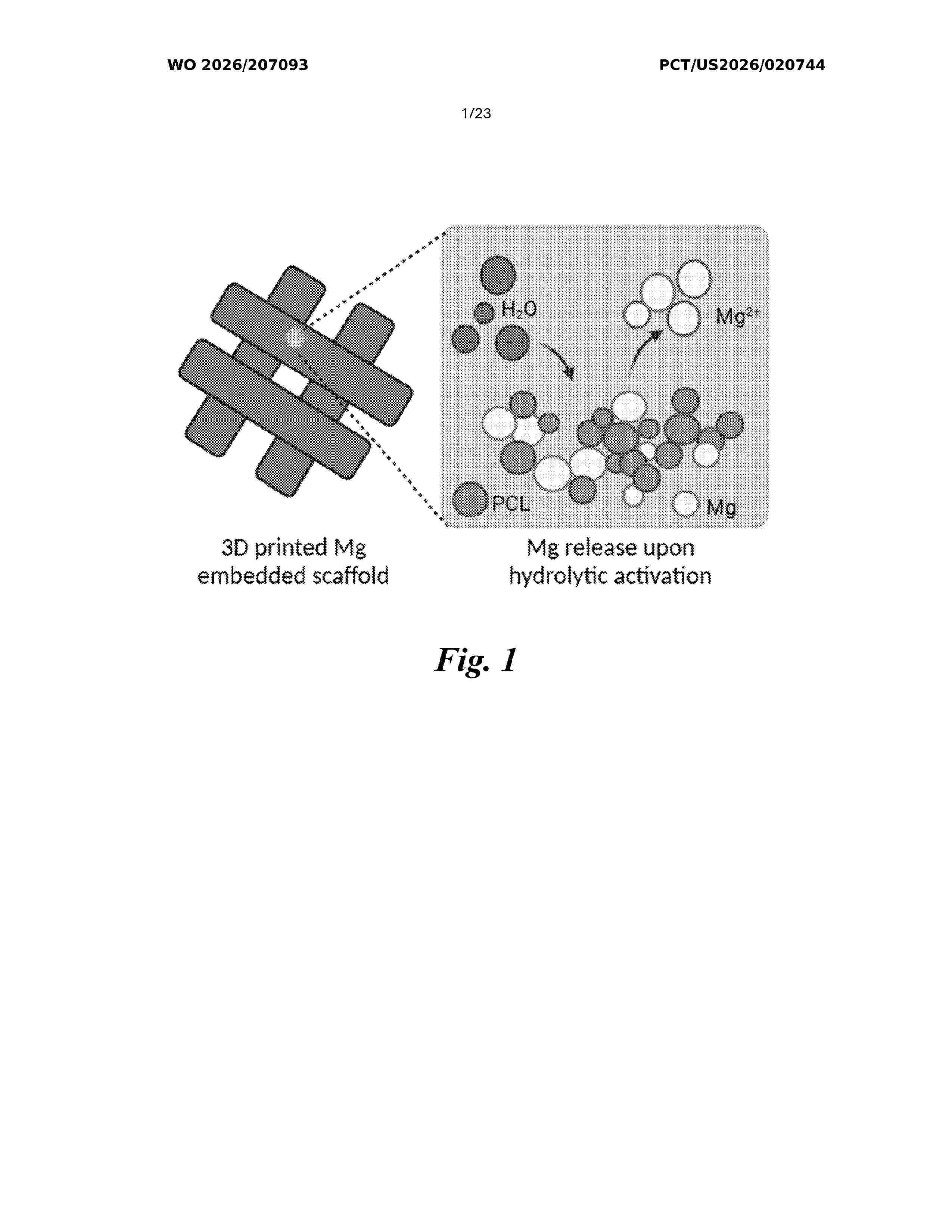
A bioactive composite filament for fused-deposition modeling comprises 10 vol% to about 60 vol% biodegradable metal powder (about 10 µm to about 100 µm particle size) and about 40 vol% to about 90 vol% biocompatible thermoplastic polymer. The filament exhibits controlled rheology (viscosity ≤ 5.5×10⁵ Pa·s at 120 °C), enhanced mechanical properties (up to 3-fold increase in tensile modulus), and printability with high fidelity (print score ≥ 0.85). Three-dimensional scaffolds manufactured from the filament demonstrate pH-neutral degradation, composition-tunable metal ion release, and sustained bioactivity with greater than 95% cell viability over 21 days. The manufacturing process involves cryogenic milling, inert-atmosphere blending, screw extrusion, and layer-by-layer deposition to produce porous scaffolds or non-porous implants for bone tissue engineering and orthopedic applications.
Resumen de: US20260295189A1
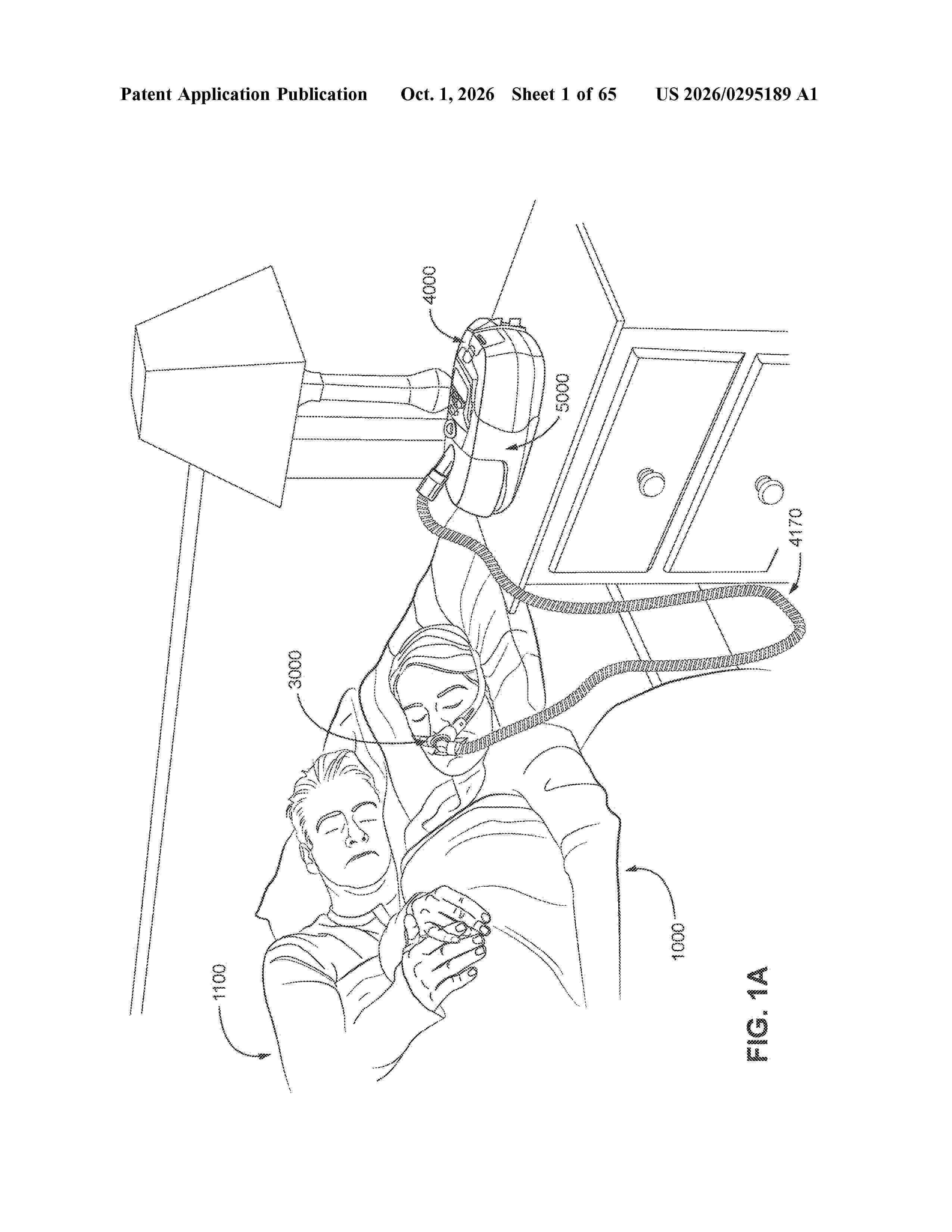
A patient interface comprising a plenum chamber, a seal-forming structure constructed and arranged to form a seal with a region of the patient's face surrounding an entrance to the patient's airways, and a vent to allow a continuous flow of gases exhaled by the patient from an interior of the plenum chamber to ambient. The seal-forming structure may comprise a cushion, the cushion being deformable and resilient and at least partially formed by a lattice structure.
Resumen de: US20260298906A1
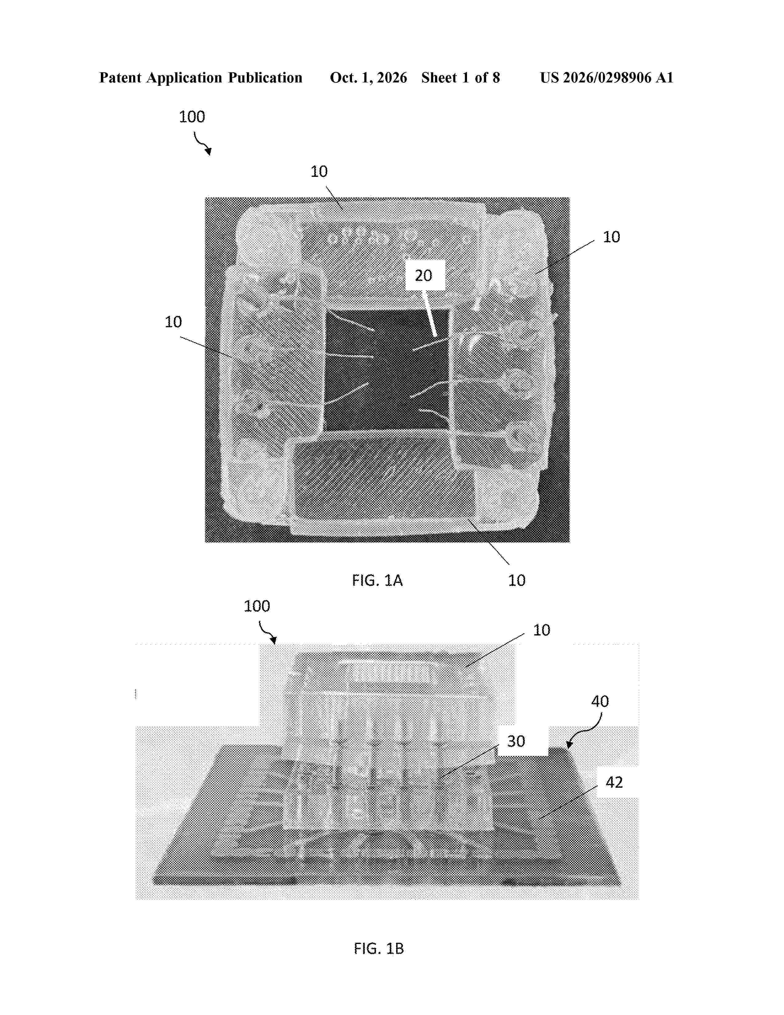
An apparatus for three-dimensional sensing is disclosed. The apparatus comprises an electrode housing, comprising: two or more walls capable of encompassing a 3D model, wherein at least two walls are detachably connected to each other, forming at a connected state an angle greater than 0 degrees, between surfaces of each of the two or more walls; and wherein each wall comprises an electrode array, extending out from the wall towards the 3D model, configured to sense electrical activity of the 3D model.
Resumen de: US20260294653A1
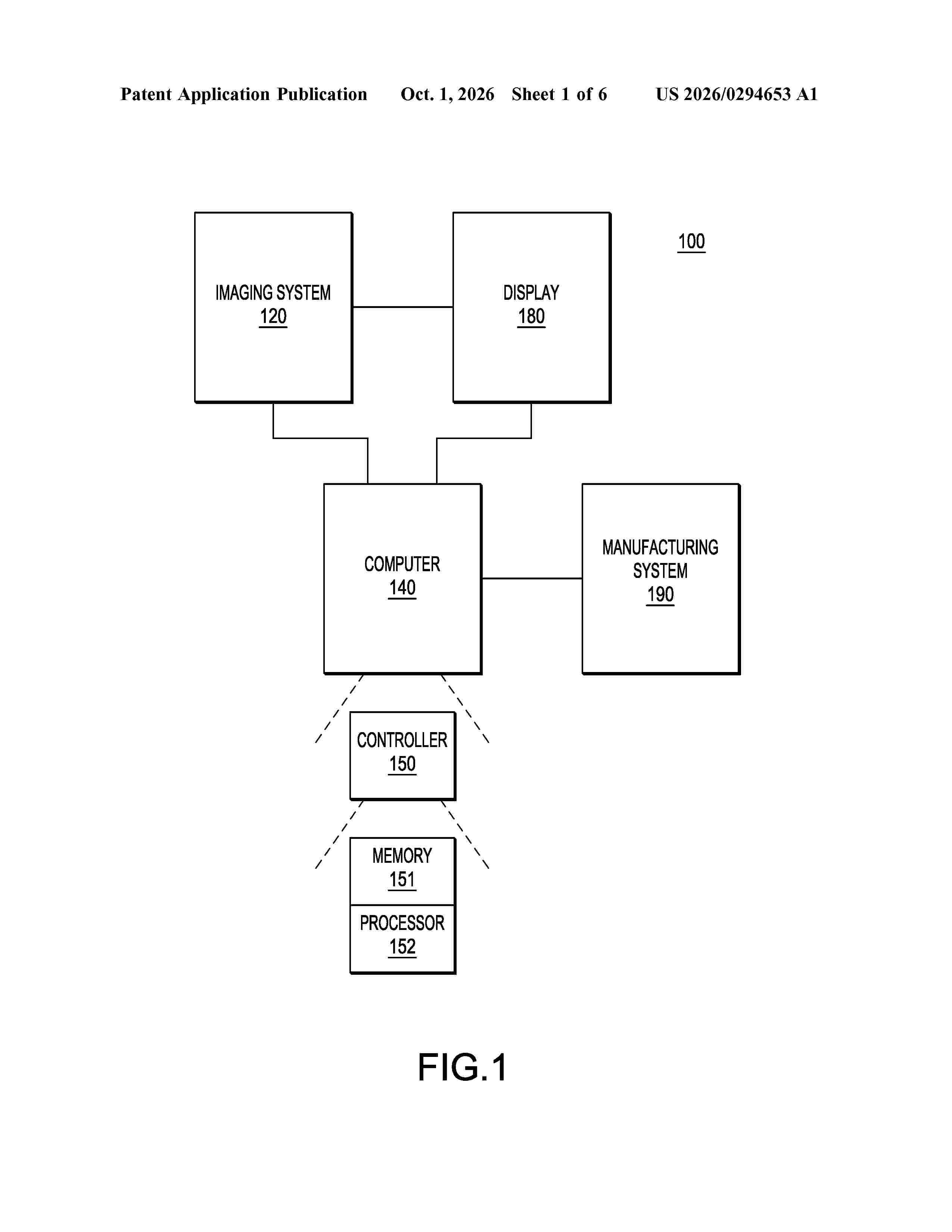
A method for additive manufacturing of a medical stent includes generating, by a processor (152) executing instructions, a model of anatomy of a subject; obtaining a computer (140)-aided design (CAD) model of a stent; simulating deployment of the stent in the anatomy of the subject; determining whether the model of the stent satisfies an optimization metric based on the simulating, and optimizing the model of the stent by repeating obtaining and the simulating until the model of the stent satisfies the optimization metric. The stent is manufactured by additive manufacturing in response to the model of the stent being optimized.
Resumen de: US20260297142A1
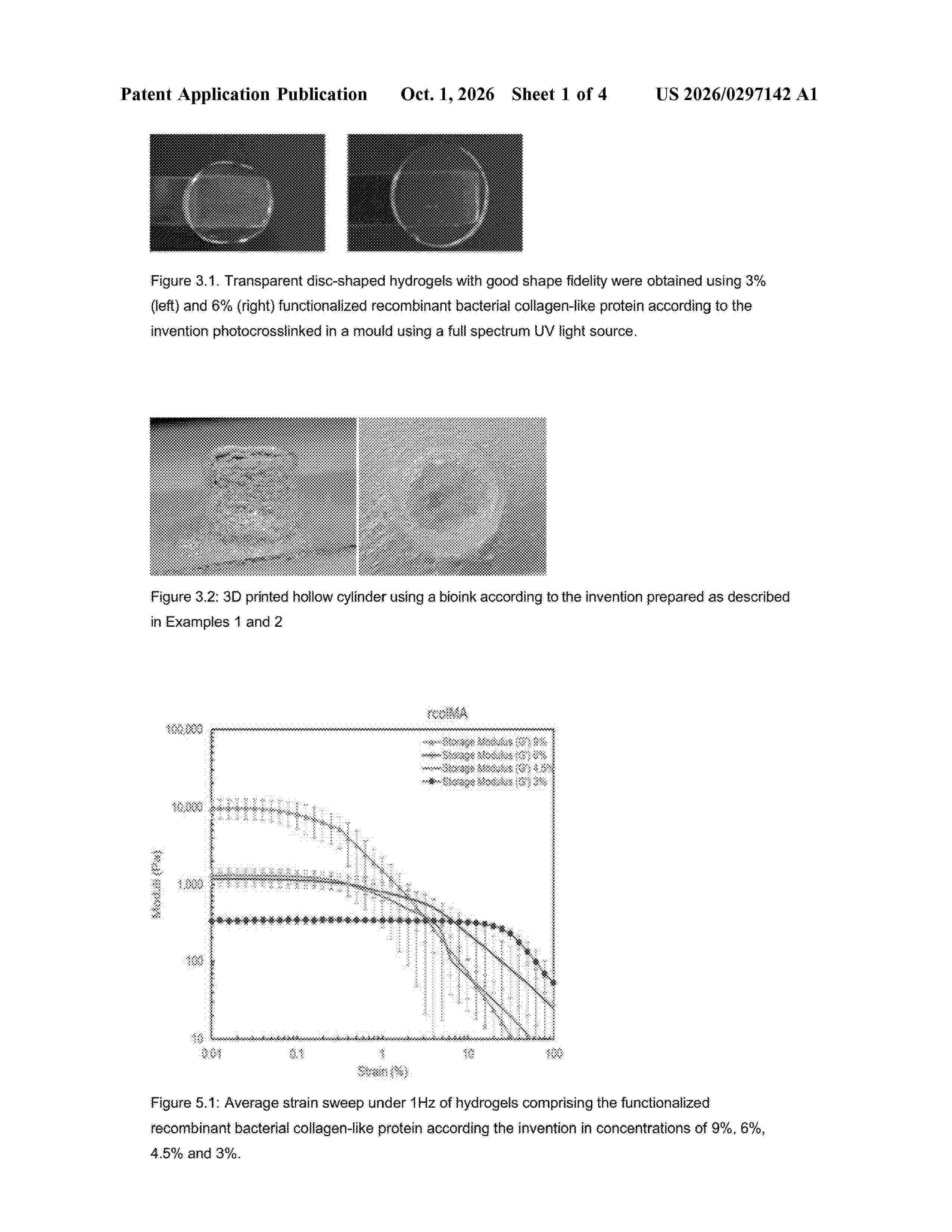
0000 Recombinant bacterial collagen-like protein with an amino acid sequence that is at least ≥60% identical to the amino acid sequence of SEQ ID NO:1 characterized in that the amino acid sequence comprises a deletion of at least 38 amino acids at the N-terminus of the amino acid sequence of SEQ ID NO:1 and that the recombinant collagen-like protein is functionalized with ethylenic unsaturated groups.
Resumen de: WO2026202082A1
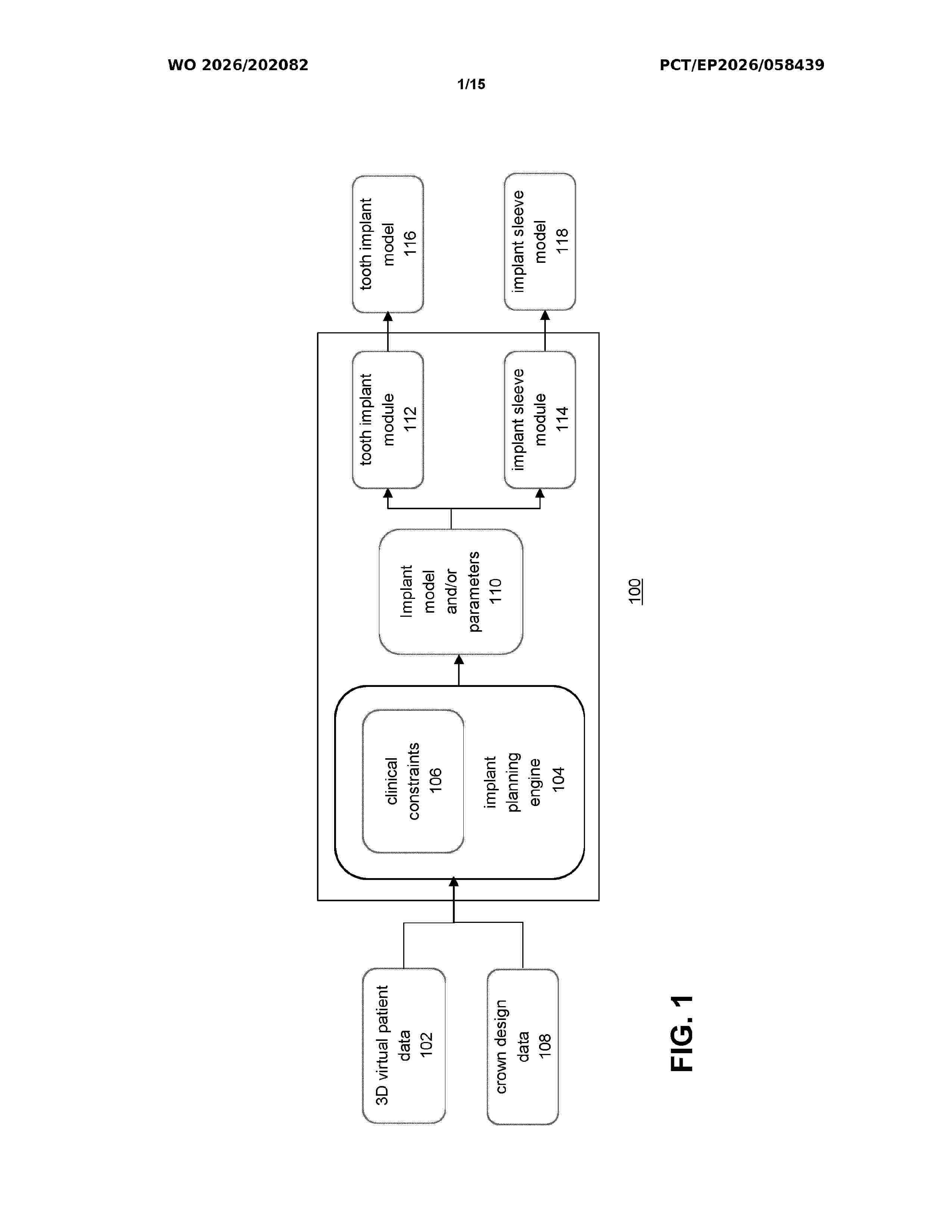
A computer-implemented method for automated planning of a tooth implant in a dento-maxillofacial complex is described, wherein the method includes receiving or determining 3D virtual patient data representing a dento-maxillofacial complex comprising anatomical structures and at least one area of interest for placing the tooth implant, the 3D virtual patient data including 3D representations of anatomical structures within a vicinity of the at least one area of interest; determining a tooth implant model and/or one or more implant parameters defining a tooth implant model to be positioned at the at least one area of interest, using an implant planning engine, the implant planning engine being configured to receive the 3D representations of anatomical structures within a vicinity of the area of interest and to determine the tooth implant model and/or one or more implant parameters defining a tooth implant model based on clinical constraints, the clinical constraints including distance requirements of the tooth implant with regard to at least part of the 3D representations of the anatomical structures; and, rendering the implant model on a display or transforming the implant model into a data format for controlling a manufacturing apparatus, such as a 3D printer, to manufacture the tooth implant.
Resumen de: WO2026205644A1
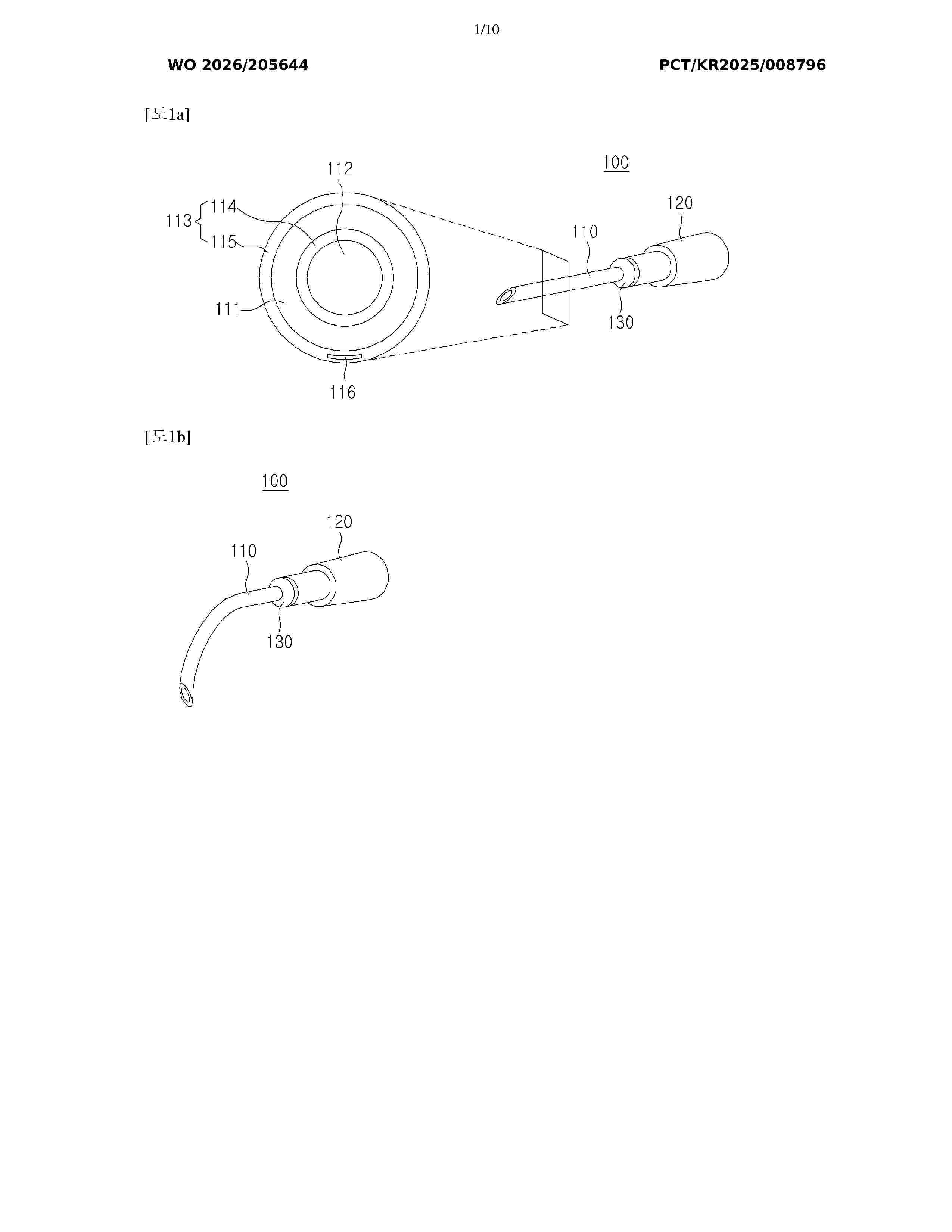
The present disclosure relates to a manufacturing method for the mass production and recycling of variable-stiffness intravenous needles of various sizes that become softened by body temperature, the method comprising the steps of: preparing a mold, which is manufactured by 3D printing and has a plurality of elongated channels aligned therein, and a plurality of elongated rods, which can be coaxially arranged in the respective channels; applying a material having variable stiffness according to ambient temperature to the mold so as to encompass each of the rods in the channels; curing the material, and removing the mold and the rods so as to leave a plurality of hollow body members; and forming coating members having biocompatibility so as to cover the respective outer surfaces of the body members, thereby leaving a plurality of variable-stiffness intravenous needles.
Resumen de: WO2026207469A1
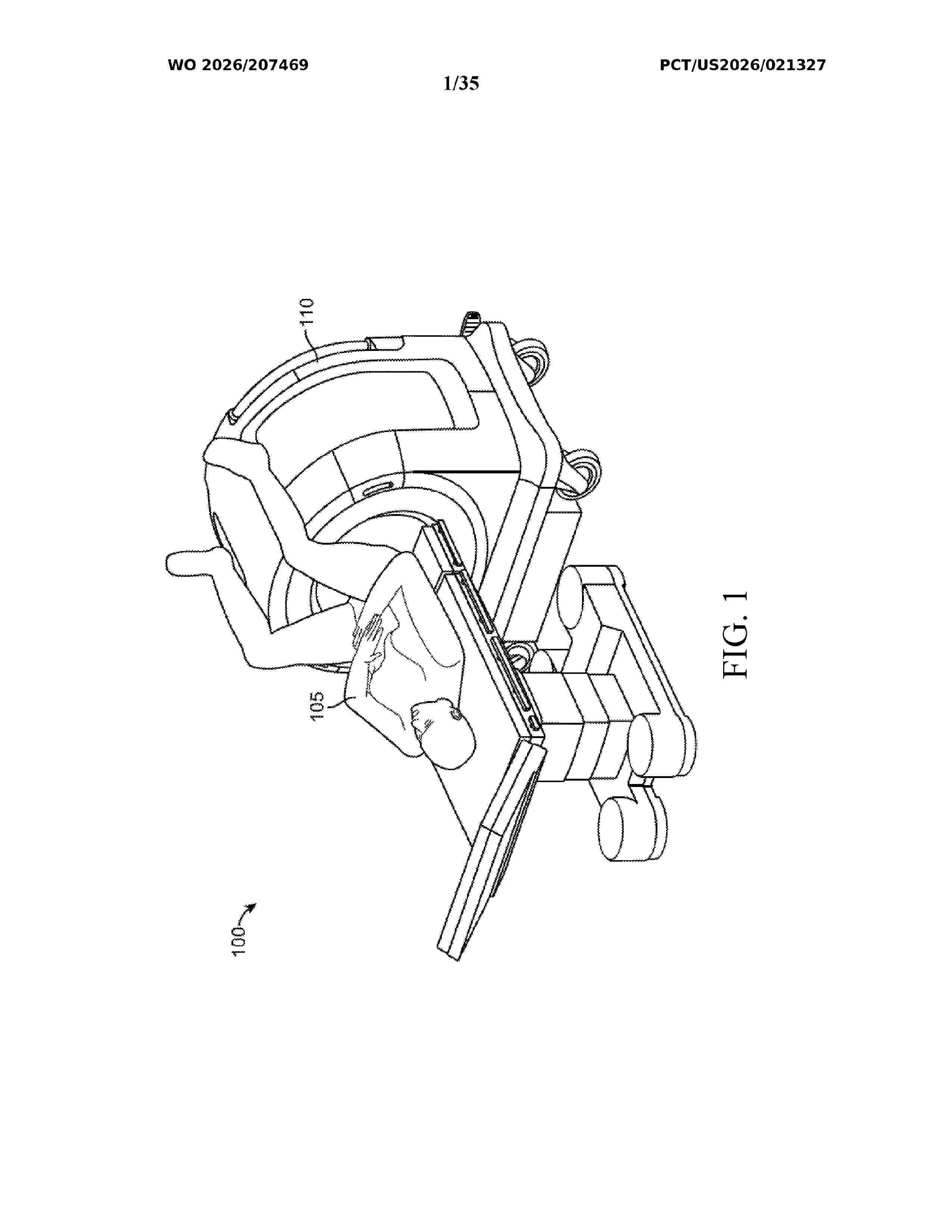
Systems and methods for three-dimensional localization of an interventional device within a magnetic resonance imaging (MRI) system are disclosed. The method includes acquiring a baseline MR signal dataset of a target region and a subsequent dataset of the region following introduction of the interventional device using a hybrid encoding pulse sequence. The hybrid sequence applies spatiotemporal encoding in a first plane and Fourier encoding along an orthogonal axis to acquire highly undersampled projection data. A difference dataset is generated by mathematically evaluating the subsequent dataset against the baseline. A processing module determines the precise 3D spatial location of the interventional device by analyzing localized signal anomalies within the difference dataset. The approach circumvents fully sampled Cartesian k-space acquisitions, enabling rapid, intra-procedural tracking of magnetic and non-magnetic devices for manual or robotic guidance across diverse MRI environments, including, but not limited to, open, low-field, and single-sided systems.
Resumen de: WO2026202089A1
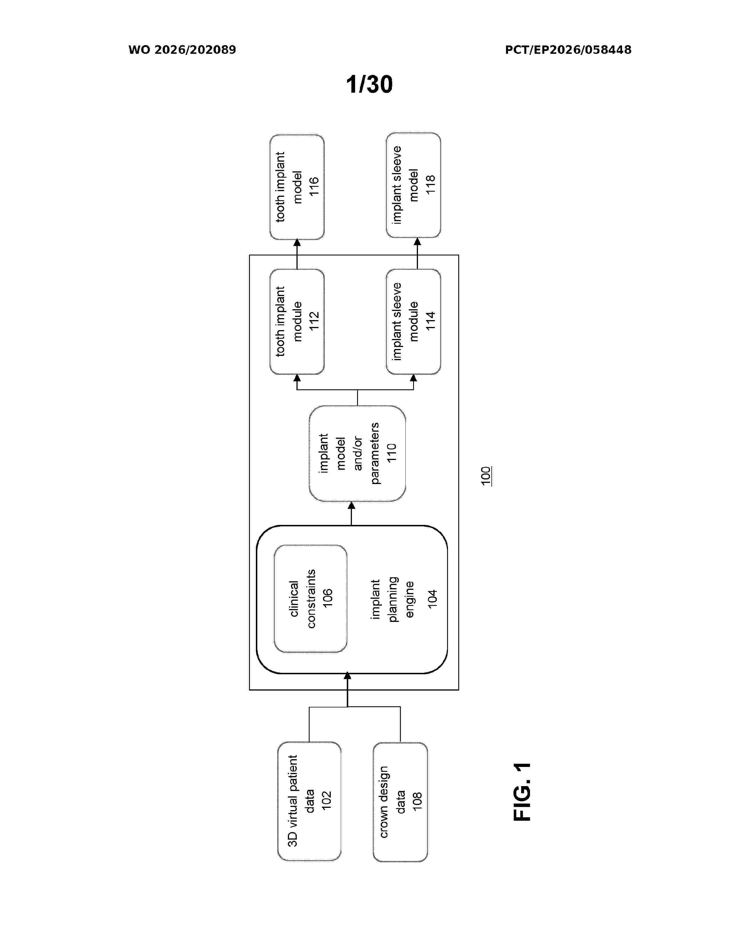
A computer-implemented method for determining a tooth implant and an associated surgical guide is described, wherein the method comprises: determining at least one tooth implant model or one or more parameters defining at least one tooth implant model using an implant planning engine, the at least one tooth implant model being associated with an implant orientation and an implant position, the implant planning engine being configured to receive 3D virtual patient data including 3D representations of anatomical structures within a vicinity of the implant position, and to determine the at least one tooth implant model or the one or more parameters defining the at least one tooth implant model based on clinical constraints associated with the anatomical structures within the vicinity of the implant position; determining an implant sleeve model based on the tooth implant model or the one or more parameters defining the tooth implant model; and, determining a surgical guide model based on the 3D virtual patient data and the implant sleeve model, the determining including forming a dental template model based on at least part of the 3D virtual patient data and forming the implant sleeve model in the dental template model so that the implant sleeve model is positioned at the implant position and so that the orientation of the implant sleeve model is parallel to the implant orientation associated with the tooth implant model.
Resumen de: WO2026202090A1
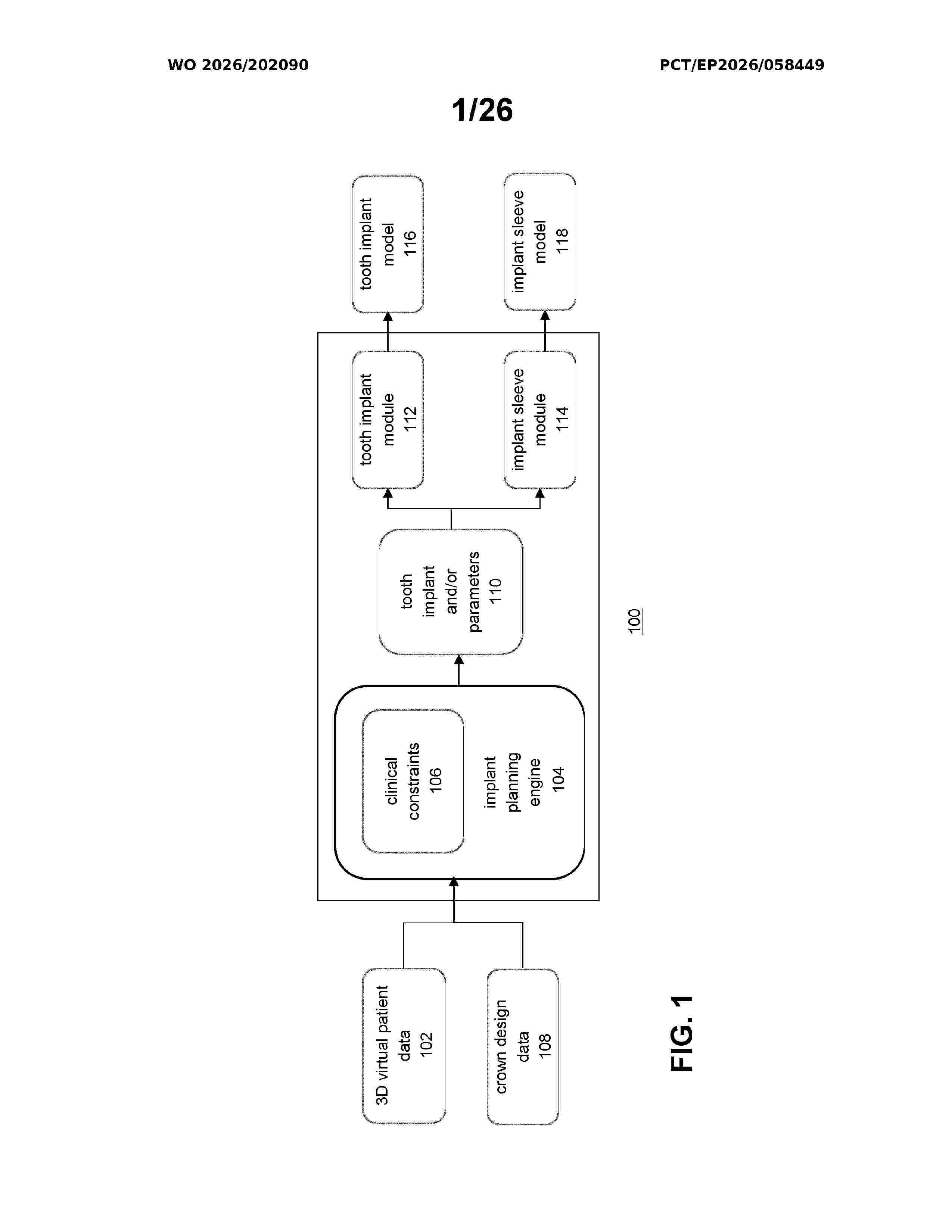
The disclosure relates methods and systems for automated planning of a tooth implant in a dento-maxillofacial complex, the method comprising: determining a tooth implant or one or more implant parameters defining a tooth implant using an implant planning engine, the tooth implant being associated with an implant orientation and an implant position in the dentition, the planning model being configured to receive 3D representations of dental objects and, optionally, tissue, of the dentition and to determine the tooth implant or the one or more implant parameters defining a tooth implant based on clinical constraints associated with the position of the tooth implant and the dental objects in the dentition; and, displaying the tooth implant and, optionally, at least part of the 3D representations of dental objects and a user interface UI associated with the displayed tooth implant, wherein the UI is configured receiving user input for modifying one or more modifying the one or more implant parameters and/or the one or more clinical constraints.
Resumen de: WO2026207112A1
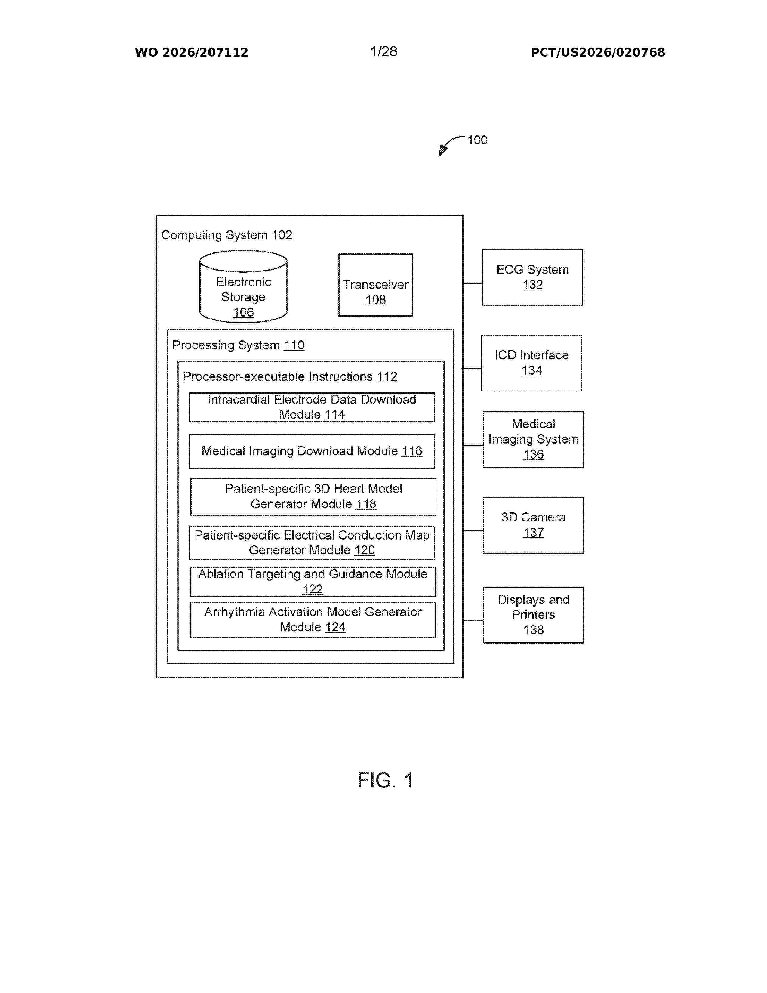
Various embodiments include a method of using information from medical electrodes positioned in a patient's heart to identify and display arrhythmia localization. The method may include obtaining electrical signals and location information from electrodes positioned within the patient's heart, generating a patient-specific 3D heart model, generating a patient-specific electrical conduction map identifying arrhythmia initiation site localization, displaying the 3D localization for physician use, and conducting a cardiac ablation procedure using the displayed localization to identify ablation locations. In some embodiments, the medical electrodes may be electrodes on electrophysiology catheters temporarily positioned with the heart. In some embodiments, the medical electrodes may be electrodes of an implanted cardiac defibrillator (ICD) permanently positioned in the heart.
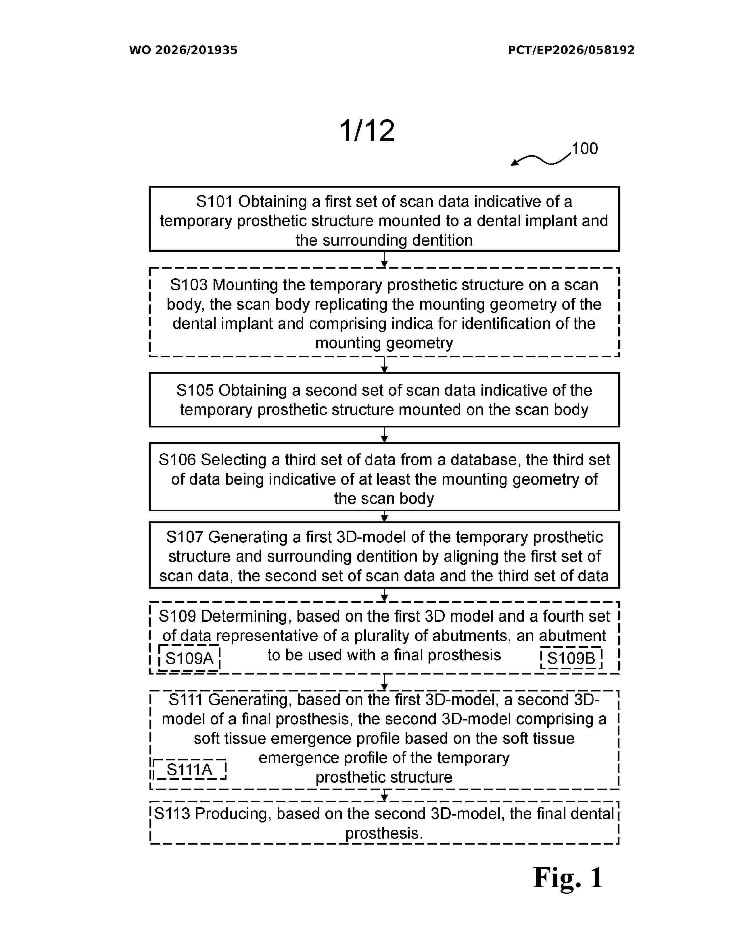
Resumen de: WO2026201935A1
A computer-implemented method for creating a digital model is provided. The method comprises obtaining a first set of scan data indicative of a temporary prosthetic structure (33) arranged on a dental implant embedded in a jaw of a patient. The method comprises obtaining a second set of scan data indicative of the temporary prosthetic structure arranged on a scan body (1) comprising indica (3) indicative of a mounting geometry of the dental implant. The second set of scan data includes a soft tissue emergence profile (31) of the temporary prosthetic structure and the indica. The method comprises selecting a third set of scan data indicative of at least the mounting geometry of the scan body from a database, based on the indica. The method comprises generating a first 3D- model (200) of the temporary prosthetic structure and surrounding dentition, by aligning the first set of scan data, the second set of scan data and the third set of data.
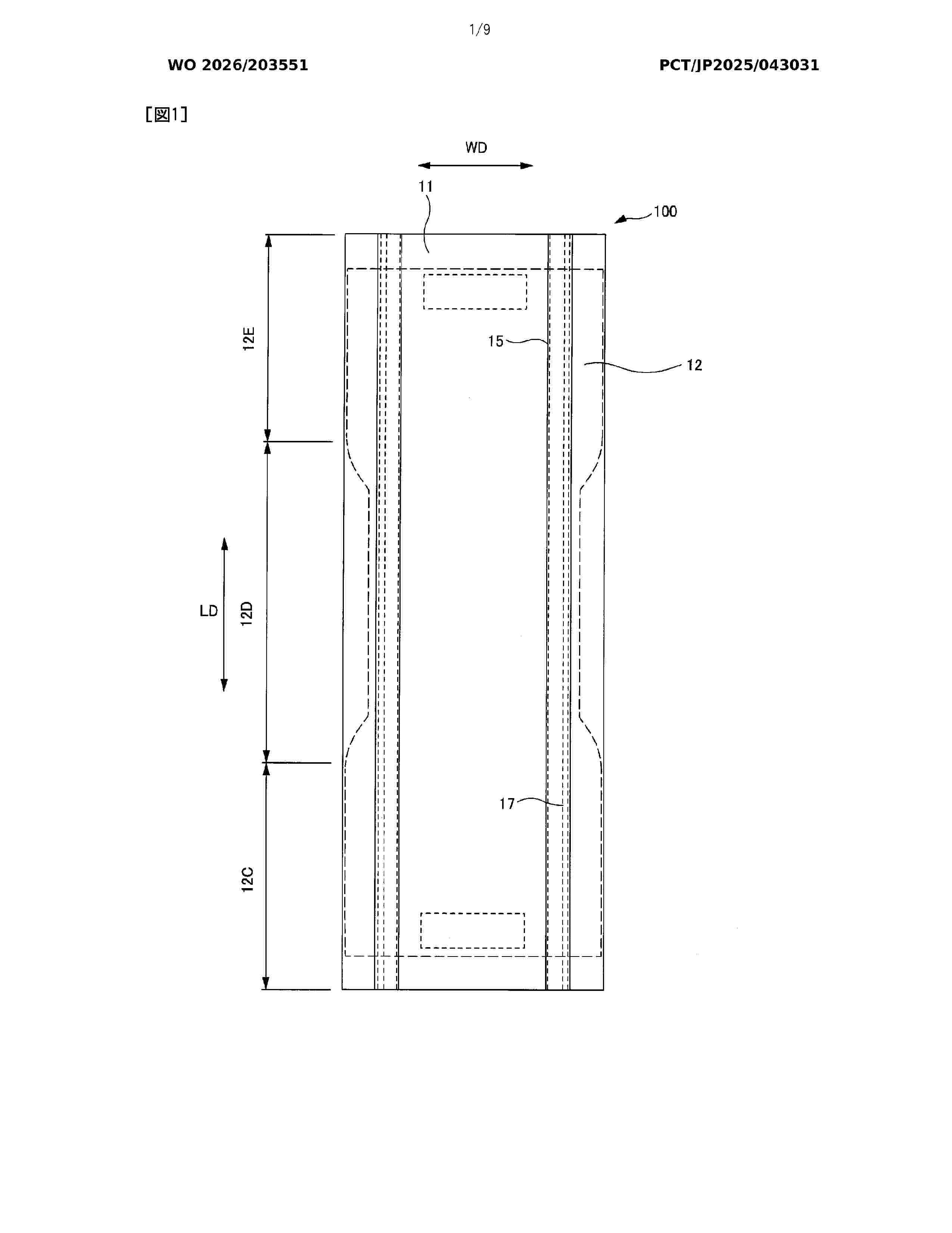
Resumen de: WO2026203551A1
Problem To provide a pad in which is formed a large space for retaining excrement such as urine in the pad, and which makes it possible to prevent excrement that could not be absorbed by an absorbent body from leaking to the outside past a three-dimensional gathering. Solution A pad provided with a liquid-permeable top sheet, a liquid-impermeable back sheet, and an absorbent body between the top sheet and the back sheet, wherein: three-dimensional gatherings that extend in the front-rear direction are fixed at both side parts of the top sheet in the width direction; the absorbent body is formed with a high-stiffness region and a low-stiffness region that is more easily deformable than the high-stiffness region; the low-stiffness region is provided surrounding the outer periphery of the high-stiffness region; and the thickness of the low-stiffness region is greater than the thickness of the high-stiffness region.
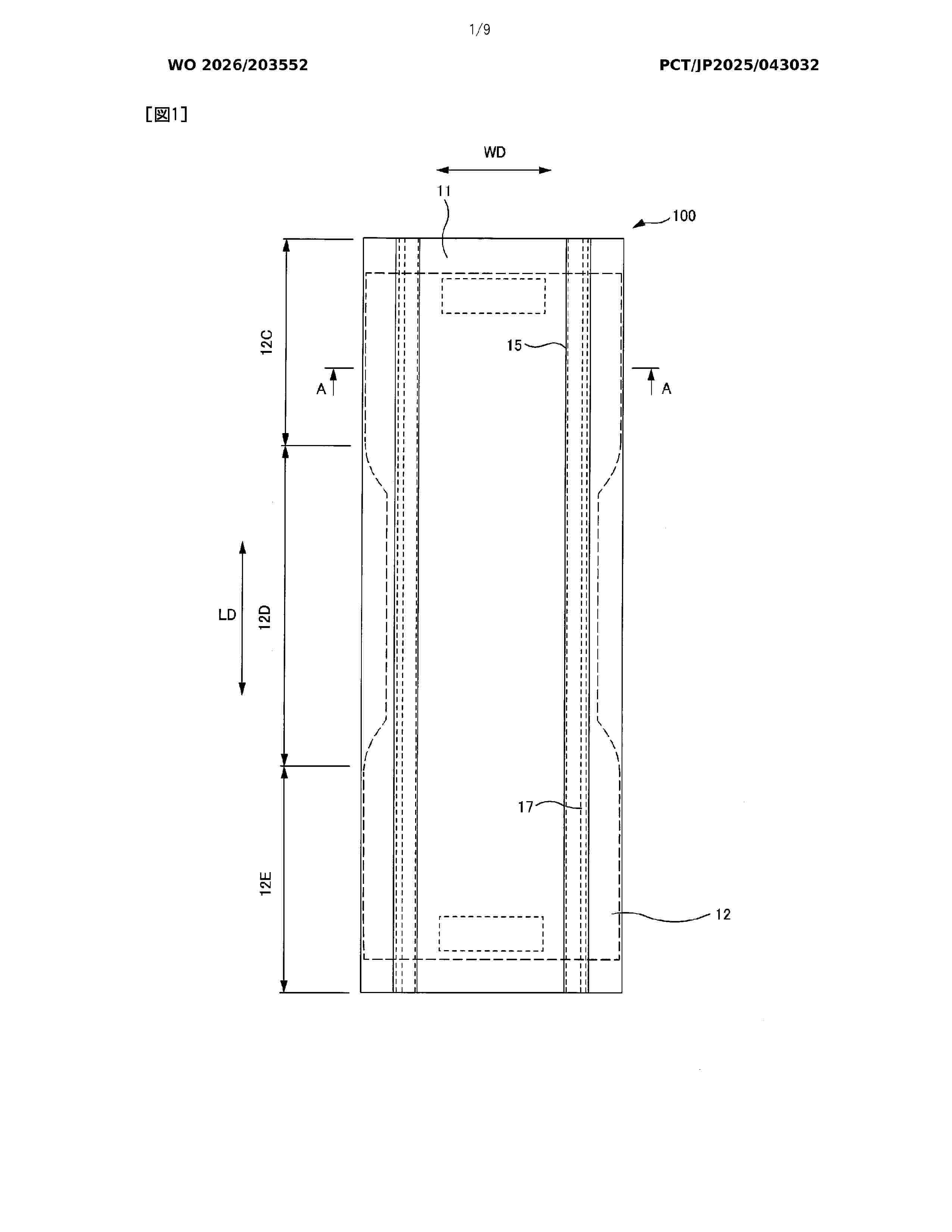
Resumen de: WO2026203552A1
Problem To provide a pad that keeps soft stool that travels along fold lines in the pad from passing through base parts of three-dimensional gathers and leaking to the outside from respective side parts of the pad. Solution According to the present invention, a liquid-permeable second sheet that has a greater width in the left-right direction than a top sheet is provided between the top sheet and an absorption body, base parts of three-dimensional gathers that extend in the front-rear direction are fixed to respective side parts in the left-right direction of the second sheet, the absorption body and the side parts in the left-right direction of the second sheet extend further in the left-right direction than the base parts, and first embossing regions are formed at respective side parts of the base parts at fold parts of the second sheet such that the length in the front-rear direction of the first embossing regions is longer than the length of the fold parts.
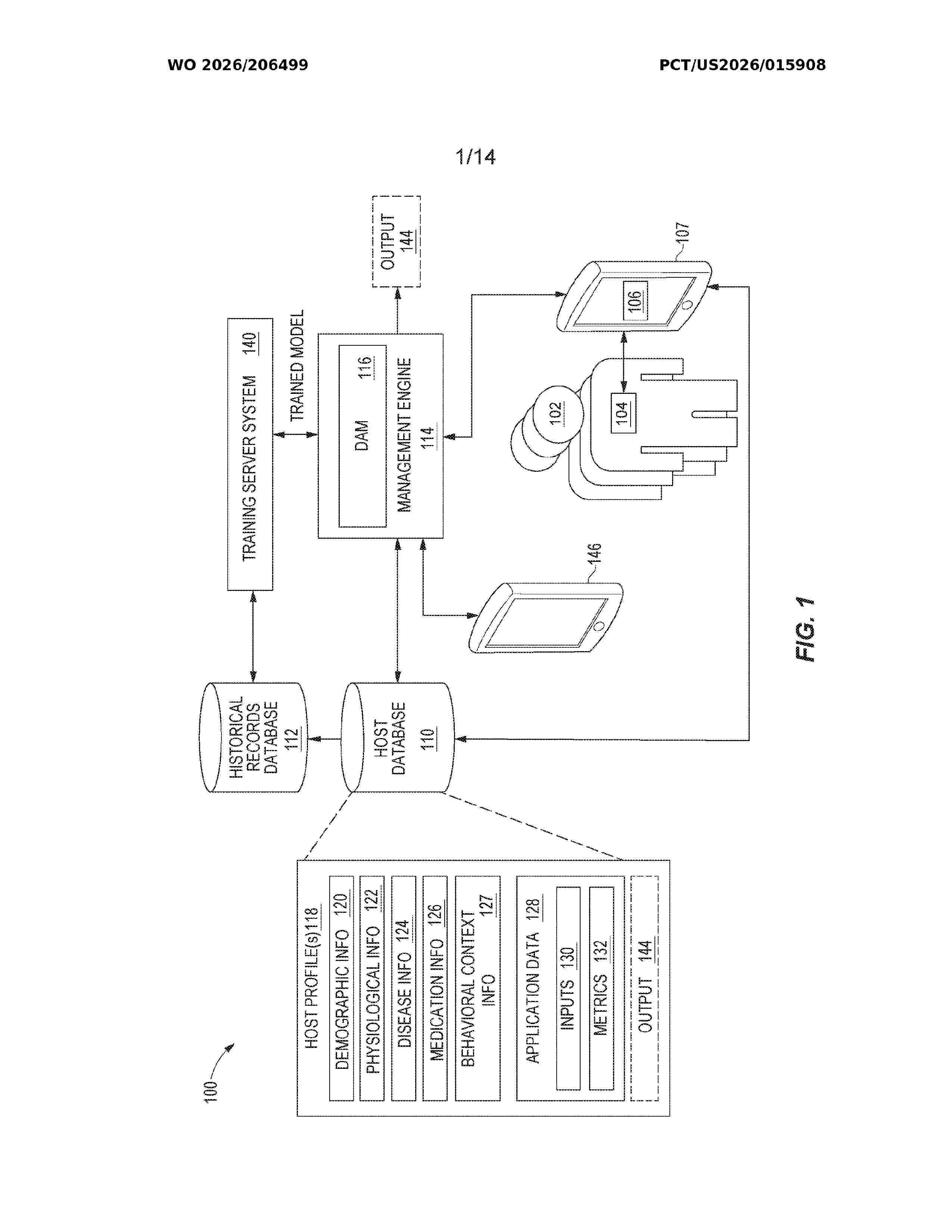
Resumen de: WO2026206499A1
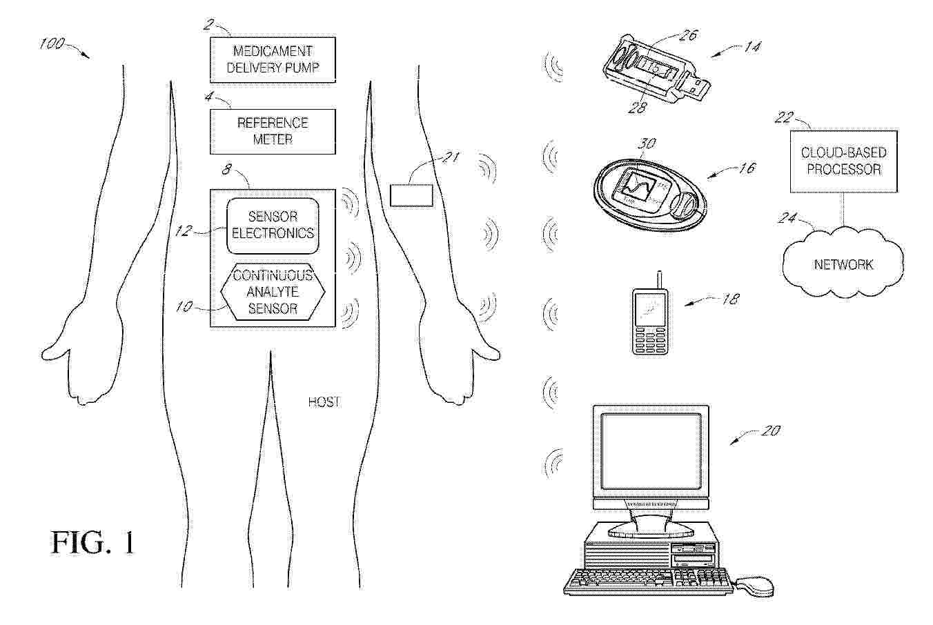
The present disclosure describes an analyte monitoring system that establishes backup or alternative communication channels that may be used when certain components or connections in the analyte monitoring system become unavailable. The analyte monitoring system may form direct connections between components of the analyte monitoring system and/or select replacement devices when certain components or connections become unavailable.
Resumen de: CN118615036A
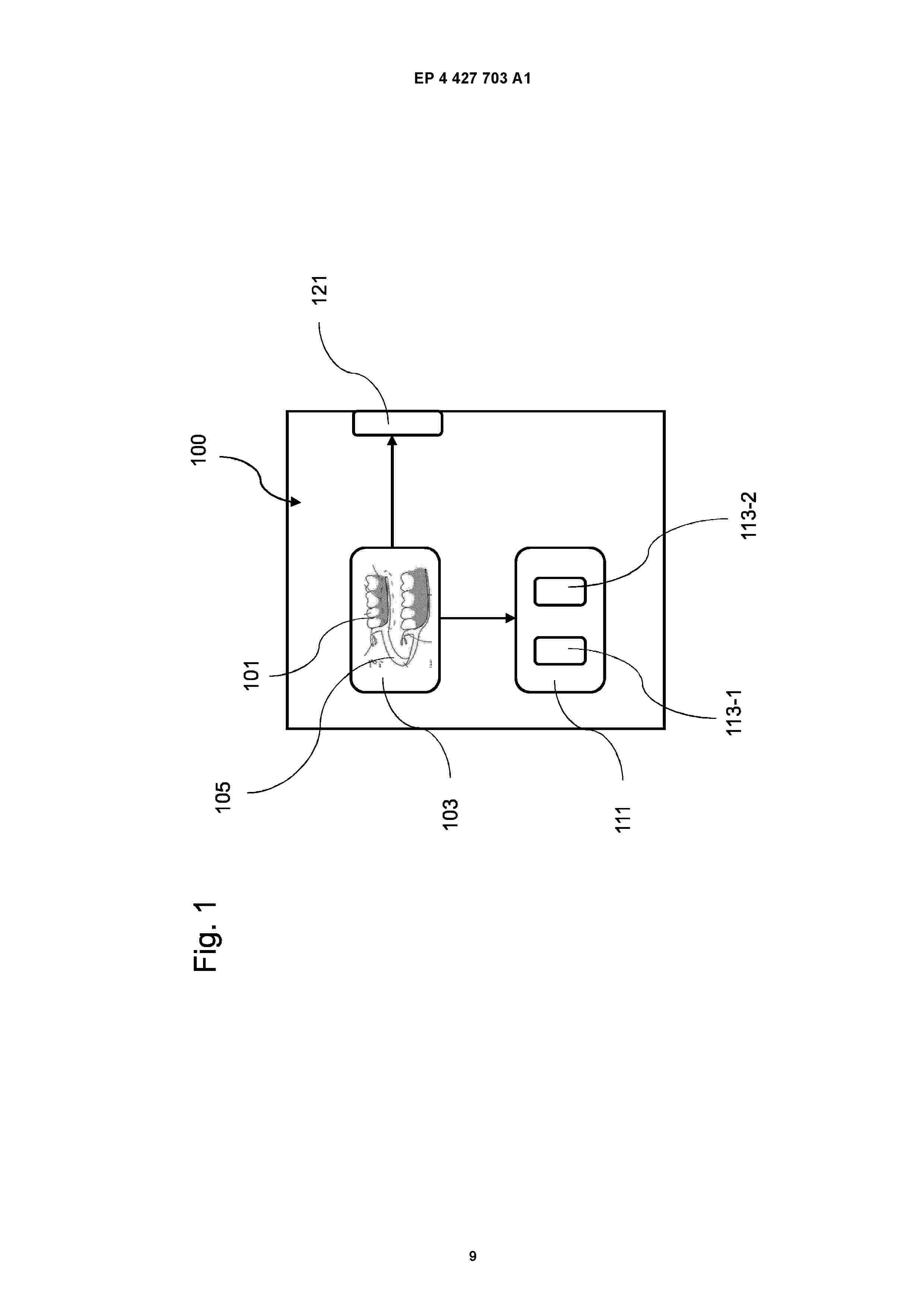
The invention relates to a production system (100) for producing a local prosthesis (101), said production system (100) having a user interface (103) for the digitized design of a prosthesis frame (105), a prosthesis base (107) and a prosthesis tooth (109), the prosthesis frame (105) having a coupling element (119) coupled to remaining dental hard tissue.
Resumen de: ZA202008033B
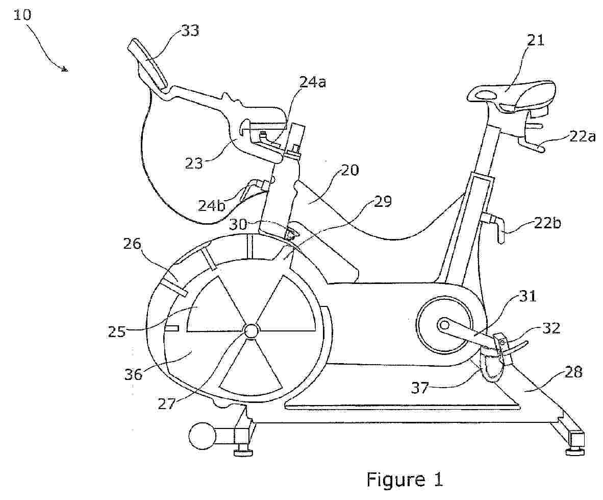
A method of monitoring user effectiveness during cyclic operation of an exercise machine (10) having a pair of human limb-operable drive members (31,32) coupled for cyclic movement. The method comprises the steps of: (i) measuring and monitoring the drive force applied to the drive members (31,32) during cyclic movement of the drive members (31,32) and generating one or more force signals indicative thereof; (ii) using the one or more force signals to derive for each cycle of movement of the drive members (31,32) at least one maximum user effectiveness coefficient signal indicative of variation of force relative to a maximum force value measured for that cycle of movement and at least one minimum user effectiveness coefficient signal indicative of variation in force relative to a minimum force value measured for that cycle of movement; (iii) using the maximum and minimum user effectiveness coefficient signals derived in step (ii) to generate a user effectiveness score signal that is indicative of user effectiveness in driving cyclic movement of the drive members; and (iv) using the user effectiveness score signal in recording, displaying, printing, storing, downloading, uploading or transmitting one or more indicia representative of the user effectiveness score signal.
Resumen de: EP4814978A2
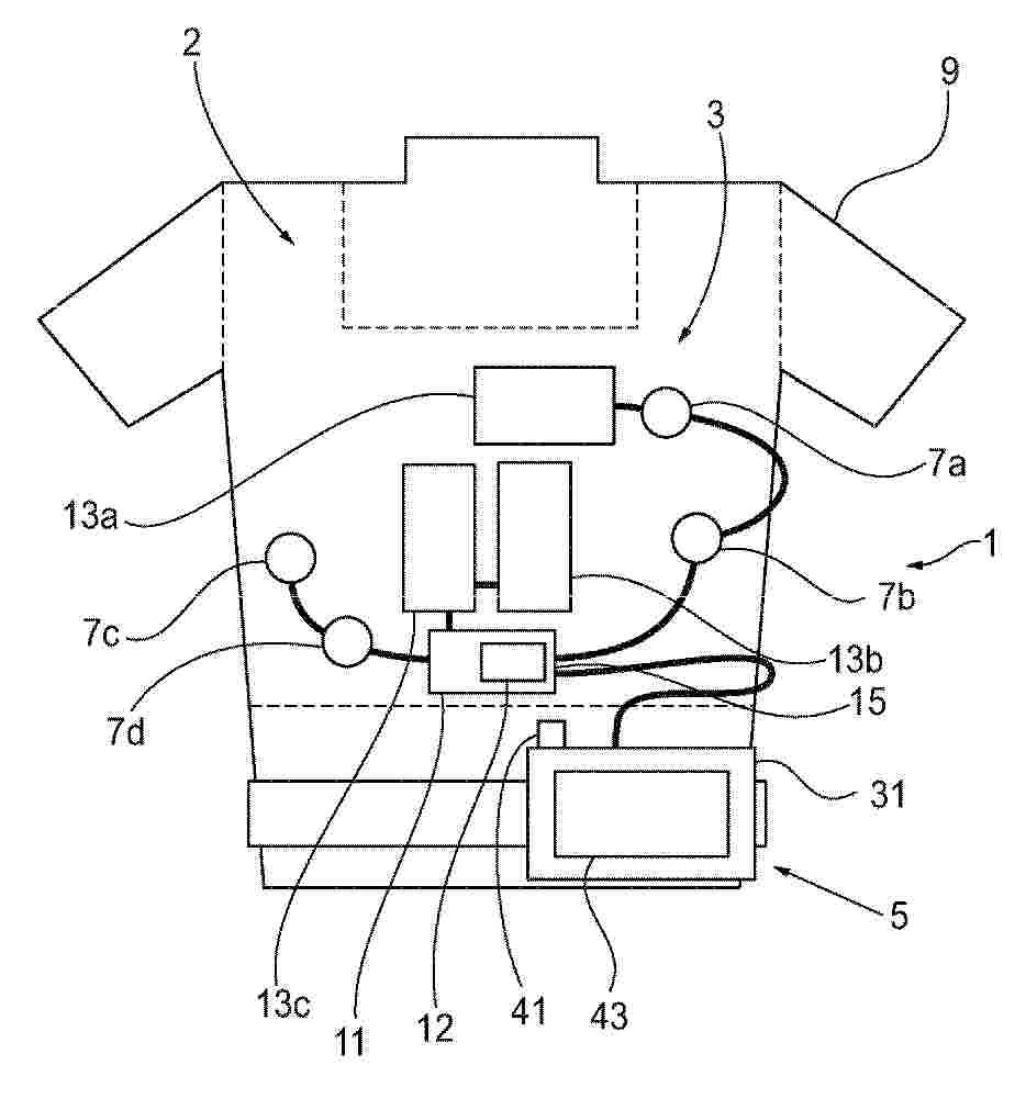
0001 A wearable medical monitoring device can include a monitor and an update manager. The monitor is configured to monitor a physiological parameter of a subject. The update manager is configured to receive a software update corresponding to the at least one software module for the monitor, and cause an installation of the software update such that the at least one software module installed on the monitor is updated. A wearable medical monitoring device can also include at least one processor, and a supervisory circuit configured to provide redundant booting. The supervisory circuit is configured to monitor a state of the processor when the processor is configured to boot from a current drive. The supervisory circuit is further configured to control the processor to boot from an alternative drive based on the state of the at least one processor or if there is an error caused by the current drive.
Resumen de: EP4813342A2
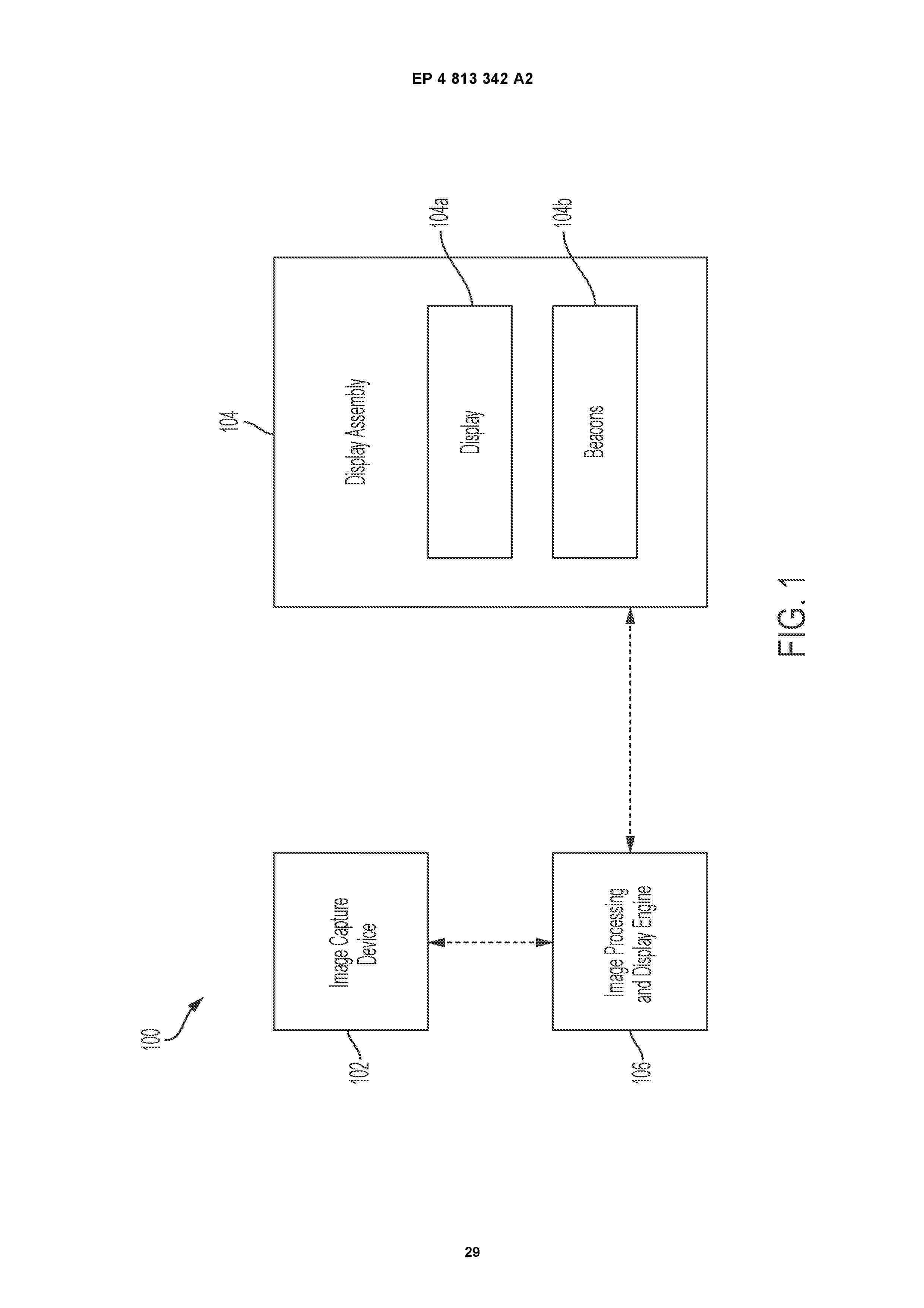
0001 A surgical image capture and display system includes a handheld image capture and pointing device and a display assembly. An image is captured by an image sensor of the handheld device and displayed on the display assembly. The image sensor detects light emitted by one or more beacons of the display assembly. The system determines, based on the light emitted by the one or more beacons, a position or orientation of the handheld device relative to the display assembly. The system updates display of a graphical user interface comprising the image on the display assembly in accordance with the determined position or orientation of the handheld device.
Nº publicación: EP4814988A2 30/09/2026
Solicitante:
DEXCOM INC [US]
DexCom, Inc.
Resumen de: EP4814988A2
The present embodiments provide systems and methods for, among others, tracking sensor insertion locations in a continuous analyte monitoring system. Data gathered from sensor sessions can be used in different ways, such as providing a user with a suggested rotation of insertion locations, correlating data from a given sensor session with sensor accuracy and/or sensor session length, and providing a user with a suggested next insertion location based upon past sensor accuracy and/or sensor session length at that location.
 BOPI
BOPI
 Sede Electrónica