Si deseas distinguir tus productos, servicios o ambos de los de otra empresa, es posible que necesites una marca o nombre comercial. Descubre qué son, en qué consiste su procedimiento de registro y qué implica.

Información sobre los plazos de presentación de solicitudes de transformación de marcas de la Unión Europea en marca nacional española. Más información


Si tienes un nuevo dispositivo, producto o procedimiento que resuelva un problema técnico o tenga una ventaja práctica, existen distintas formas de protegerlo en España y en otros países. Descubre cómo hacerlo.

¿Tu innovación reside en la estética, la ornamentación o la apariencia de tu producto? Protégela mediante un diseño industrial. Descubre qué derechos confiere el registro y cómo realizar la tramitación.

Las indicaciones geográficas protegen el nombre de un producto originario de una zona geográfica, a la cual le debe una determinada calidad, reputación u otra característica. Descubre qué son, en qué consiste su procedimiento de registro y qué beneficios conceden.

Las patentes publicadas en todo el mundo son una valiosa fuente de información científica, técnica y comercial.

Si eres emprendedor/a o una empresa y quieres potenciar y mejorar la rentabilidad de tu negocio protegiendo de forma adecuada los activos intangibles de tu organización, en este espacio encontrarás lo necesario.


 568
resultados
568
resultados
 Última actualización
17/10/2026 [07:24:00]
Última actualización
17/10/2026 [07:24:00]


 Resultados 375 a 400 de 568

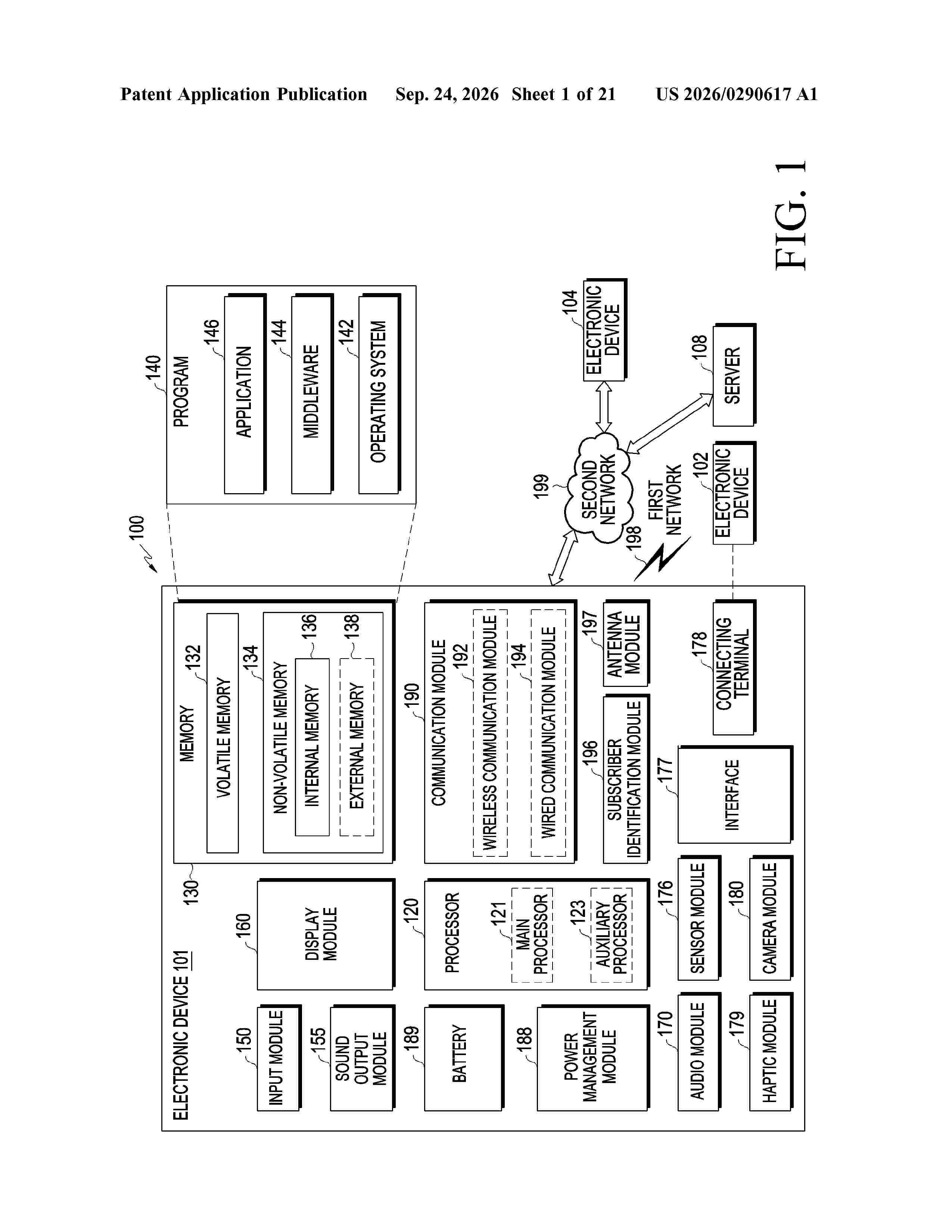
Resumen de: US20260290617A1
An electronic device may include: a touch screen; a communication circuit; a first memory for storing instructions; at least one processor; and a second memory. The instructions, when executed by the at least one processor, cause the electronic device to: store, in the second memory as medication information, information about a medicine administered to a patient and information about medication timing acquired via the touch screen; store, in the second memory, first data related to the patient received from an external device; store, in the second memory, second data related to the patient received from the external device; identify the amount of change in biometric information about the patient based on the first data and the second data; and provide a notification based on the medication information stored in the second memory and the identified amount of change in biometric information.

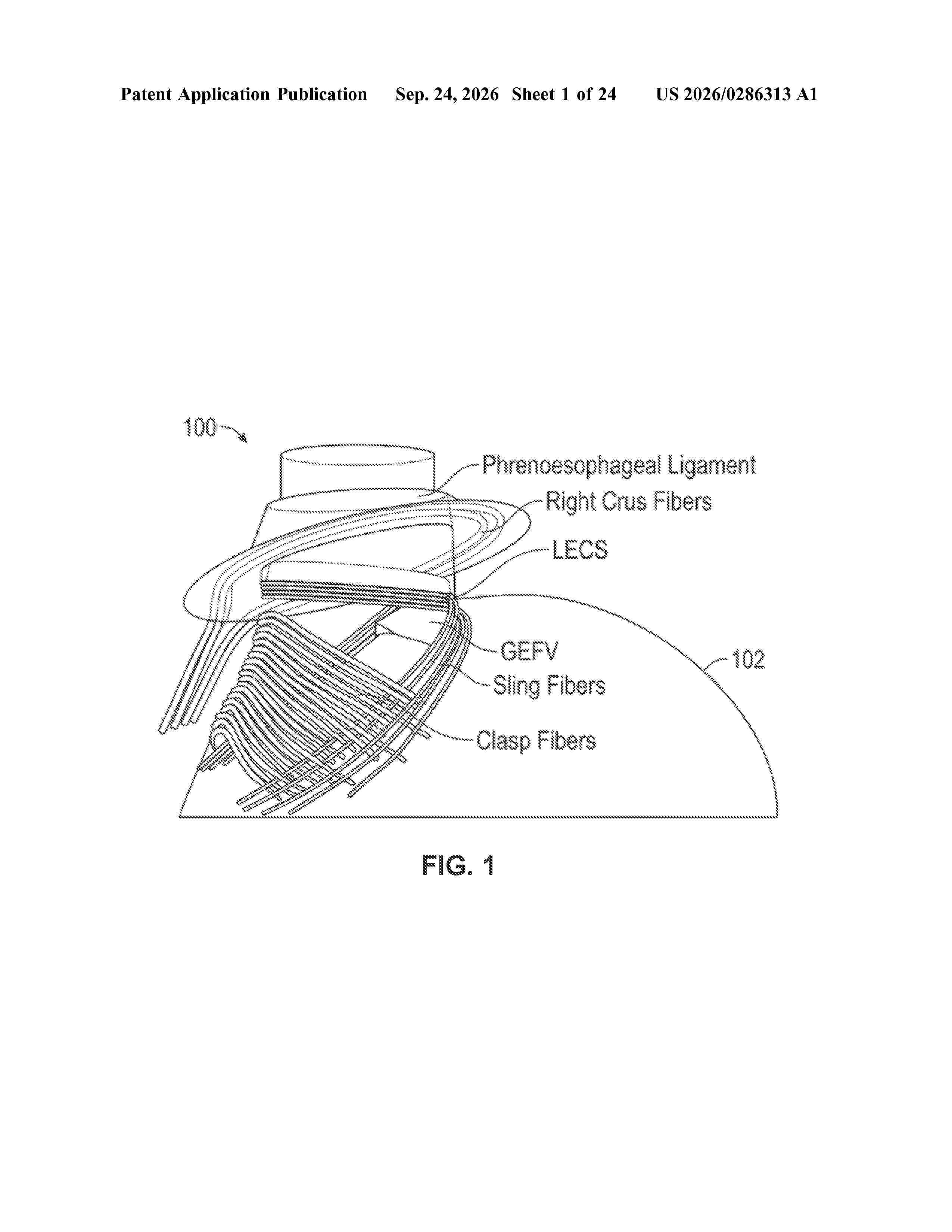
Resumen de: US20260286313A1
0000 Devices, systems, and techniques are described for creating pre-aligned cells in fibers to be wound into tissue constructs or microtissues that can be printed into larger tissue constructs. For example, a method includes dispensing a biological fiber into a solution, wherein the biological fiber comprises a plurality of cells within a hydrogel. The method may then include spooling, under tension, the biological fiber from the solution and onto a spool structure. In some examples, the method may also include winding the biological fiber around a tissue frame to create a tissue construct or cutting the biological fiber matured into a pre-aligned microtissue from the spool structure and used in a bioink for printing tissue constructs.
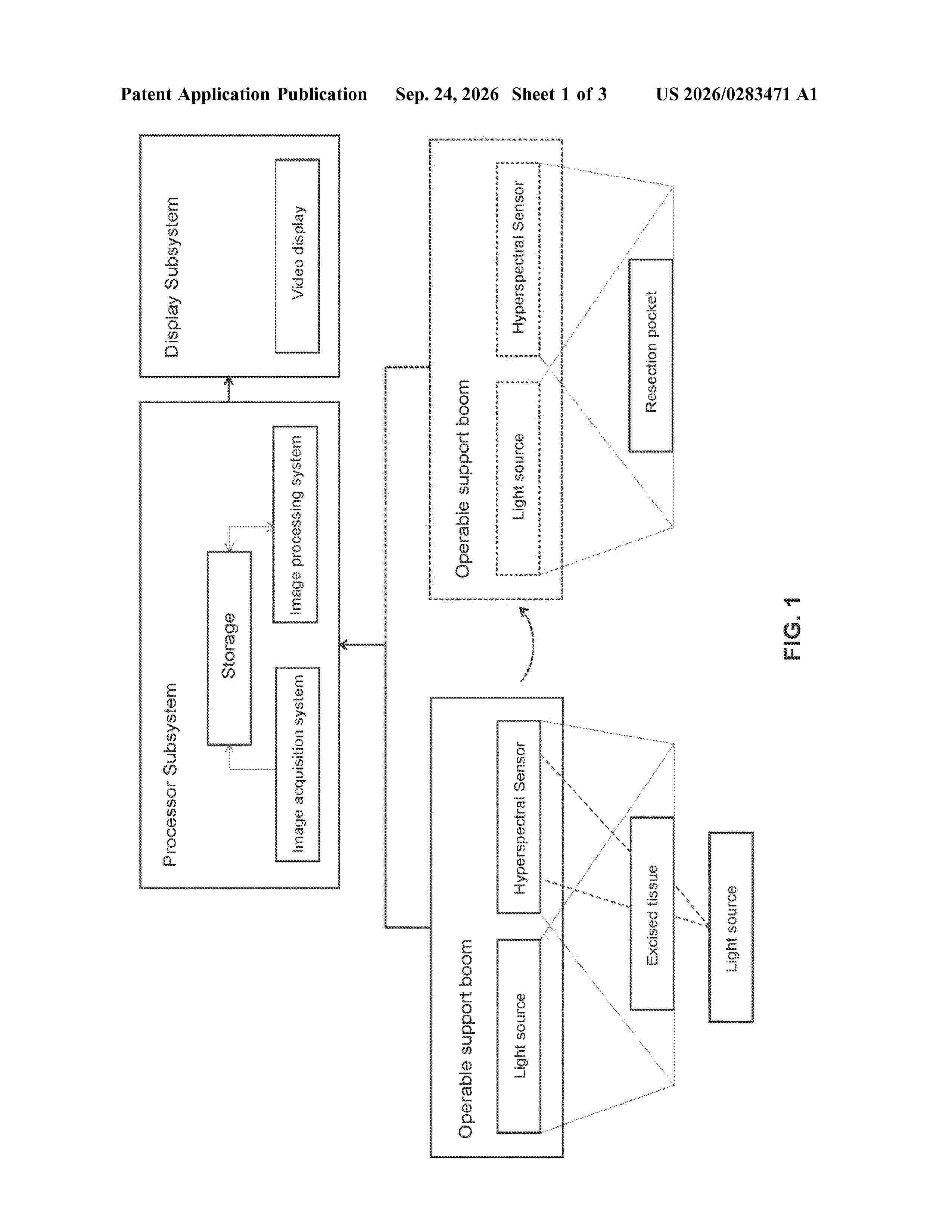
Resumen de: US20260283471A1
Systems and methods for generating hyperspectral medical images capable of producing and processing hyperspectral images of in vivo or ex vivo tissue samples. In processing hyperspectral images, the systems and methods are capable of producing pseudocolor or color-coded images, indicating various tissue types present in the image. The systems and methods may be especially useful for surgical margin assessment, including intraoperative surgical margin assessment, for example, for determining surgical margins during tumor resection.
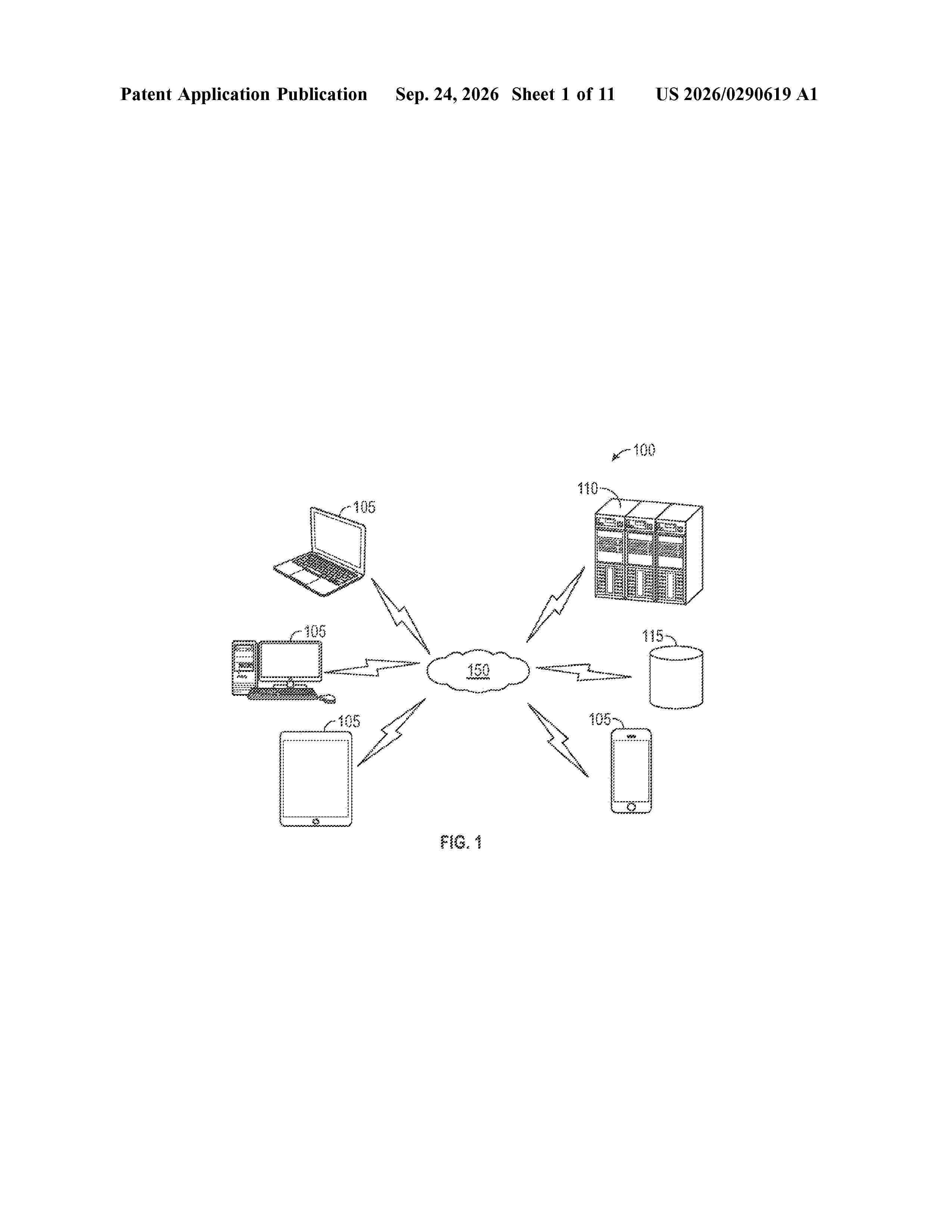
Resumen de: US20260290619A1
A system and method for facilitating virtual medical checkups is provided. Generally, a user may activate a display device of a medical charging/fueling station, which may then identify the user via a security means, such as logging into a user profile via a computing device operably connected to the display device, scanning a predefined pattern, and/or a secondary security device. A computer readable signal containing login credentials may be sent to the display device, allowing the user to access various applications thereon, including an application that may connect the user with healthcare personnel. In some embodiments, the system may allow a user to connect medical devices to the charging station to collect vitals of the user. In other embodiments, the system may provide additional security for electronic devices of a vehicle to reduce the likelihood of a security breach.
Resumen de: US20260283598A1
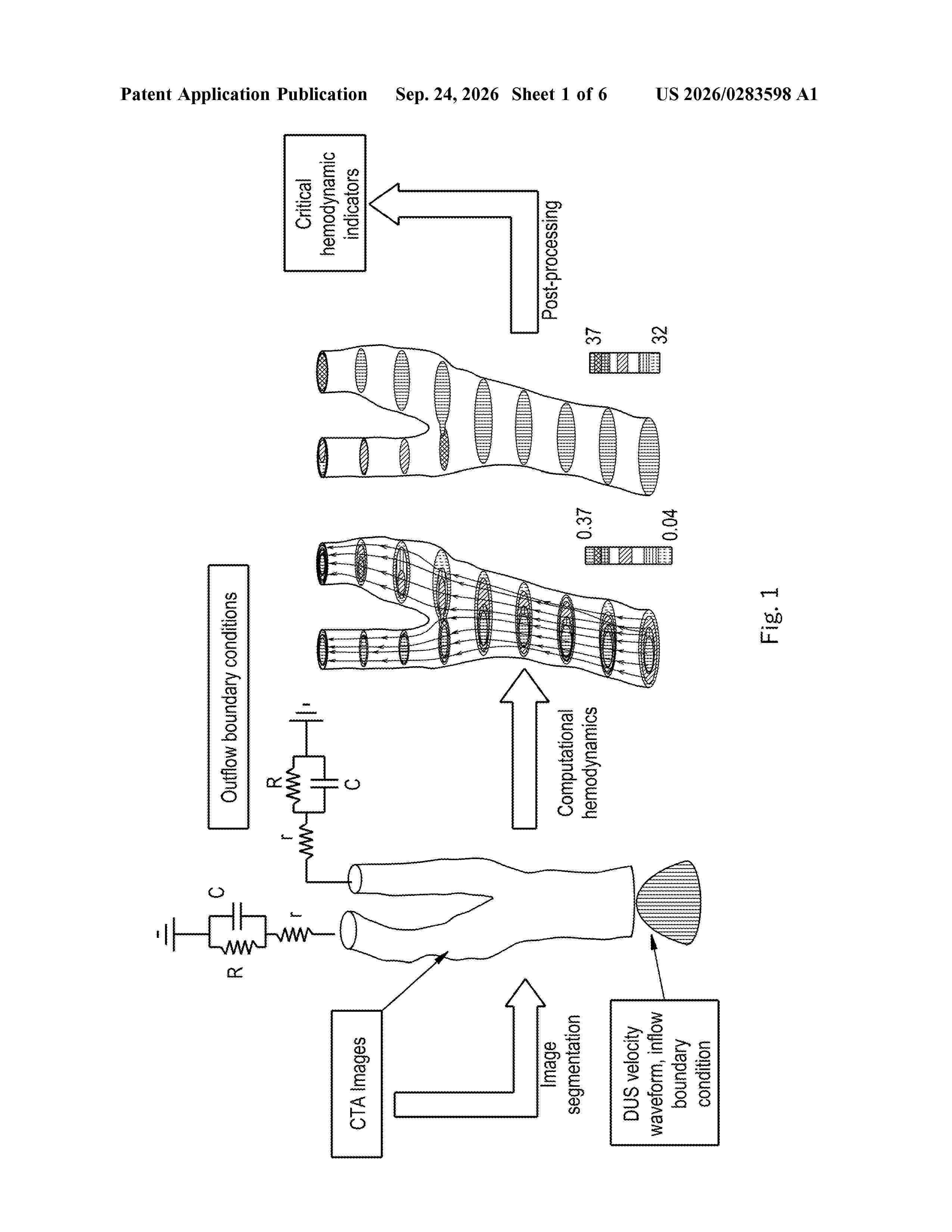
Methods of constructing three-dimensional arterial morphological geometries from two-dimensional images produced by duplex-ultrasound brightness-mode processes. Ultrasound cine looping is used in order to produce multiple images from multiple directions, including longitudinal and transverse directions of an artery, and three-dimensional geometries are produced from the aforementioned images by an image-based deep-learning model.
Resumen de: US20260283597A1
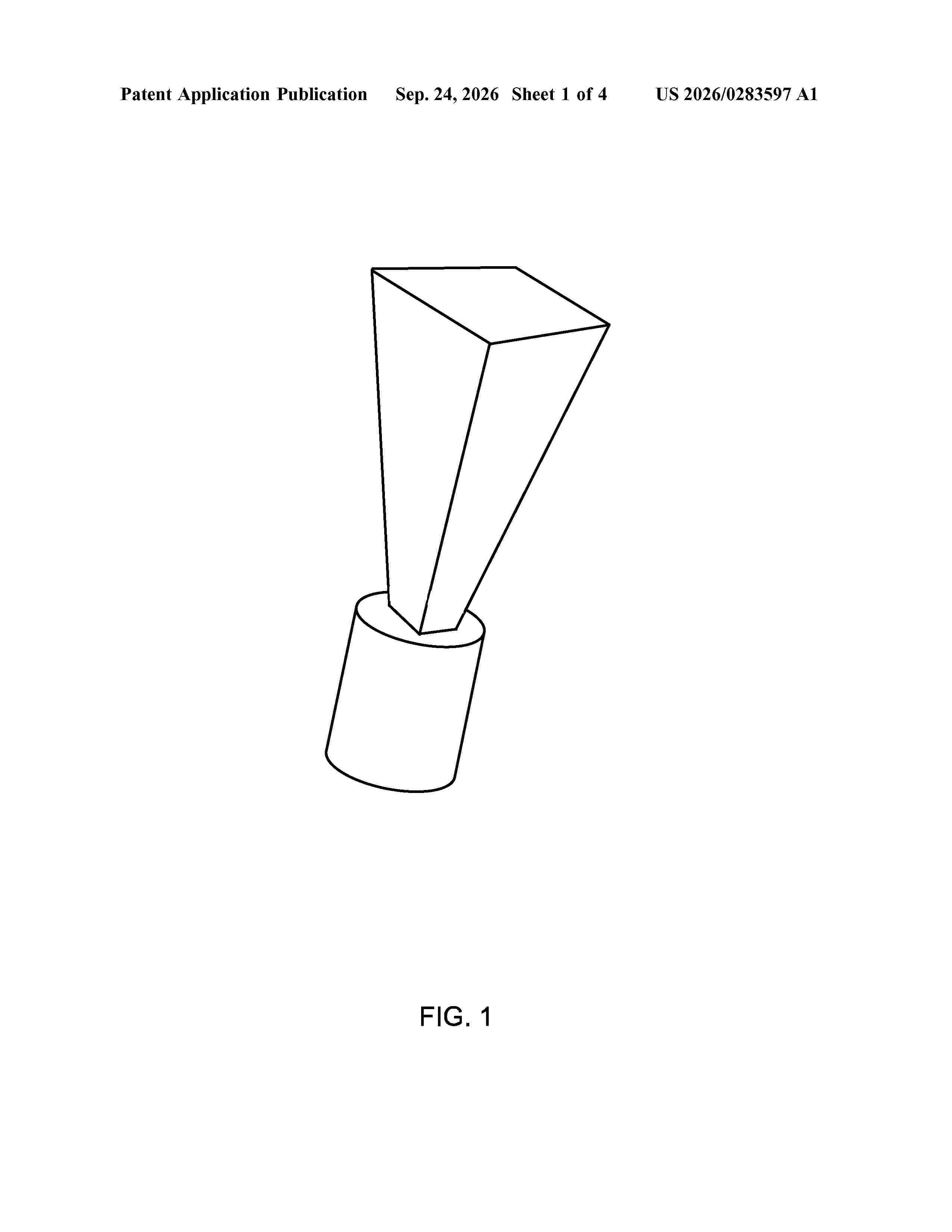
Proposed concepts aim to provide schemes, solutions, concepts, designs, methods and systems pertaining to assessing acquisition quality of a scan of a subject by a 3D ultrasound scanner. Specifically, assessment of image quality in 3D is achieved based on a coherence value of the image data generalized in an elevation dimension (rather than an azimuth dimension used for 2D ultrasound data). Thus, image quality values are determined for different parts of the 3D region based on the coherence factor of the associated ultrasound image data. From the image quality values, a volume of the 3D region that is adequately scanned may be determined. In this way, feedback regarding the acquisition quality of the 3D ultrasound scan may be provided.
Resumen de: US20260283753A1
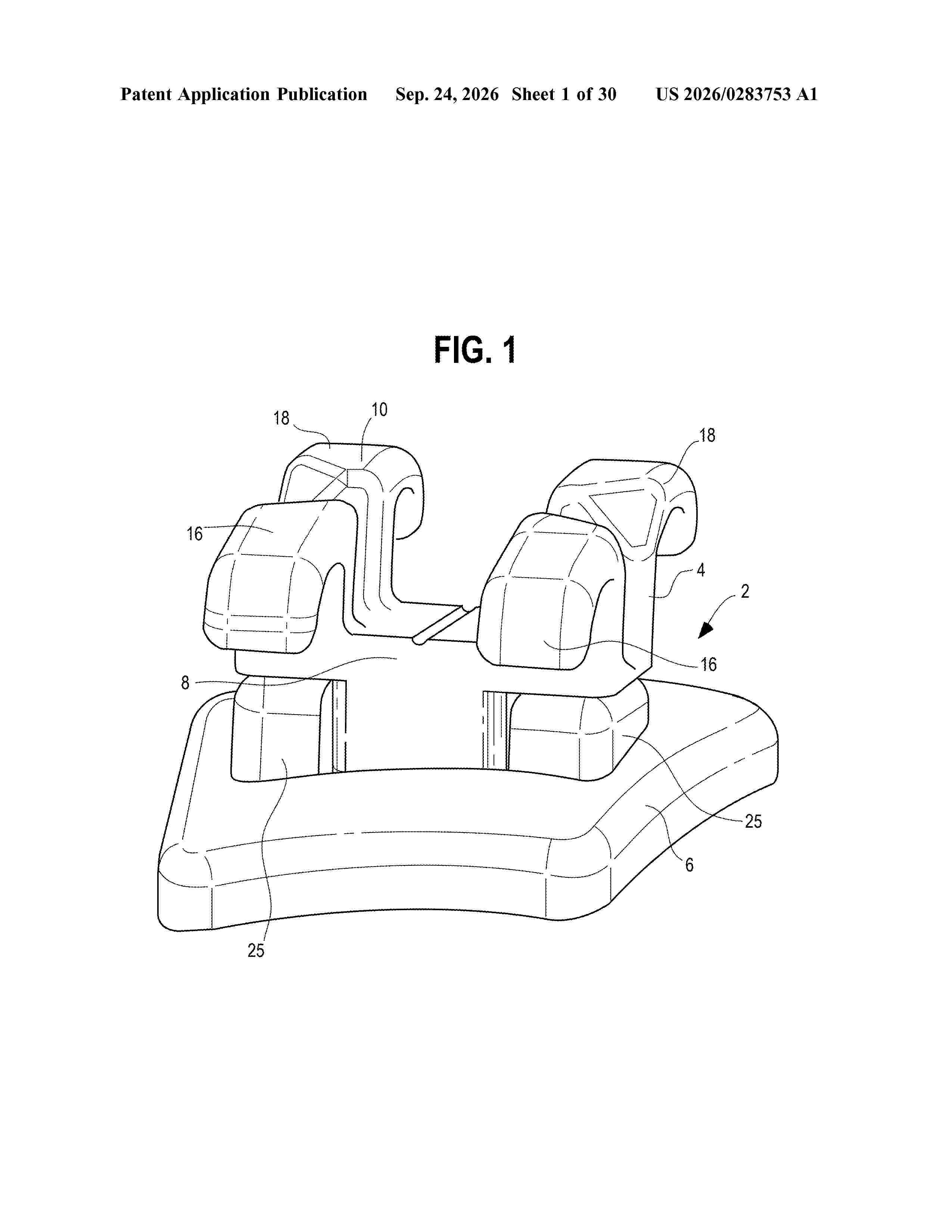
An orthodontic bracket assembly includes a bracket portion and a base portion. The bracket portion includes a body including a first side and a second side. The base portion includes a first side and a second side. The second side of the base portion can include a base contour configured to contact a surface of a patient's tooth. The bracket portion can be affixed to the base portion with the first side of the bracket portion facing away from the first side of the base portion. An orthodontic prescription for the patient's tooth can be incorporated into the orthodontic bracket assembly. A process of manufacturing an orthodontic bracket assembly can include forming a base portion by an additive manufacturing process, providing a bracket portion, and affixing the bracket portion to the base portion.
Resumen de: US20260284615A1
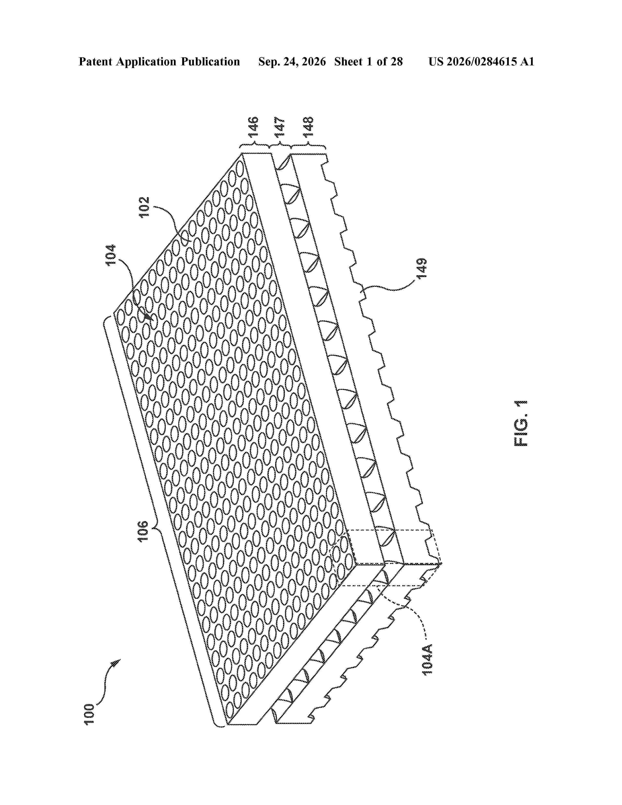
A device 100 for producing nanoparticles includes a microplate 102 which includes a plurality of fluidic mixing units 104 arranged in an array. Each fluidic mixing unit is configured to produce nanoparticles. The device enables high throughput lipid nanoparticle testing and development, and is configured to generate large numbers of novel or unique nanoparticle formulations.
Resumen de: US20260283888A1
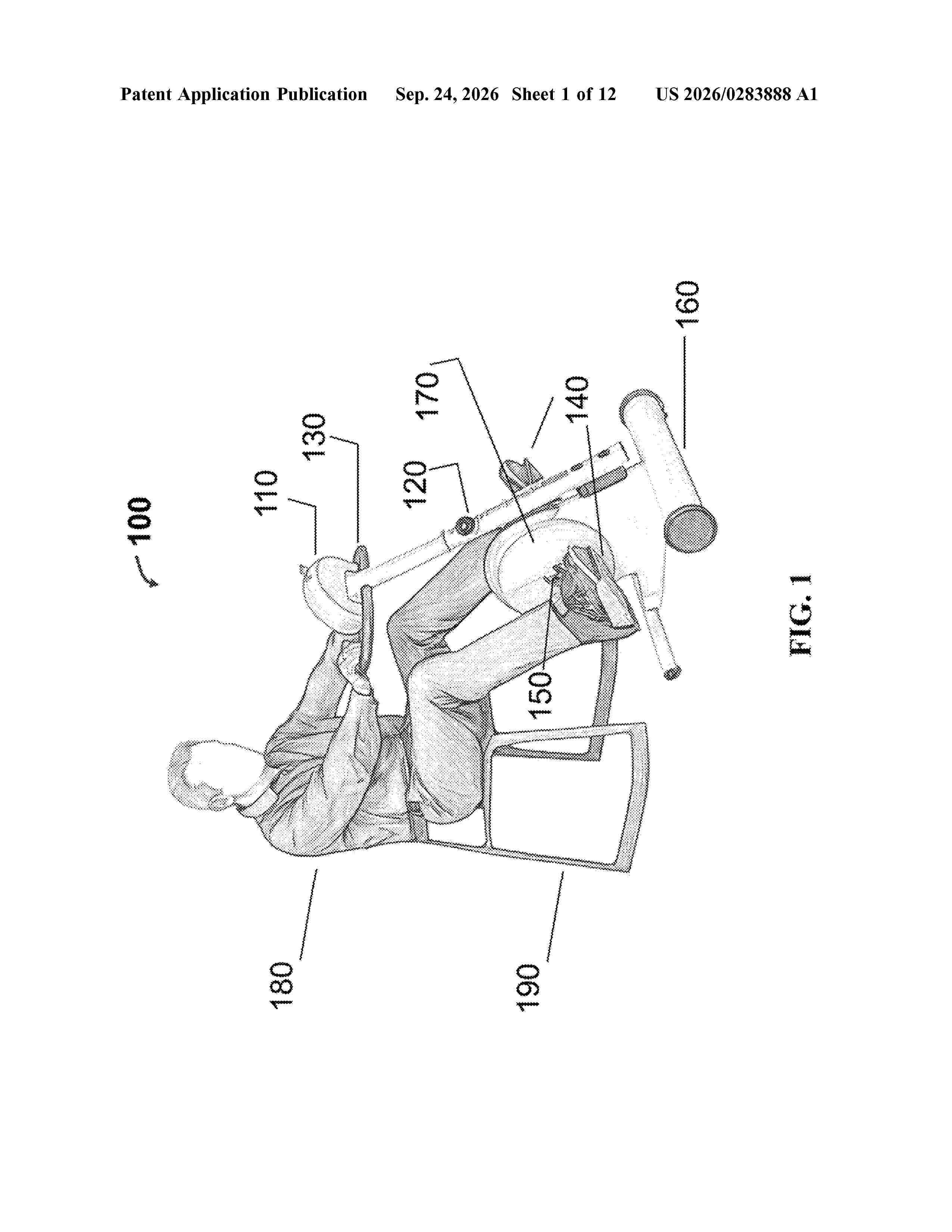
0000 A motorized stationary exercise apparatus includes a motor-driven pedal assembly, at least one sensor configured to measure rider interaction, a personalized exercise planning system, and a control system configured to dynamically personalize operation of the apparatus. The control system generates and continuously updates a rider-specific adaptive performance model that predicts future rider performance. Target motor operating parameters for each exercise session are determined by the personalized exercise planning system to optimize a rider performance objective function, and the electric motor dynamically adjusts pedal speed and torque during and across exercise sessions. The system may implement model-based predictive control, model-free estimation including artificial neural networks, or reinforcement learning or extremum seeking control. The adaptive model may estimate latent rider state variables including fatigue, endurance, strength, or neuromotor capability. The apparatus supports rehabilitation, neurological therapy, performance training, neuromotor entrainment, remote monitoring, multi-device coordination while incorporating safety detection and automatic torque reduction mechanisms.
Resumen de: US20260284300A1
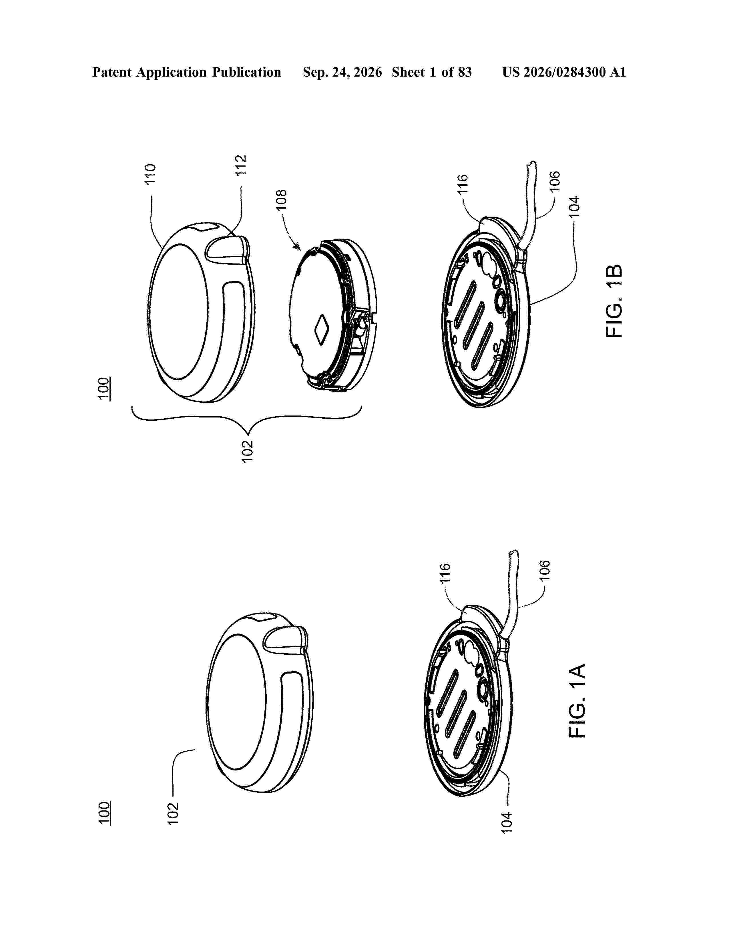
A medical device system is disclosed. The medical device system includes a first medical device and a second medical device. A remote interface including a touch screen is also included. The remote interface is in wireless communication with the first medical device and the second medical device. The remote interface is configured to provide a user interface to the first medical device and the second medical device. The remote interface is configured to receive user input through a touch screen. Also, a charging device is included. The charging device is configured to receive at least the first medical device and the remote interface and the charging device is configured to recharge a first medical device battery and the charging device is configured to recharge an interface battery in the remote interface. The charging device is connected to a personal computer wherein the personal computer provides information to the remote interface.
Resumen de: WO2026198934A1
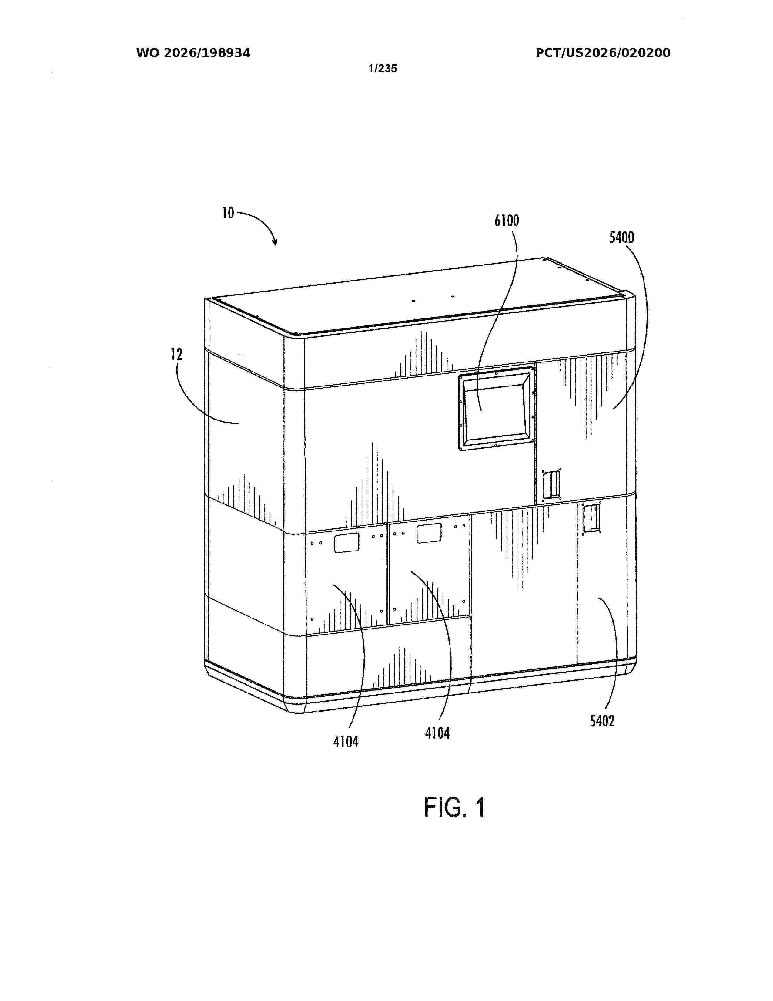
An example dispenser of medical reservoirs may comprise a rigid frame defining a central fenestration. The dispenser may further comprise a shell coupled to the frame. The dispenser may further comprise a removable cover retained within the fenestration. The cover may include a panel with a port and a breathable microbial barrier extending across the port coupled to the panel. The frame, shell, and cover may define an interior dispenser volume. The dispenser may further comprise a frangible coupled to and at least partially covering exterior faces of the frame and the panel. The frangible may have an aperture aligned with the port. The frangible may be coupled to the exterior faces of the frame and the panel to form seals around the port, along the exterior face of the frame adjacent the fenestration, and along the exterior face of the panel adjacent the peripheral edge of the panel.
Resumen de: US20260283496A1
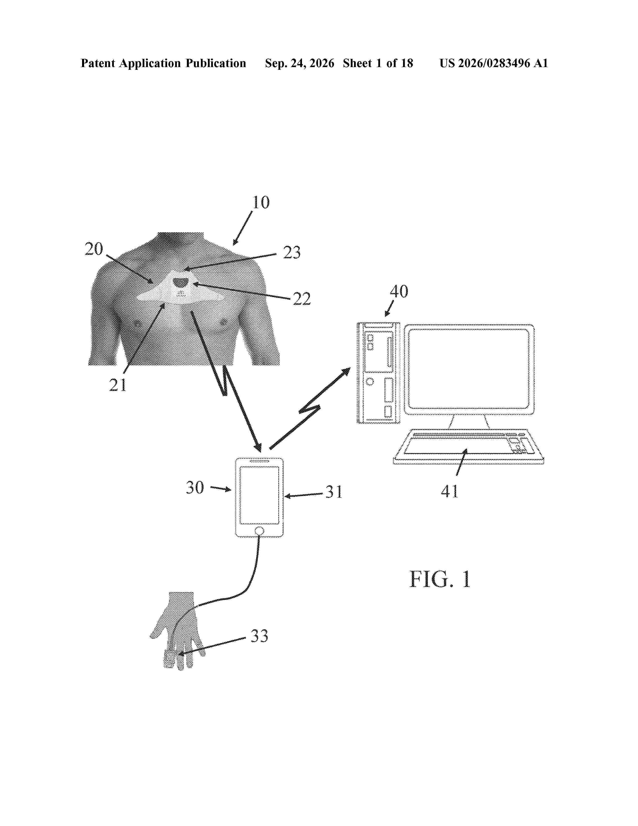
0000 Systems and methods are providing for detecting and monitoring respiratory, cardiac and/or sleep trend or status, and cardiopulmonary coupling in real time, wherein data obtained from a non-invasive electrode patch is analyzed using analysis algorithms for an electrical equivalent model that have been personalized for a patient's physiologic characteristics, medical condition and/or historical medical information using machine learning trained on a dataset representative of a large and diverse patient population. The systems and methods provide a simple, real-time, highly sensitive and specific, non-invasive, bedside solution for fluid level assessment, checking for increased air trapping, and ventilation assessment. The methods described include a variety of use cases for the inventive system and methods.
Resumen de: AU2025232484A1
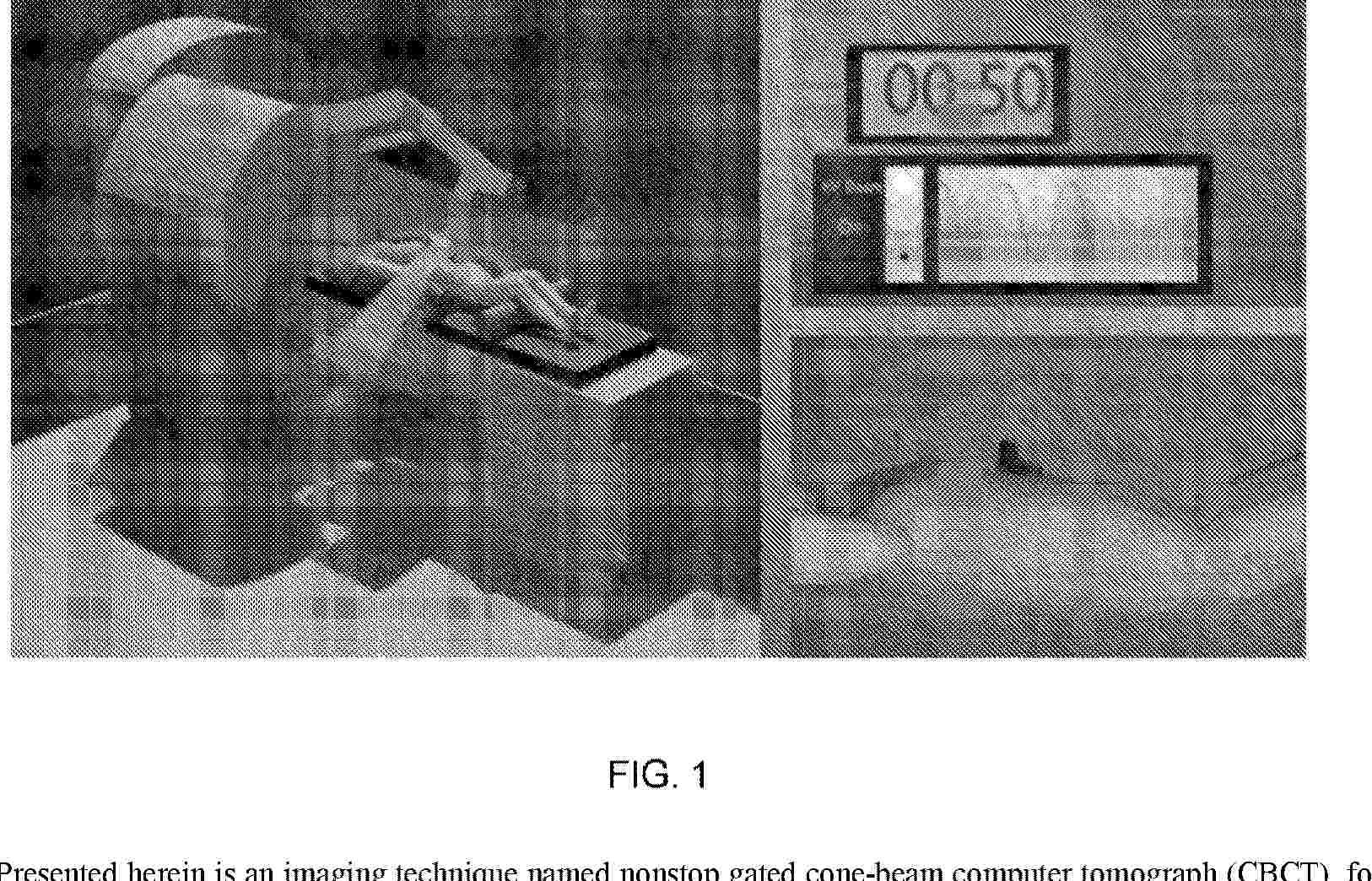
Presented herein is an imaging technique named nonstop gated cone-beam computer tomograph (CBCT), for guiding free-breathing respiratory gating radiotherapy of thoracic and abdominal cancer patients (e.g., lung, liver, pancreas). The nonstop gated CBCT technique reduces the data acquisition time to 1 minute and decreases the imaging dose by >40% while simultaneously retaining high quality images was developed. This is done by allowing the gantry to rotate continuously and having the kV x-ray beam on when the patient breathing signal is within the respiratory gate and the x-ray beam off when it is out of the respiratory gate. A machine learning model can be used to reconstruct high-quality CBCT images from the acquired data.
Resumen de: US20260287432A1
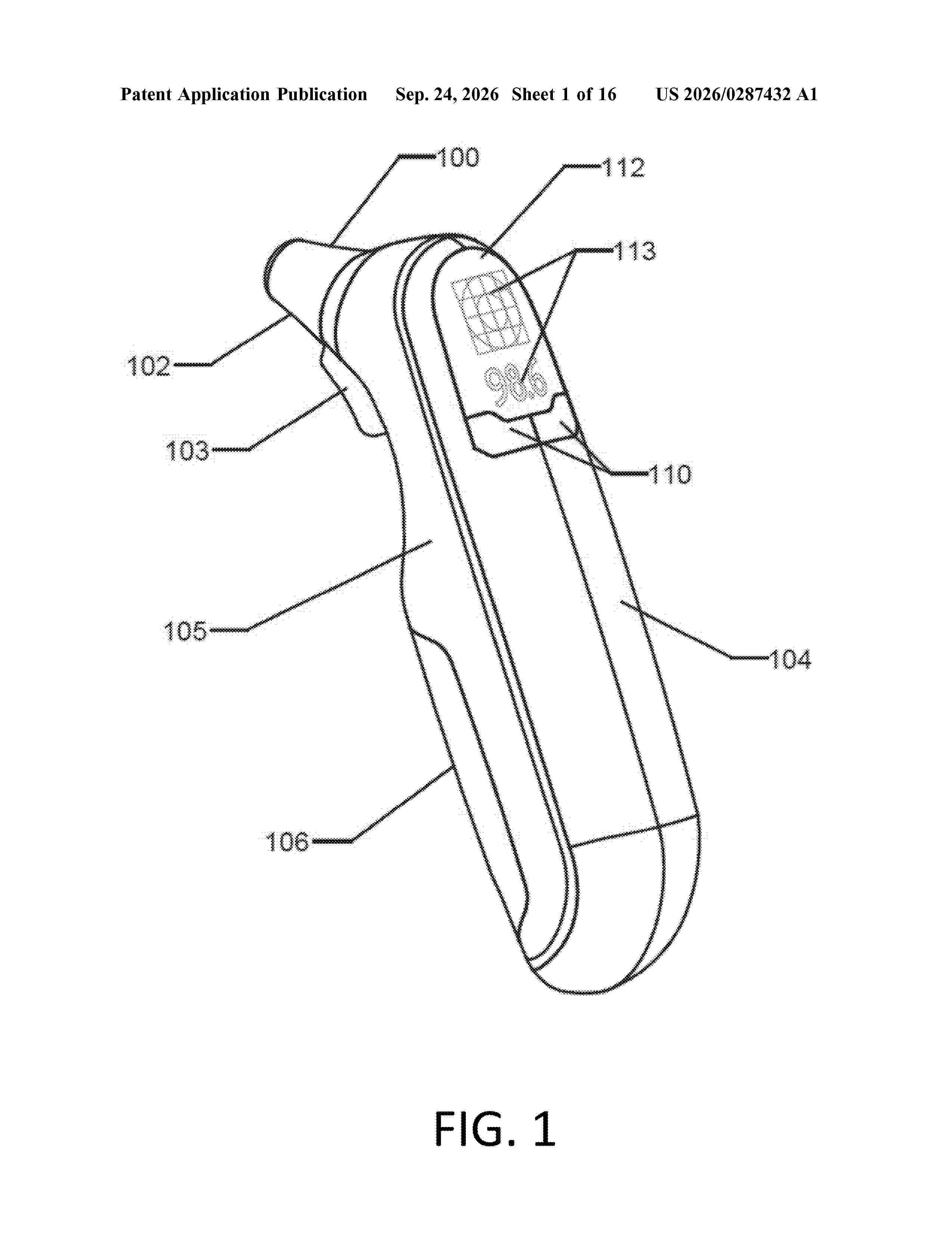
0000 The disclosed advanced ear infection detection device enhances diagnostic accuracy and efficiency, particularly for acute otitis media. A grid array infrared sensor offers both contact and non-contact options while integrating traditional temperature measurements. The device employs algorithmic calibration and data analytics to refine detection accuracy, ensuring precise, real-time assessments. Its wide-surface infrared scanning improves data collection, reducing diagnostic errors and streamlining the process. With rapid, reliable readings in seconds, it minimizes patient discomfort and seamlessly integrates into various healthcare settings, including pediatrics and emergency care. By addressing the limitations of conventional methods, this innovation sets a new standard in medical diagnostics, leveraging technology to improve.
Resumen de: US20260283479A1
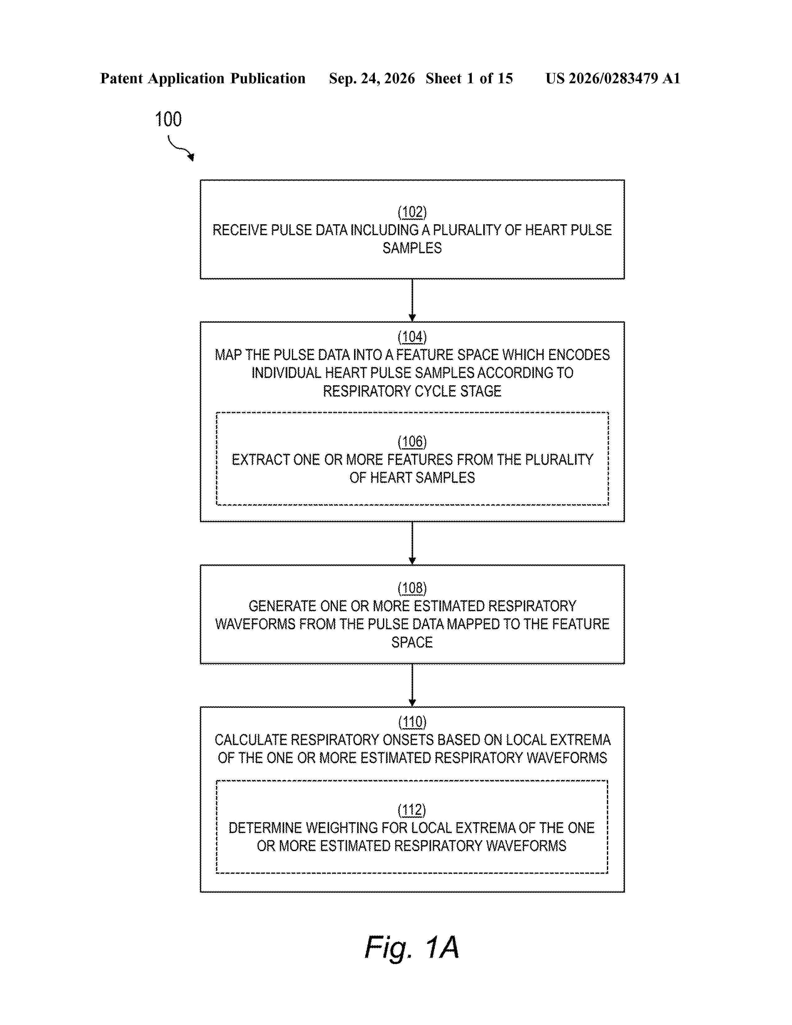
0000 Pulse data including a plurality of heart pulse samples of a user during a time window may be received from a wearable physiological monitor worn by the user. The pulse data may be mapped into a feature space that encodes individual heart pulse samples according to respiratory cycle stage. A first estimated respiratory waveform of the user during the time window may be generated from the pulse data mapped to the feature space and a plurality of respiratory onsets for the user during the time window calculated based on a plurality of local extrema of the first estimated respiratory waveform. Motion data may also be used, either alone or in a multi-model system, to evaluate user respiration.
Resumen de: WO2026195199A1
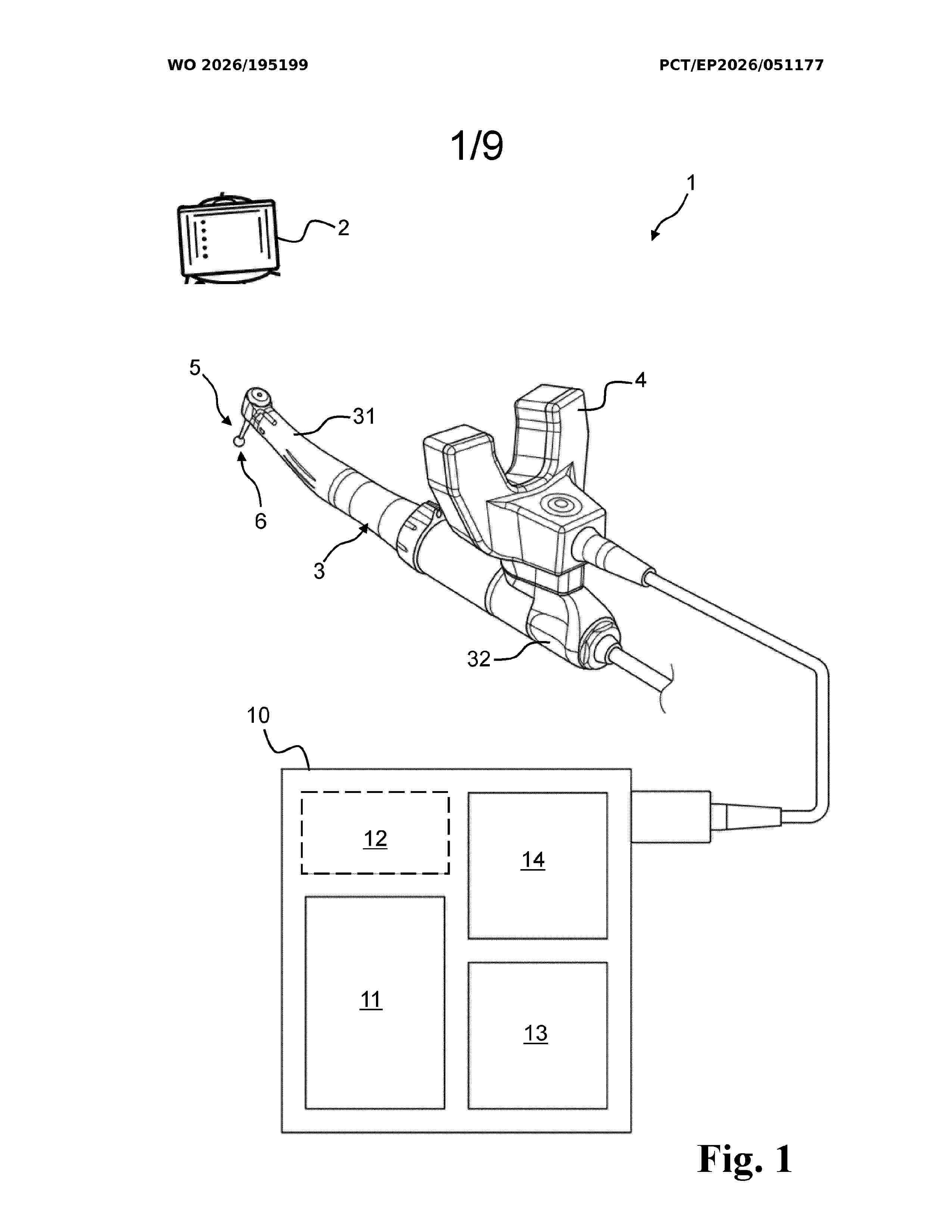
A method for determining a position of a marker member for alignment of a surgical handpiece is provided. The surgical handpiece has an imaging unit releasably attached thereto and being configured to detect the marker member. The surgical handpiece further has a probe comprising a probe tip arranged at a defined position relative to the imaging unit. The method comprises providing the marker member at a fixed position relative to a dental and/or cranio-maxillofacial structure of a patient. The method comprises obtaining a data set indicative of a shape of a landmark and a position of the landmark relative to the marker member. The method comprises determining whether the data set meets one or more data collection criteria. The method comprises, upon the data set meeting the one or more data collection criteria, matching the data set to a digital model of the dental and/or cranio-maxillofacial structure. The method comprises mapping the position of the marker member to the digital model of the dental and/or cranio-facial structure based at least in part on the data set.
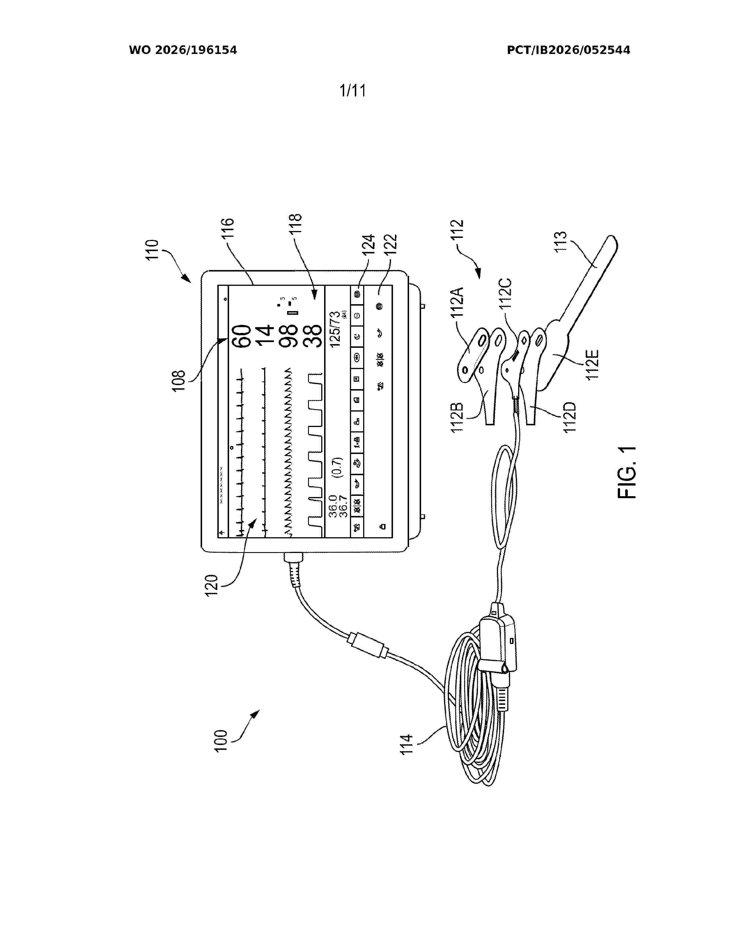
Resumen de: WO2026196154A1
The techniques of this disclosure generally relate to multi-site medical sensors that can be repositioned to multiple sites on a patient. A multi-site sensor includes a flexible substrate with physiologic sensing elements responsive to physiologic characteristics of the patient, and a deflection sensor responsive to deflection of the sensor about a folding axis. The measured deflection is used to determine the location of the sensor on the patient.
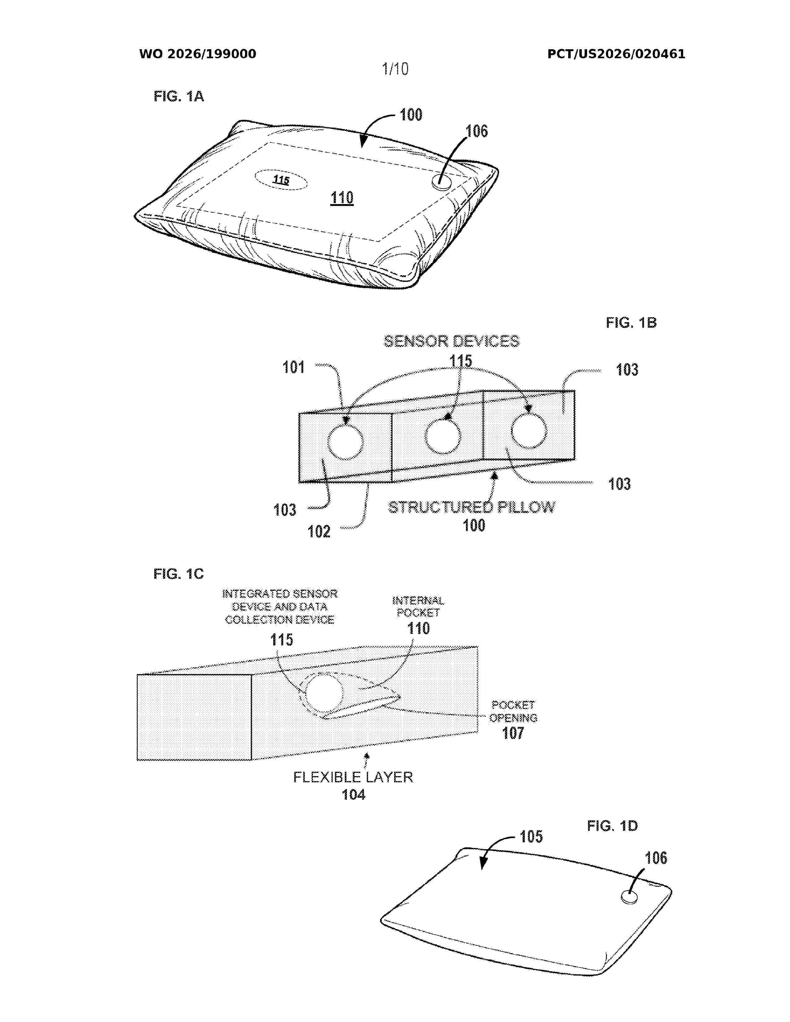
Resumen de: WO2026199000A1
A polysomnography system comprises a pillow with a sensor and a fluid-filled chamber that dampens mechanical vibrations from a subject's environment. The sensor includes an accelerometer that converts vibrations into electrical ballistocardiogram signals representing head movement caused by heartbeats. A computing device with processors and memory receives these signals via a communication interface. The device applies signal processing to extract features indicative of respiratory events and uses a trained neural network to estimate an apnea-hypopnea index by detecting apnea and hypopnea events. Based on this index, the system transmits data to a remote server or stores it in a database.
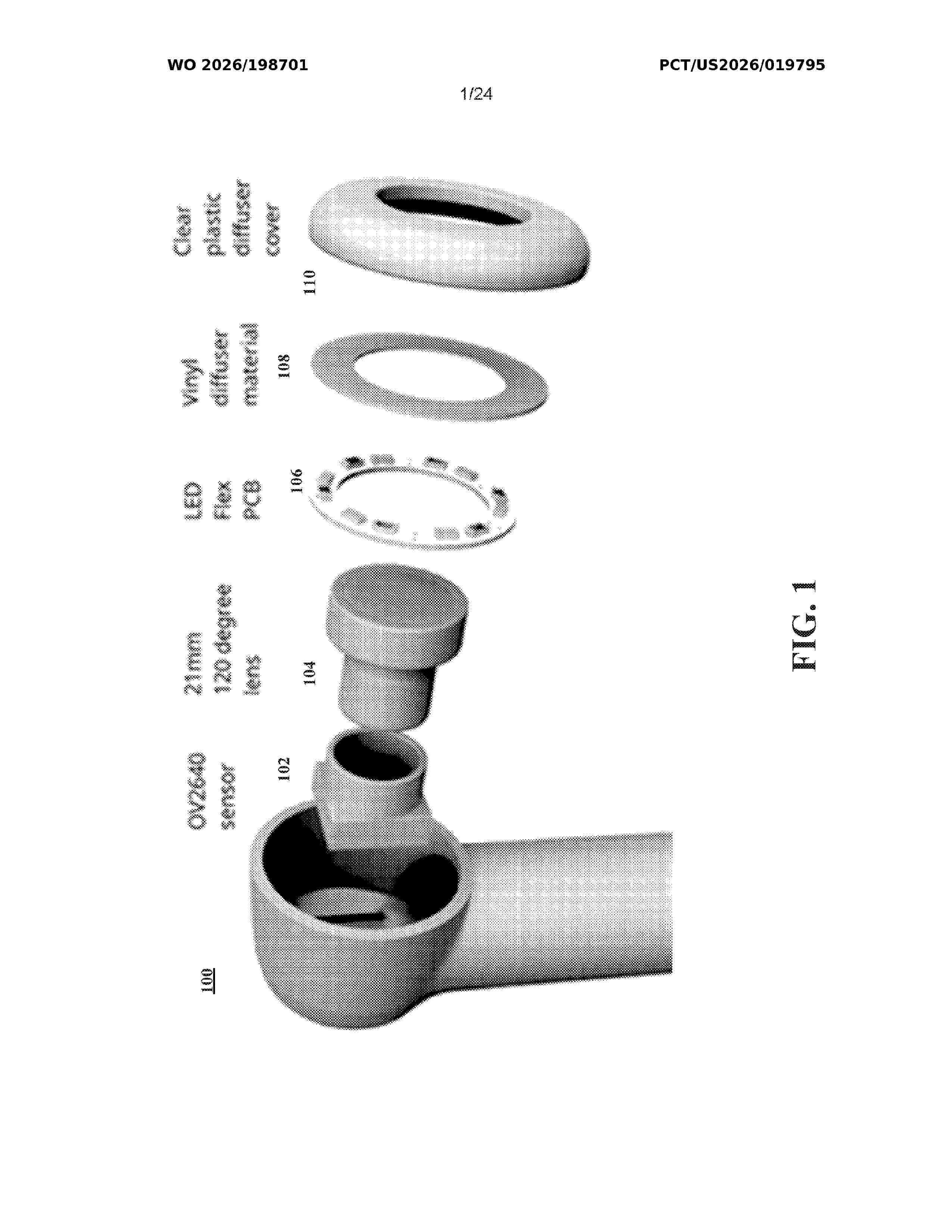
Resumen de: WO2026198701A1
A health monitoring system is disclosed that includes a handheld intraoral imaging device, a mobile computing device executing a mobile application, and a multi-layer artificial intelligence (AI) analysis framework. The handheld intraoral imaging device includes an image sensor, lens assembly, illumination sources, wireless communication module, processor, and memory, and is configured to capture intraoral image data and transmit the image data to the mobile computing device. The AI analysis framework is executed by at least one of the handheld intraoral imaging device and the mobile computing device. The AI analysis framework filters non-relevant or low-quality images, identifies anatomical regions within retained images, associates retained images with specific teeth or oral regions, and generates longitudinal condition indicators by comparing historical and current image data. The mobile application presents the longitudinal condition indicators to a user to support oral health monitoring and risk-awareness.
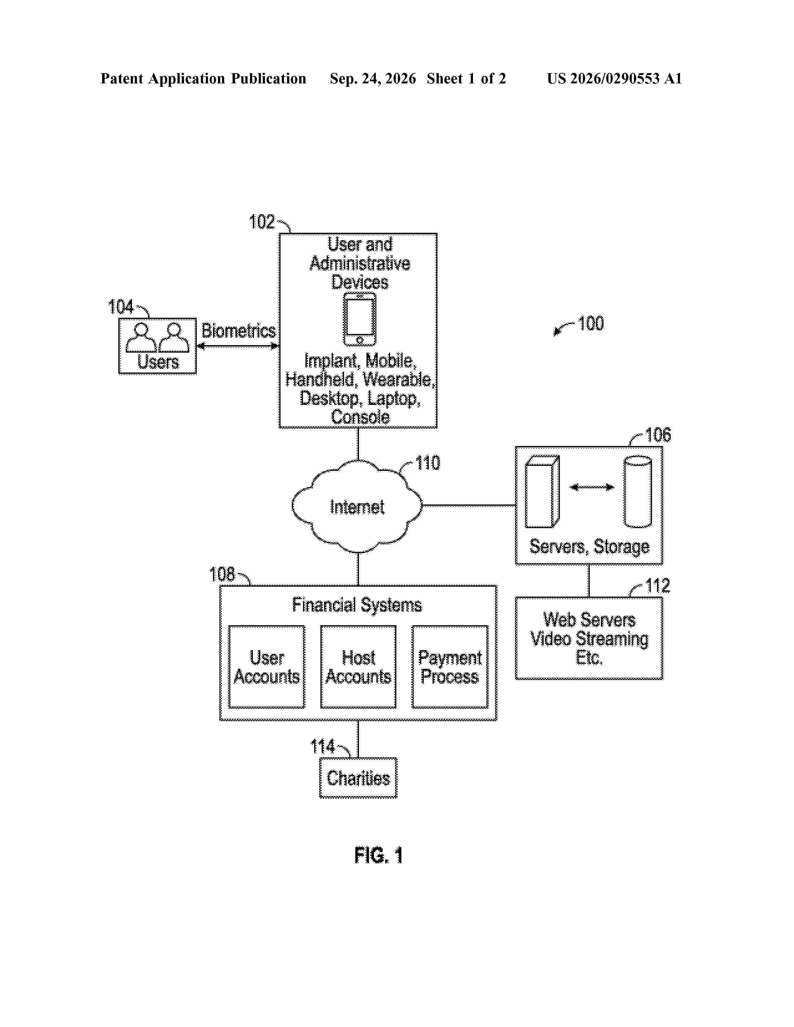
Resumen de: US20260290553A1
Systems and methods for incentivizing a user to satisfy a goal for attending twelve step meetings. The method includes the steps of: providing the user with a mobile device; detecting the presence of the mobile device at a first location and at a first time selected by the user; comparing the first time and the first location to a database of approved meetings, wherein the comparing is substantially contemporaneous with the first time; and providing a reward to the user upon confirming that: i) the first time and the first location correspond to an approved meeting; and ii) the user is proximate the mobile device at the first time.
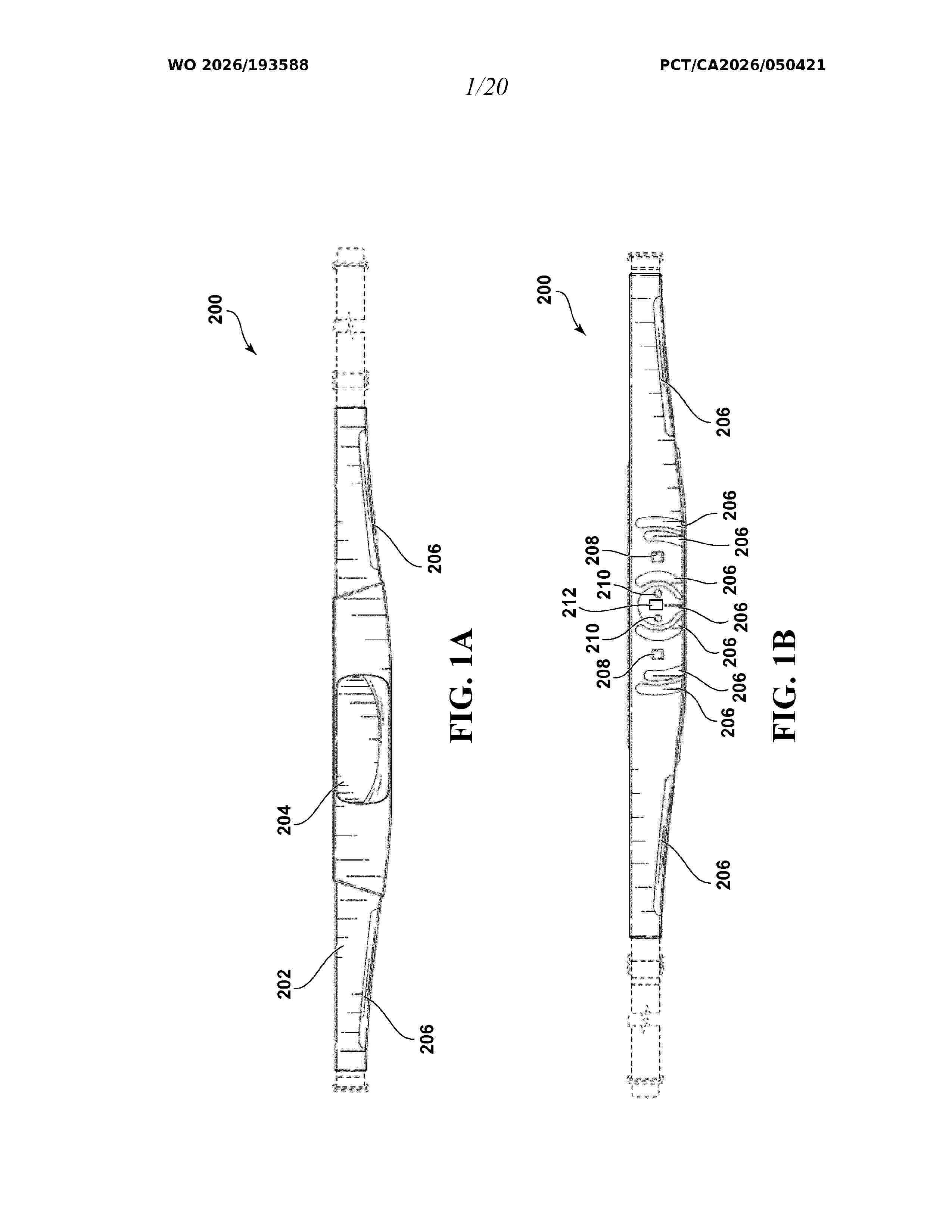
Resumen de: WO2026193588A1
According to an aspect, there is provided a flexible wearable band comprising: a plurality of EEG sensors configured to measure electrophysiological signals, at least one light emitter positioned within a center the central EEG sensor, at least one optical sensor to measure optical signals in close proximity to the at least one light emitter, and an electronics module connected to the band. The at least one optical sensor and at least one of the plurality of EEG sensors are arranged in close proximity for co-located measurement. In some embodiments, the EEG sensors and/or the at least one optical sensor are positioned on the left and right hemispheres for measuring brain states associated with symmetric indexes.
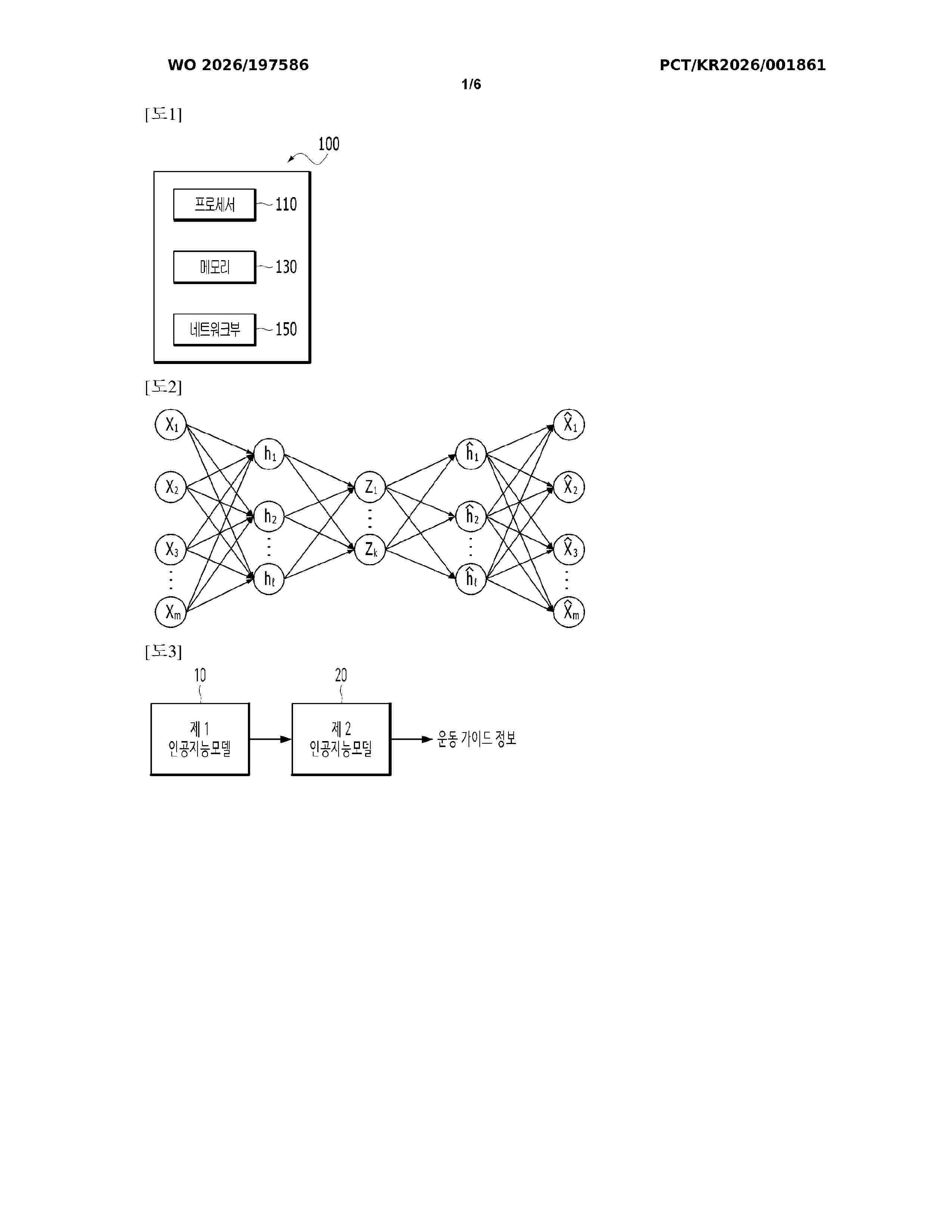
Resumen de: WO2026197586A1
According to an embodiment of the present disclosure, disclosed is a method performed by a computing device and generating exercise guide information by using an artificial intelligence model. The method may comprise the steps of: acquiring at least one among force information related to a user's body, appearance information about the user's body, or movement information about the user's body; on the basis of at least one among the force information related to the user's body, the appearance information about the user's body, or the movement information about the user's body, generating an embedding by using a first artificial intelligence model; and generating, on the basis of the embedding, exercise guide information for the user by using a second artificial intelligence model.
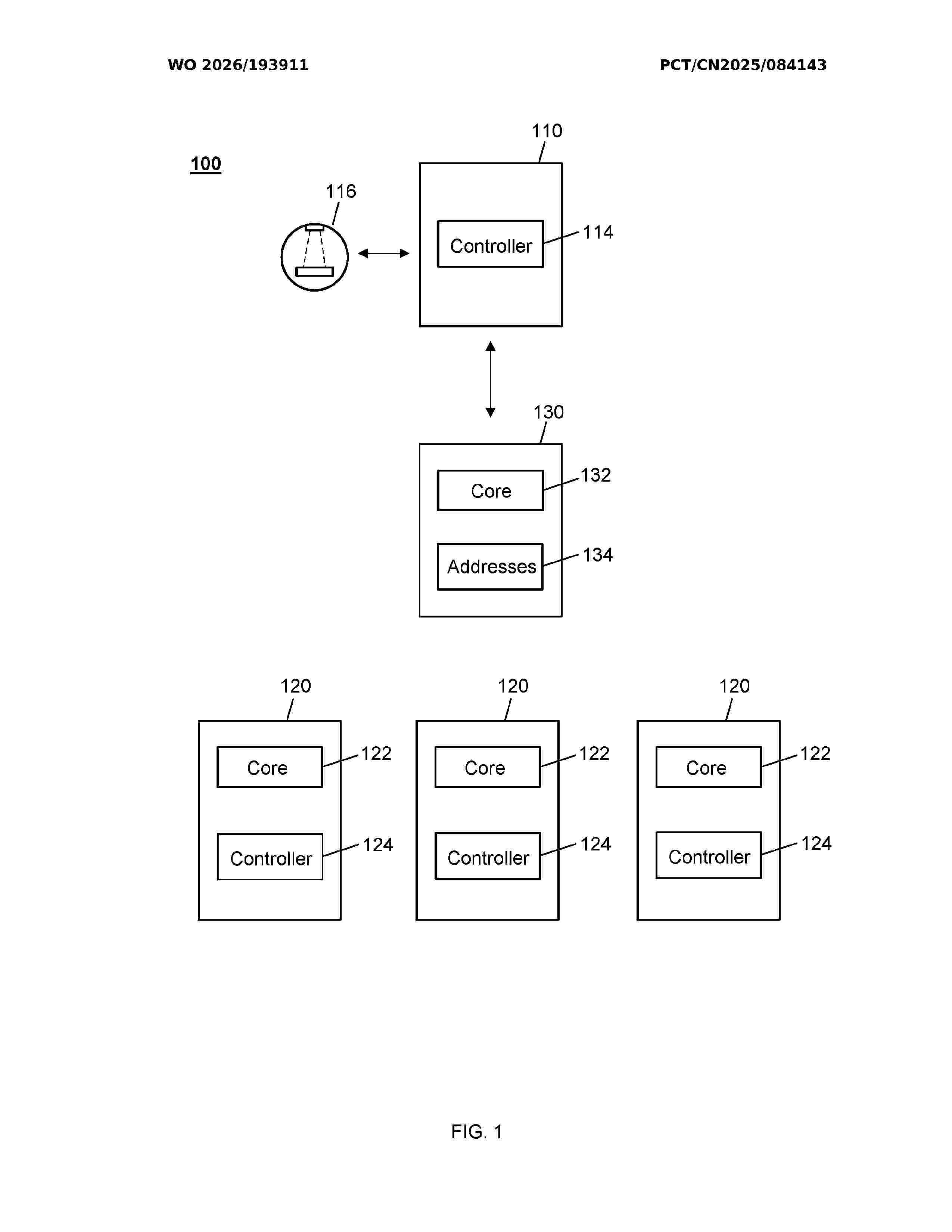
Resumen de: WO2026193911A1
A computer-implemented method performed by a radiotherapy treatment-planning client device, the method comprising: receiving, from a load-balancing server, address information for one or more available radiotherapy treatment-planning processing servers; in a first mode of operation: upon receiving a user command to perform a radiotherapy treatment-planning task, communicating with the load-balancing server to select one of the one or more available radiotherapy treatment-planning processing servers; in a second mode of operation: upon receiving a user command to perform a radiotherapy treatment-planning task, selecting one of the one or more available radiotherapy treatment-planning processing servers based on the received address information; and in the first and second modes of operation, communicating with the selected radiotherapy treatment-planning processing server to perform the radiotherapy treatment-planning task.
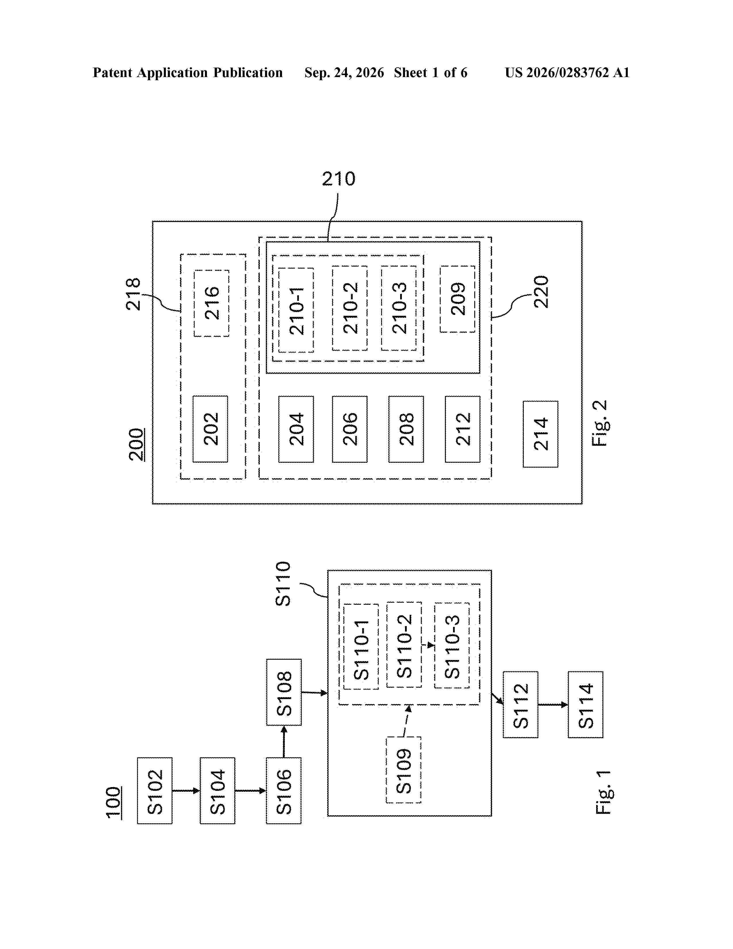
Resumen de: US20260283762A1
0000 A method for defining an orientation of an origin of a model of an anatomy of a patient's dental arch (302; 402) within a global reference frame comprises receiving a dataset comprising a surface mesh, defined by a plurality of polygon, representing the surface of the dental arch (302; 402). For each polygon, a exterior normal vector is determined. A vector sum of exterior normal vectors defines a normal vector, and thereby an orientation, of an occlusion plane (412). A first rotation angle is determined for aligning the normal vector of the occlusion plane (412) with a first direction of the reference frame. An anterior side (504) of the dental arch (302; 402) is defined based on geometric properties of the surface mesh. A second rotation angle is determined for aligning an anterior-posterior direction of the dental arch (302; 402) with a second direction of the reference frame.
Nº publicación: WO2026193582A1 24/09/2026
Solicitante:
THE SLEEP HEALTH ORGANIZATION INC [CA]
THE SLEEP HEALTH ORGANIZATION INC.
Resumen de: WO2026193582A1
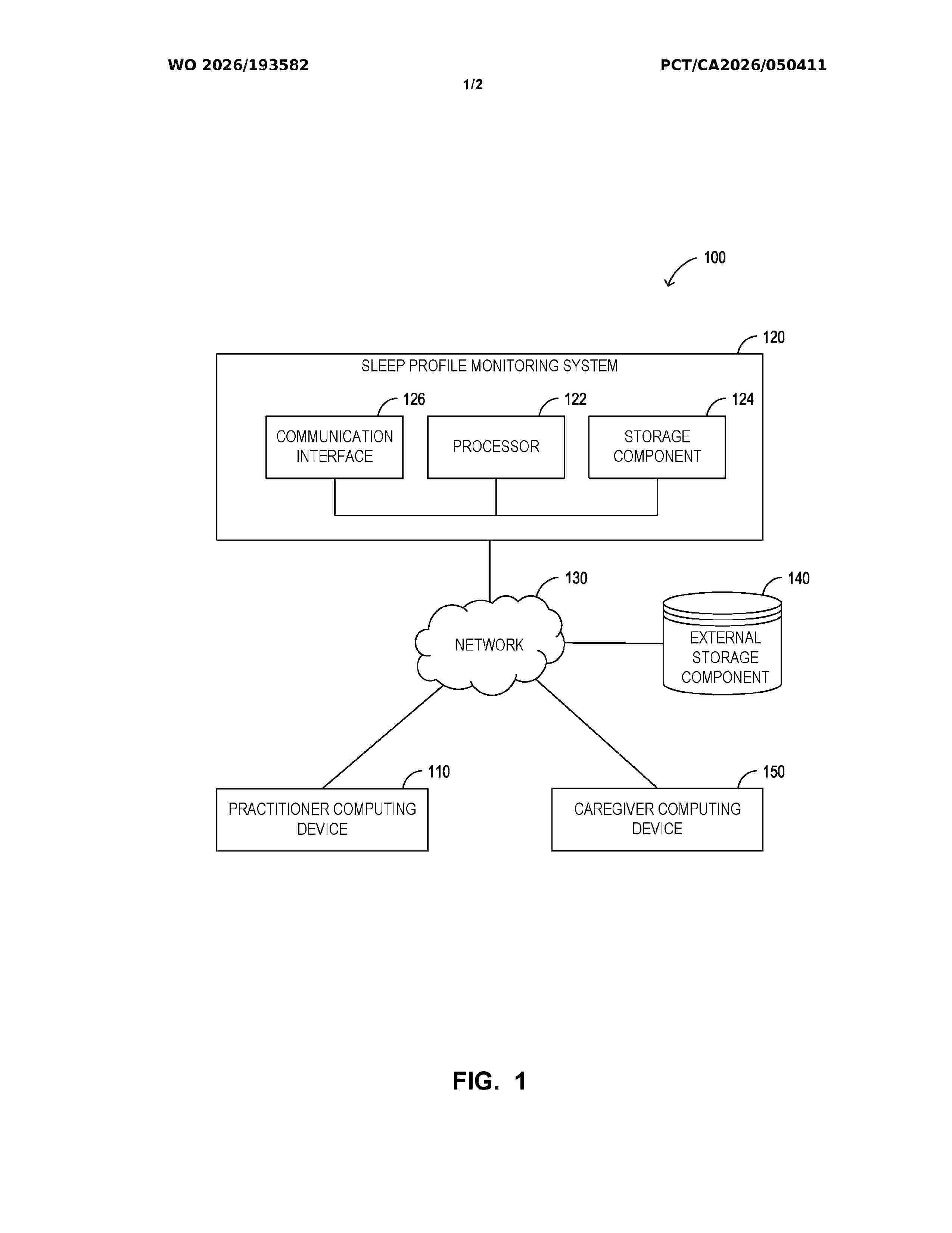
Systems and methods for monitoring sleep profiles are disclosed. A processor is operable to: obtain, from a practitioner computing device, initial sleep profile data for a patient; apply a sleep profile classification model to the initial sleep profile data to generate a proposed sleep classification; receive, from the practitioner computing device, an initial sleep classification in response to the proposed sleep classification being displayed at the practitioner computing device; based on the initial sleep classification, identify a sleep characteristic to monitor. The processor is operable to receive monitoring data related to the identified sleep characteristic; automatically generate current sleep profile data; generate an updated proposed sleep classification; compare the proposed sleep classification with the initial sleep classification; in response to detecting the updated proposed sleep classification does not match the initial sleep classification, automatically generate a progress update; and transmit the progress update to the practitioner computing device.
 BOPI
BOPI
 Sede Electrónica