Si deseas distinguir tus productos, servicios o ambos de los de otra empresa, es posible que necesites una marca o nombre comercial. Descubre qué son, en qué consiste su procedimiento de registro y qué implica.

Información sobre los plazos de presentación de solicitudes de transformación de marcas de la Unión Europea en marca nacional española. Más información


Si tienes un nuevo dispositivo, producto o procedimiento que resuelva un problema técnico o tenga una ventaja práctica, existen distintas formas de protegerlo en España y en otros países. Descubre cómo hacerlo.

¿Tu innovación reside en la estética, la ornamentación o la apariencia de tu producto? Protégela mediante un diseño industrial. Descubre qué derechos confiere el registro y cómo realizar la tramitación.

Las indicaciones geográficas protegen el nombre de un producto originario de una zona geográfica, a la cual le debe una determinada calidad, reputación u otra característica. Descubre qué son, en qué consiste su procedimiento de registro y qué beneficios conceden.

Las patentes publicadas en todo el mundo son una valiosa fuente de información científica, técnica y comercial.

Si eres emprendedor/a o una empresa y quieres potenciar y mejorar la rentabilidad de tu negocio protegiendo de forma adecuada los activos intangibles de tu organización, en este espacio encontrarás lo necesario.


 355
resultados
355
resultados
 Última actualización
01/05/2026 [08:11:00]
Última actualización
01/05/2026 [08:11:00]


Resumen de: DE102024131256A1
Die vorliegende Erfindung liegt im technischen Gebiet der additiven Fertigung und stellt ein kontinuierliches Verfahren zum Herstellen von flächigen, porösen Hydrogelen und Strukturen bereit. Ebenfalls stellt die vorliegende Erfindung eine Vorrichtung zur kontinuierlichen Herstellung von Hydrogelen bereit.

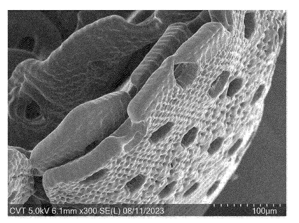
Resumen de: US20260115083A1
The invention relates to a finger orthosis for a finger exoskeleton. The finger orthosis comprises an actuating member, a second phalanx accommodation and a third phalanx accommodation. The second phalanx accommodation is freely pivotable in a first joint on the actuating member. The second phalanx accommodation is coupled to the third phalanx accommodation via a constraining guide in such a way that a fixed dependency of the relative first pivot angle between the actuating member and the second phalanx accommodation on the one hand and the relative second pivot angle between the second phalanx accommodation and the third phalanx accommodation on the other hand is given. The finger orthosis according to the invention is easy to dimension and can be specifically adapted to a patient. The invention also relates to a finger exoskeleton and a method of manufacturing a finger orthosis.
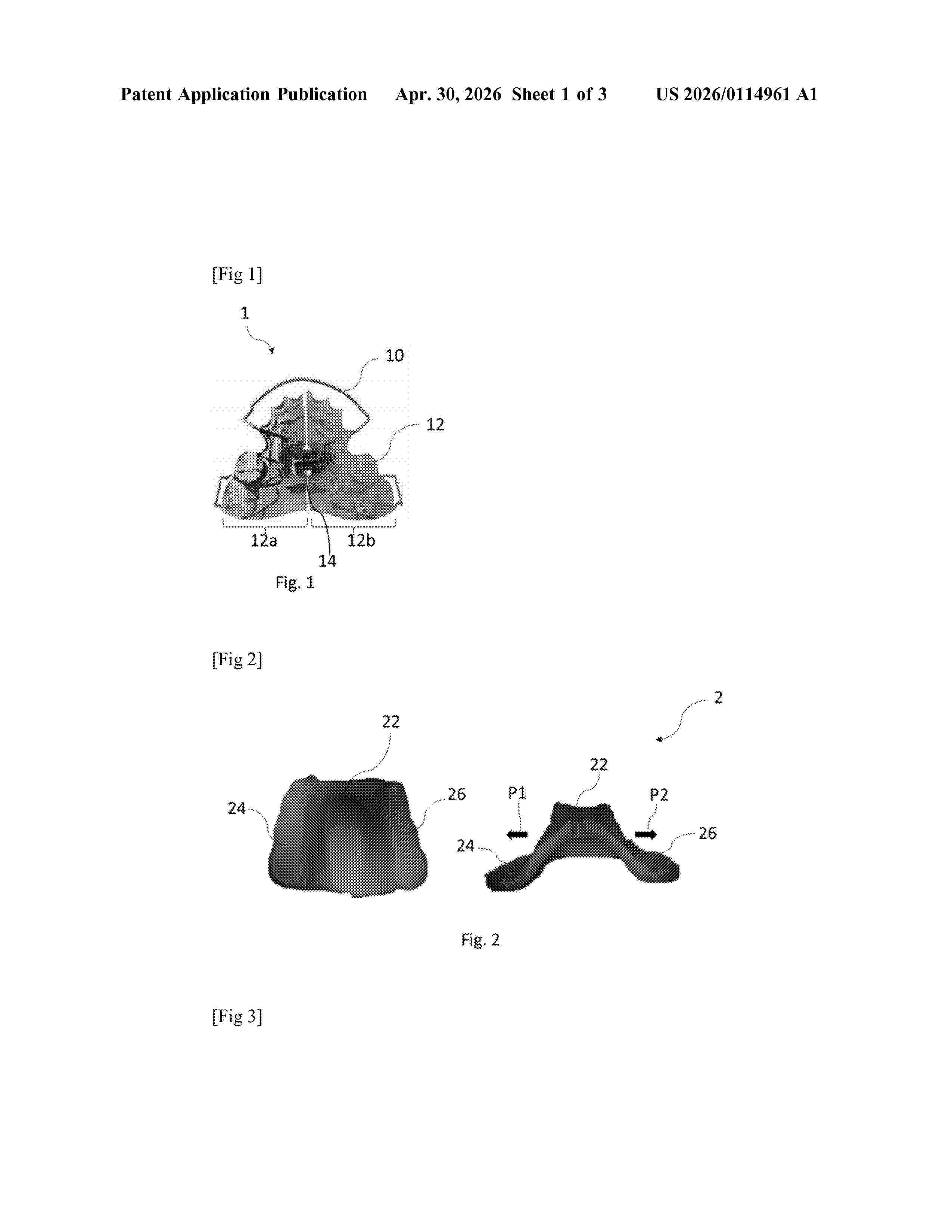
Resumen de: US20260114961A1
The invention relates to a maxillary modeller (2) intended to be worn, in a service position, on a dental arch of a user in order to modify the shape of the palate, the maxillary modeller being one-piece and removable and comprising: —a central palatal portion (22) configured to be in contact with the user's palate when the maxillary modeller is in the service position; and —a first and a second dental portion (24; 26) each comprising at least one, preferably multiple cavities configured to receive teeth of the user when the maxillary modeller is in the service position, the first and second dental portions extending on either side of the central palatal portion, the central palatal portion being configured so as to exert on the palate, in the service position, forces suitable for modifying the shape thereof.
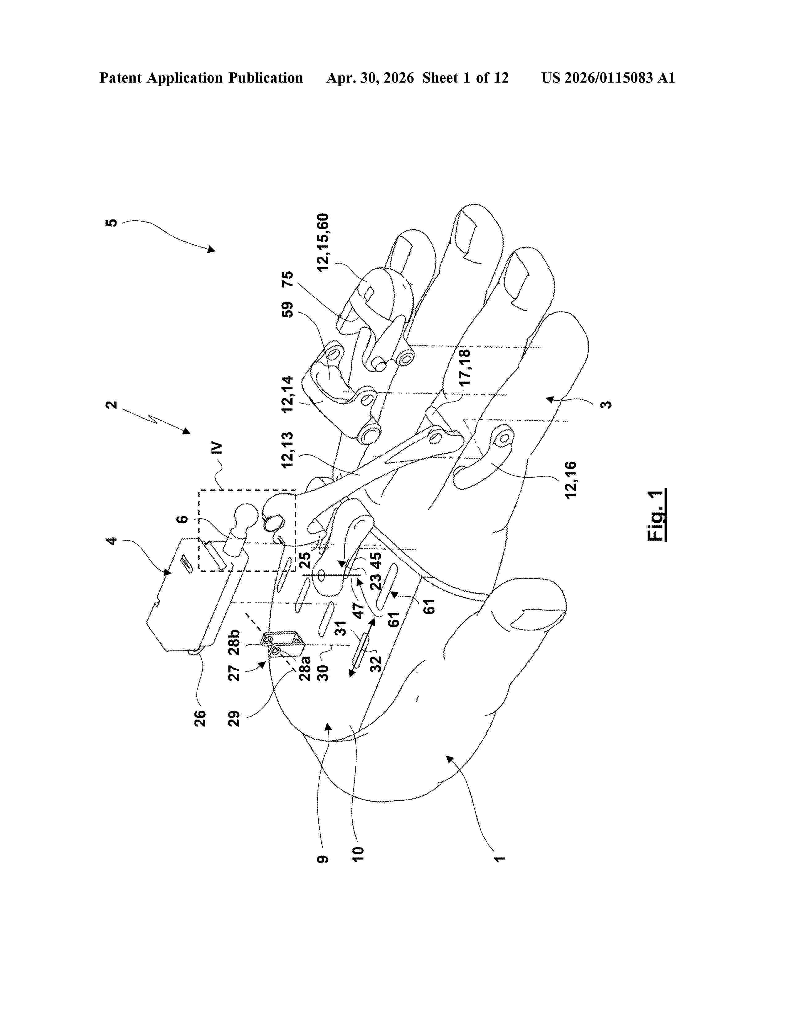
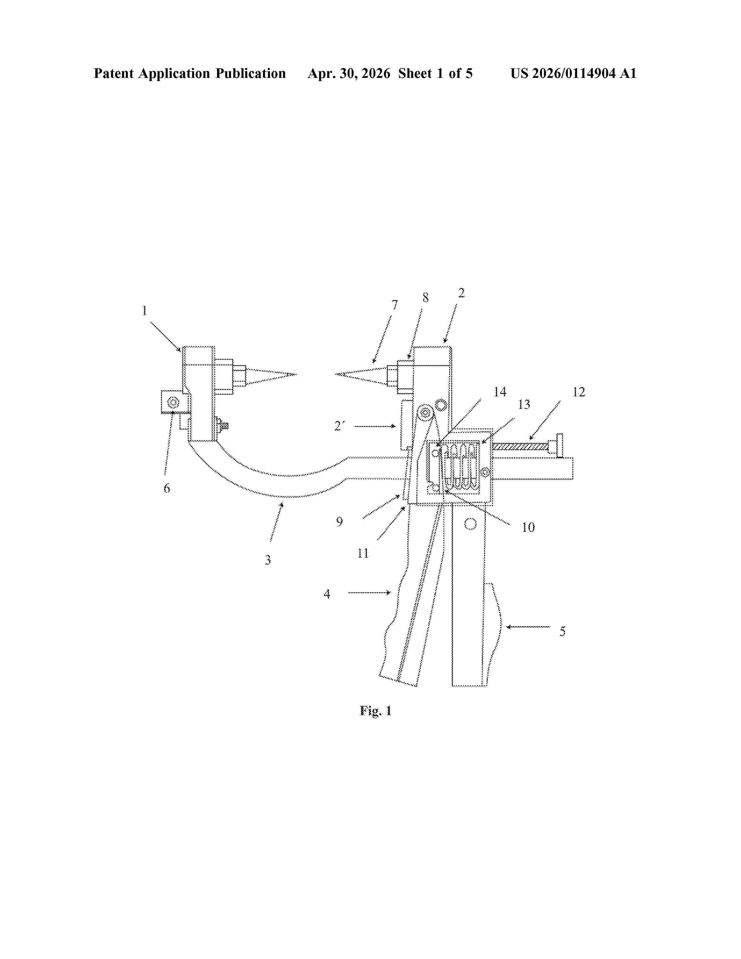
Resumen de: US20260114904A1
Reposition and penetration forceps for fractures of long bones, in particular traumatic fractures of tibia in centre line, that comprise two jaws forming a three-point system in opposition, wherein the first movable jaw (1) comprises two threaded pins (7) with tips (21) and the second firm jaw (2) comprises one threaded pin (7) with a tip (21). An external fixator for fractures of long bones, in particular traumatic fractures of tibia in centre line, which comprises at least two sets of forceps, wherein the sets of forceps are arranged identically or in a mirror image to each other.
Resumen de: US20260114758A1
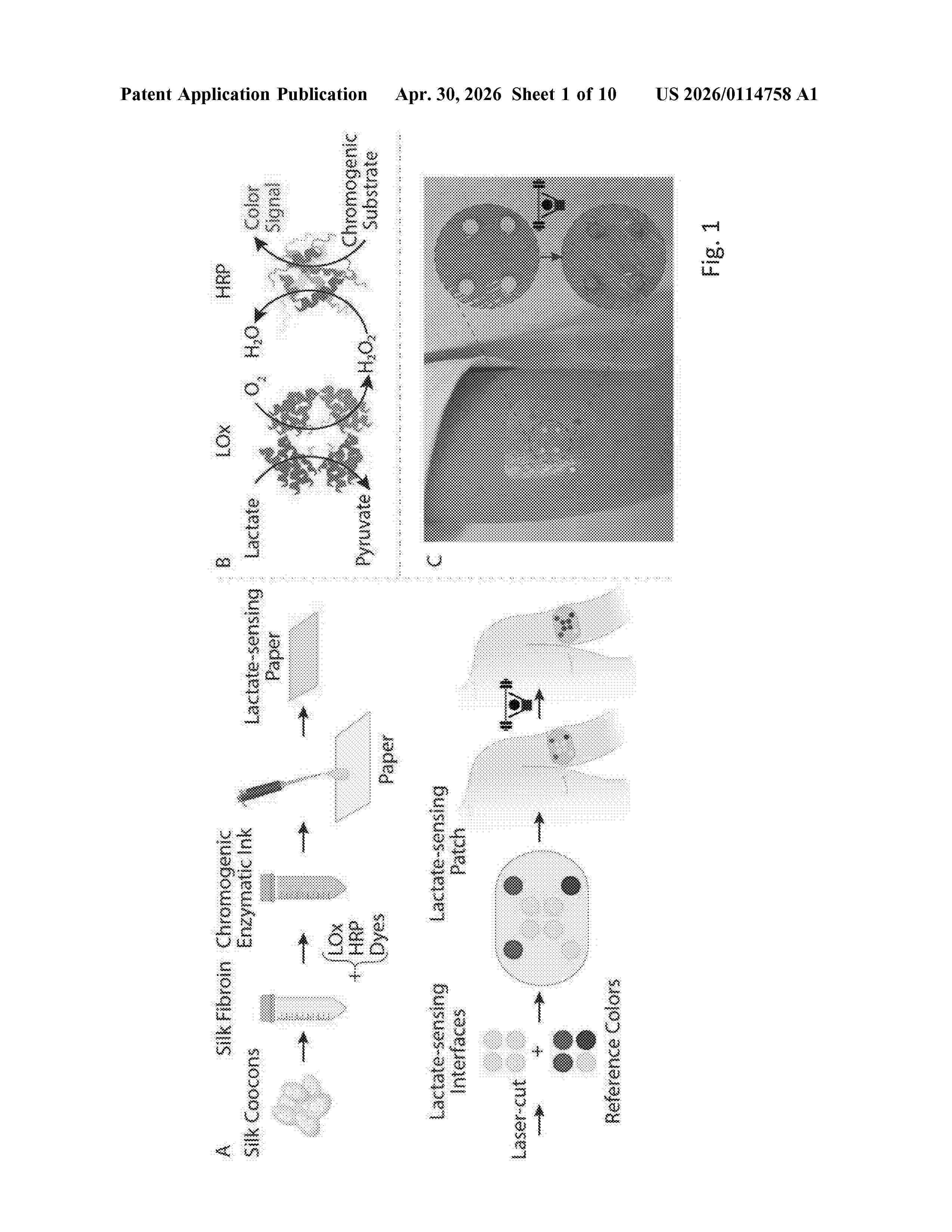
Disclosed herein is a printable liquid lactate sensor composition including silk fibroin in an amount by weight of between 0.1% and 30%, a lactate oxidase that is activated by lactate to produce hydrogen peroxide, a peroxidase that is activated by the hydrogen peroxide, and a chromogenic substrate that changes color upon activation of the peroxidase. Wearable sensors including the printable liquid lactate sensor composition are disclosed herein.
Resumen de: EP4732807A1
A computer implemented method for determining a 3D position of a surgical object relative to a region of interest in a patient's anatomy is suggested. The method comprising the steps of receiving an X-ray image and a 3D data set. The X-ray image is a 2D projection image generated with an imaging direction and defining a 3D image coordinate system. When applying the method, the 2D projection image may depict a part of a patient's anatomy and at least a part of the surgical object. The received 3D data set describes the part of the patient's anatomy and a region of interest in a 3D data coordinate system. In order to determine a spatial relation between the 3D image coordinate system and the 3D data coordinate system, the part of the patient's anatomy is localized based on the X-ray image. Subsequently, the 3D position of the surgical object relative to the region of interest is determined in the 3D data coordinate system based on (I) the determined spatial relation between the first 3D image coordinate system and the 3D data coordinate system, (II) a spatial relation between the surgical object and the first 3D image coordinate system, and on (III) information related to the geometrical aspect.
Resumen de: EP4732758A1
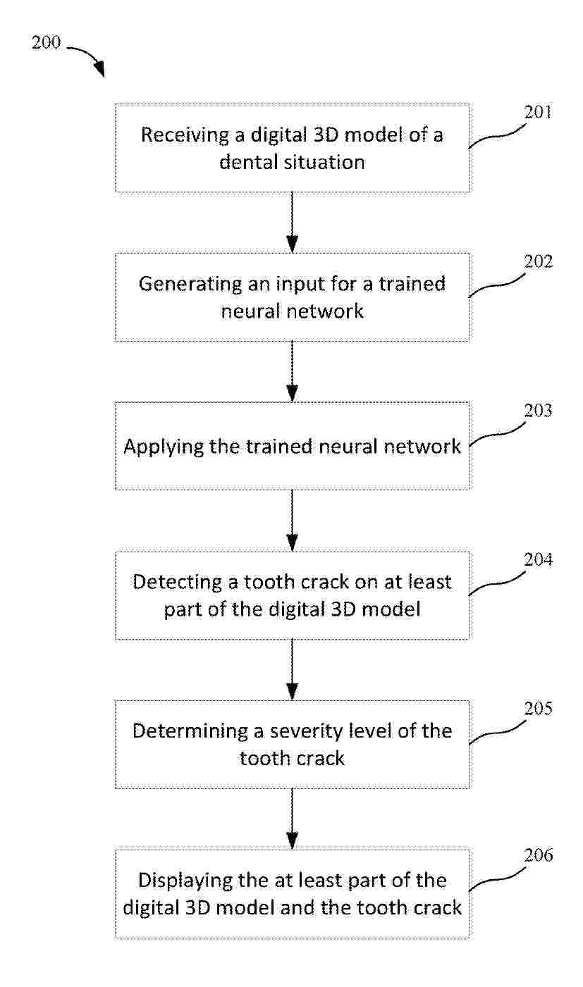
A computer-implemented method for detecting tooth cracks on a digital 3D model (501) of a dental situation is disclosed. The method comprises receiving the digital 3D model (501) of the dental situation, generating an input (301, 401) for a trained neural network (300, 400) for tooth crack detection, wherein the input (301, 401) comprises at least a part of the digital 3D model (501) with associated color information. The method further comprises detecting a tooth crack (100) on the at least part of the digital 3D model (501) by applying the trained neural network (300, 400) to the generated input (301, 401). Further, the method comprises determining a severity level of the detected tooth crack (100) based on fluorescence information associated with the at least part of the digital 3D model (501) and displaying the at least part of the digital 3D model (501), the detected tooth crack (100) and the severity level of the detected tooth crack (100).
Resumen de: EP4732777A1
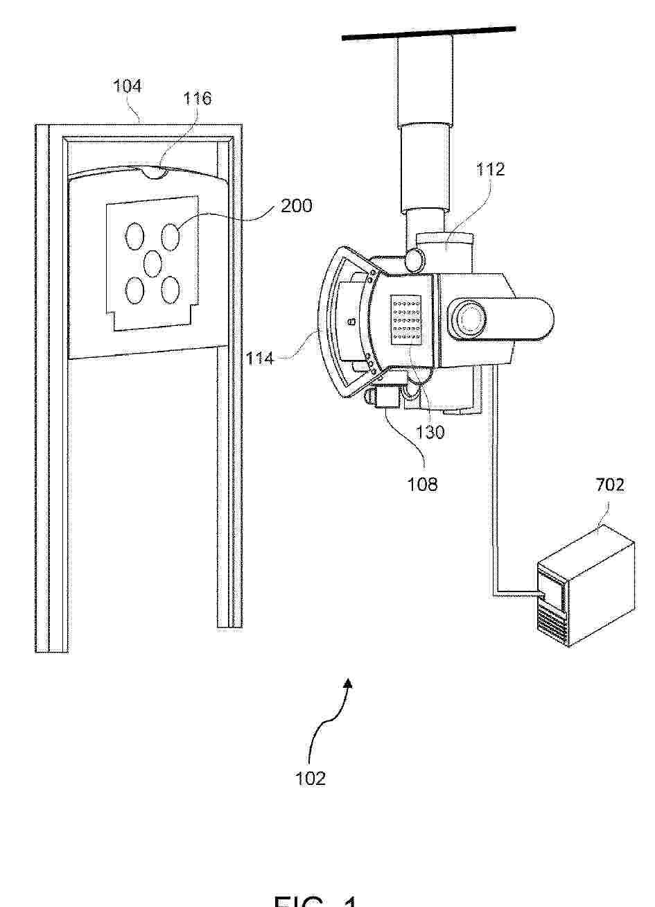
Disclosed is a method (100) for controlling an x-ray system (102), wherein the x-ray system (102) is configured to acquire x-ray images (142) of a target radiation field (134) based on imaging instructions (132) provided by an operator (500) on a display unit (130), wherein the size of the target radiation field (134) may exceed a size of a detector (116). The display unit (130) is configured to display augmented image data (120), wherein the augmented image data (128) comprises 3D projection information (124) about the position of the subject (106) as well as overlays of virtual collimation fields (136). The virtual collimation fields (136) comprise sets of virtual positions (138) of the x-ray components (110). The x-ray system (102) may then be controlled to move the x-ray components (110) to the virtual positions (138), acquiring an x-ray image (142) at each virtual position (138).
Resumen de: WO2024263974A2
This disclosure is directed to compositions of engineered cells that are incorporated into immunomodulatory substances that act as semi-permeable membranes. These compositions provide for sustained delivery of various biologic and therapeutic molecules, such as cytokines or monoclonal antibodies, for a range of diseases, including infectious diseases, cancer immunotherapy and auto-immune disorders.
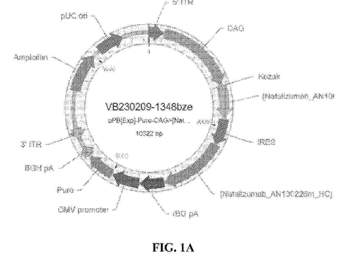
Resumen de: EP4732869A2
A plastic flange (1) for a medical container (2), said plastic flange (1) defining an opening (11), said opening (11) being surrounded by a peripheral collar (10), said collar (10) providing a support to a user's fingers, wherein the flange (1) comprises connecting means for connecting said flange (1) to an external surface (24) of a tubular barrel (20) of the medical container (2), and wherein the flange (1) further comprises a remotely readable electronic component (3) for remote identification of the medical container (2), said remotely readable electronic component (3) being embedded into the plastic flange (1).
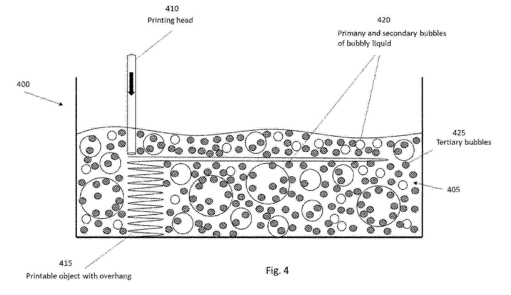
Resumen de: WO2024260561A1
Disclosed is a method for producing bubbly liquid material that is suitable for being used as a supporting material in a 3D printing system, the method comprising, producing to liquid material primary bubbles, stabilizing the primary bubbles with cross-linking enhancing compound, producing secondary bubbles to the liquid material, and using the liquid material comprising the primary and secondary bubbles as the supporting material for printing a 3D object, wherein the liquid material comprises a bubble system comprising at least the primary and secondary bubbles.
Resumen de: EP4480456A1
0001 The present disclosure relates to an implant (10) configured to be at least partially inserted into at least one opening (44) in at least one bone (46) of a patient. The implant (10) includes at least one implant body (12) made at least partially of at least one resorbable material. The implant body (12) includes at least one first region (14) and at least one second region (16). The first region (14) has a plurality of open pores and the first region (14) has a greater porosity than the second region (16). 0002 The present disclosure also relates to an instrument (42) for implanting an implant (10) into an opening (44) defined in at least one bone (46) of a patient.
Resumen de: GB2701355A
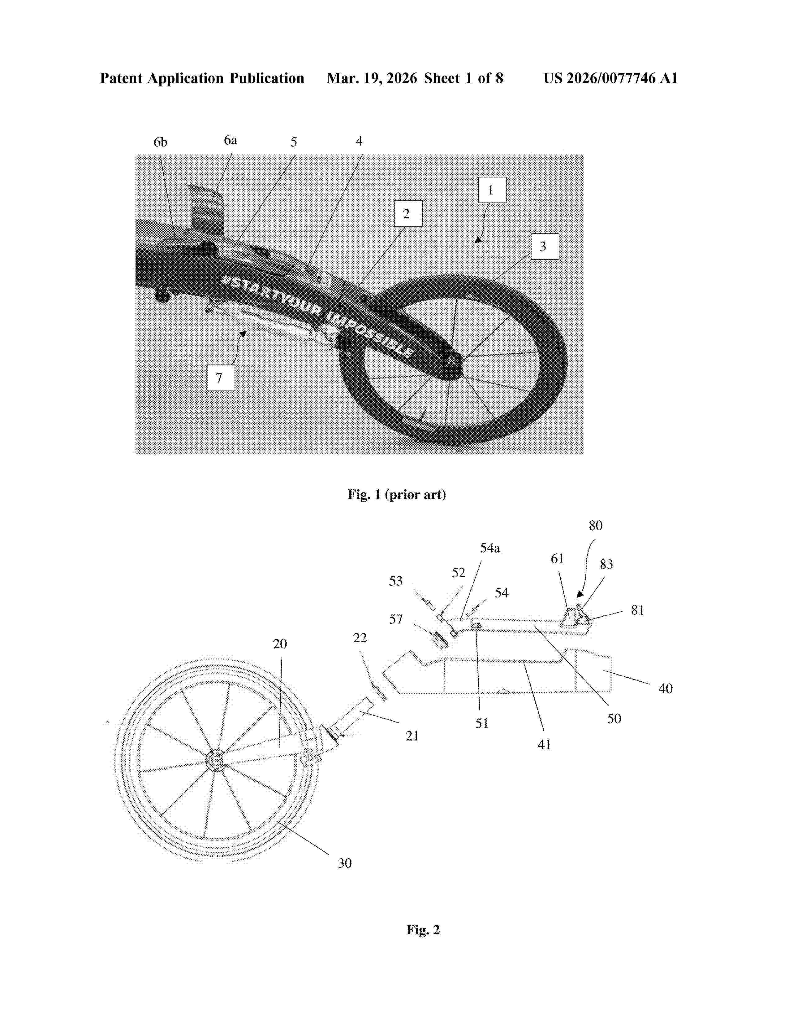
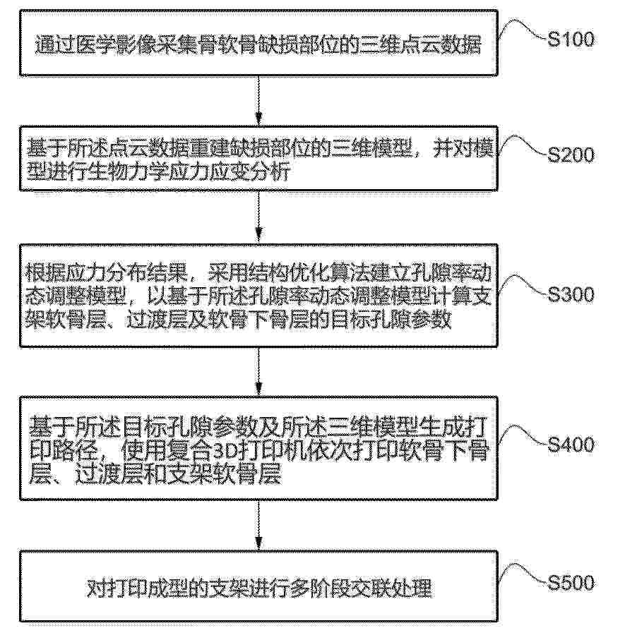
Brake assembly 80 for a racing wheelchair, comprising a brake lever 83 for actuating a brake cable (86, Fig. 6), wherein the brake lever is a 3D printed glass filled nylon unit, or a 3D printed unit of up to 30% glass filled nylon. The brake lever may be adapted to be pivotally mounted between mounting plates 81 provided by a steering arm 50 that is 3D printed of Aluminium, that is either ALSI10mg or the steering arm has a triangular cross-section with smoothed edges. The brake assembly may have a spring mount pin (85, Fig 5a) for mounting between the mounting plates and a spring 84 engaging the brake lever. The brake cable may extend from the brake lever and through the steering arm. Also disclosed is the brake assembly integrated into a steering arm, which may have steering means 61, e.g. wings. The steering arm may have a frame (55, Fig. 4j) for enforcement with brake troughs or openings (56c, Fig 4j) for the brake cable. Figure 2
Resumen de: CN121928779A
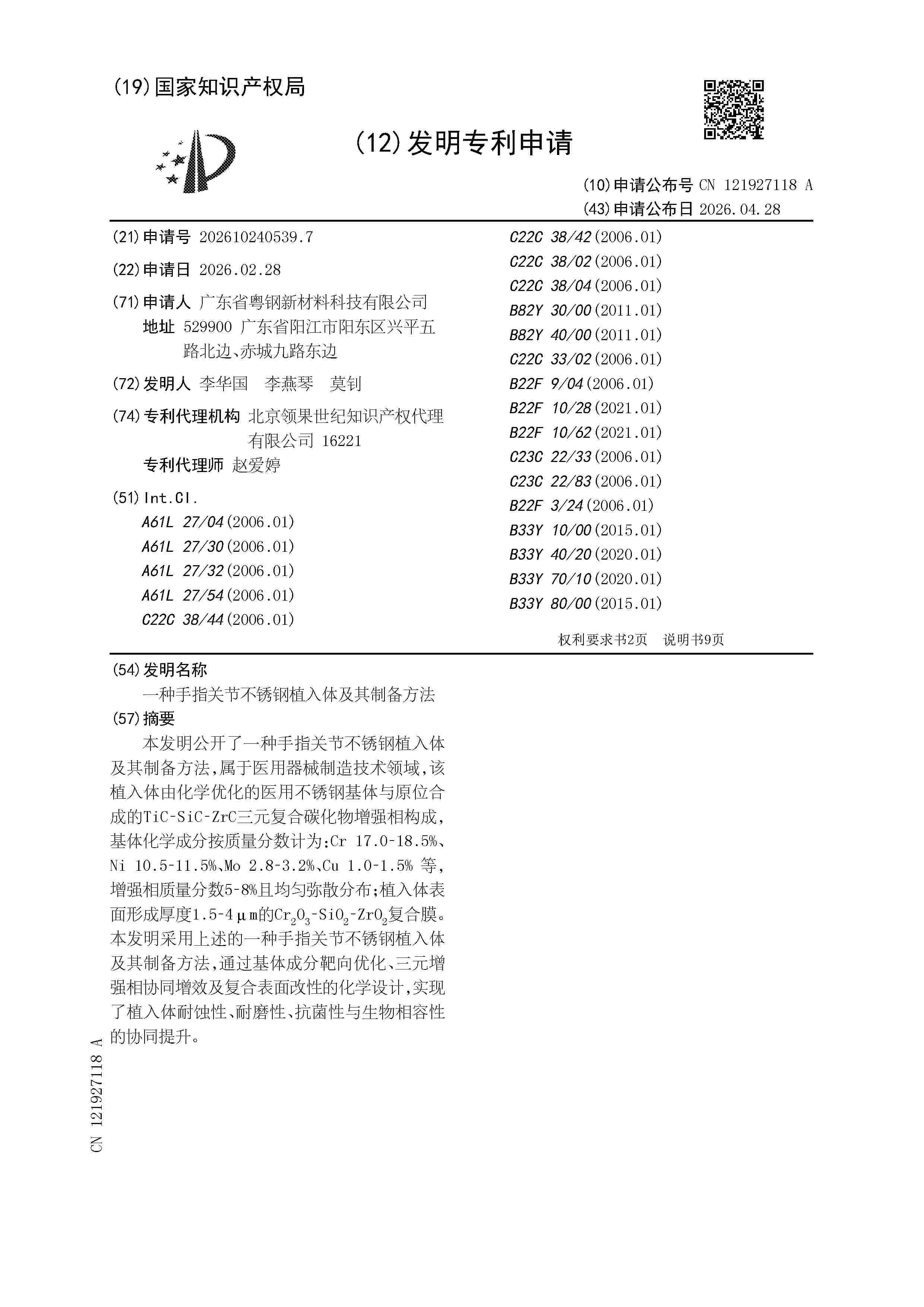
本发明提供一种骨软骨支架的3D打印方法及系统,该方法包括以下步骤:采集骨软骨缺损部位的三维点云数据;基于点云数据重建缺损部位的三维模型,并对模型进行应力应变分析;根据应力分布结果,建立孔隙率动态调整模型调整孔隙参数;基于目标孔隙参数及三维模型生成打印路径,依次打印软骨下骨层、过渡层和支架软骨层。通过医学影像数据驱动模型重建与应力分析,结合结构优化算法实现孔隙参数动态校准,并利用多材料打印与多阶段交联,通过孔隙率动态调整模型调整孔隙参数,避免应力集中导致的支架失效;梯度打印与交联协同增强了支架稳定性与生物活性离子缓释能力,从而显著提升细胞迁移效率、胶原沉积质量及骨软骨界面整合强度。
Resumen de: CN121927118A
本发明公开了一种手指关节不锈钢植入体及其制备方法,属于医用器械制造技术领域,该植入体由化学优化的医用不锈钢基体与原位合成的TiC‑SiC‑ZrC三元复合碳化物增强相构成,基体化学成分按质量分数计为:Cr 17.0‑18.5%、Ni 10.5‑11.5%、Mo 2.8‑3.2%、Cu 1.0‑1.5% 等,增强相质量分数5‑8%且均匀弥散分布;植入体表面形成厚度1.5‑4μm的Cr2O3‑SiO2‑ZrO2复合膜。本发明采用上述的一种手指关节不锈钢植入体及其制备方法,通过基体成分靶向优化、三元增强相协同增效及复合表面改性的化学设计,实现了植入体耐蚀性、耐磨性、抗菌性与生物相容性的协同提升。
Resumen de: WO2025079951A1
The present invention relates to a method for designing, using a computer, an orthodontic device on the basis of partial designation of teeth, the method being characterized by comprising the steps of: displaying the arrangement of teeth of a patient (S01); setting up and displaying all the teeth in the arrangement of teeth in the state after orthodontics (S02); designating teeth in the teeth arrangement (S03); designating specific portions in the designated teeth (S04); and inputting the thickness of the orthodontic device in contact with the specific designated portions in the designated teeth (S05).
Resumen de: CN121927122A
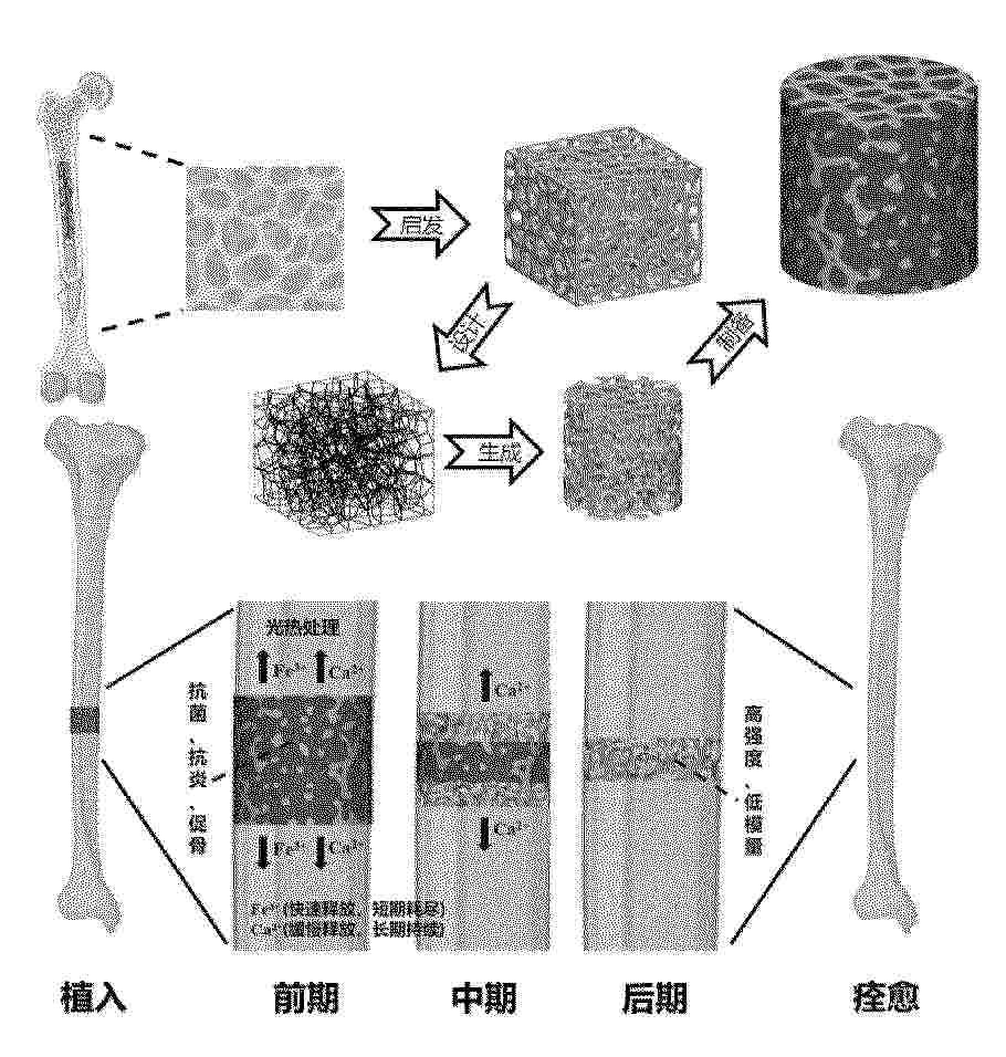
本发明公开了一种仿生Voronoi‑镍钛‑水凝胶异质结构植入物及其制备方法,涉及骨植入技术领域,借鉴人类骨小梁的结构理念,通过创新设计方法完成Voronoi多孔结构构建与仿生Voronoi‑镍钛骨架的制备,再以该仿生Voronoi‑镍钛骨架为基底,采用烘干成胶法制备仿生Voronoi‑镍钛‑水凝胶异质结构植入物。该植入物整合了镍钛合金高强度、低模量的力学优势与仿生Voronoi多孔结构高连通性、高比表面积的结构特点,有效弥补水凝胶承载薄弱的短板,同时水凝胶兼具缓冲吸能功效,可实现抗菌、抗炎、促骨再生的多功能时序递释,适配骨修复各阶段需求。
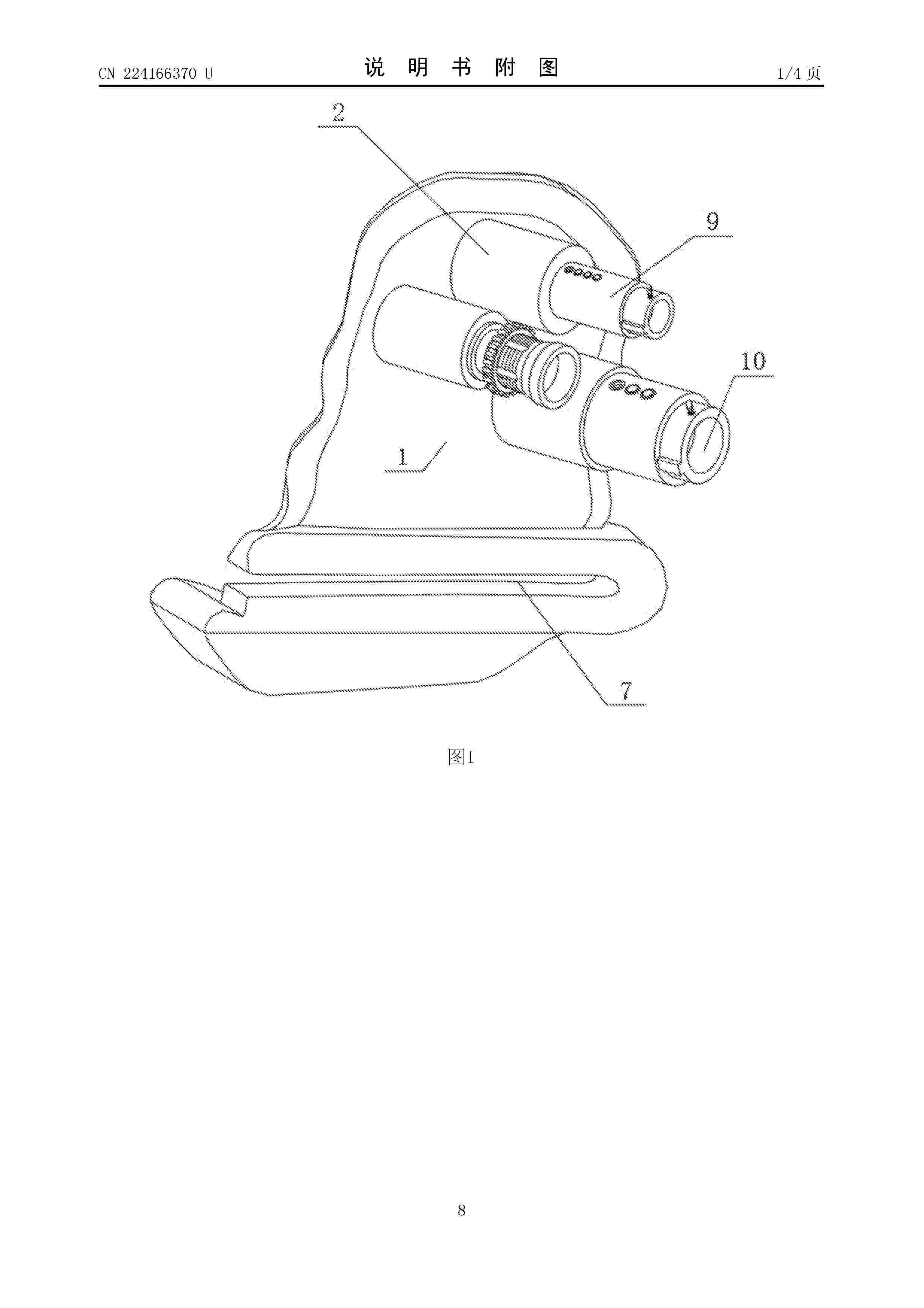
Resumen de: CN224166370U
本实用新型涉及医疗器械技术领域,且公开了单髁关节股骨3D打印导板,包括导板本体,所述导板本体的一侧开设有股骨远端假体远端锁定孔,所述导板本体的一侧开设有股骨远端假体近端锁定孔和导板固定孔、导板外侧卡压。本实用新型结构合理,通过在术前使用膝关节CT扫描并进行3D导板设计,在使用中使导板外侧卡压、导板内侧卡压和股骨后髁卡压进行模板卡压。术前准备对接模板卡压完毕后使用导板固定孔打入克氏针固定加强导板本体的稳定性,防止在截骨过程中导板本体松动,股骨后髁截骨槽截股骨后髁术中外侧使用弯撬保护内侧副韧带,内侧为封闭结构保护前后交叉韧带,该设计及可以使得截骨时摆锯有效的运动又可以防止损伤韧带及周围组织。
Resumen de: CN121927126A
本发明属于磁性材料技术领域,涉及一种基于透明质酸基和明胶基的磁性生物墨水及其制备方法与应用。本发明以甲基丙烯酰化透明质酸与甲基丙烯酰化明胶为基质,引入单宁酸修饰的四氧化三铁纳米粒子,构建具有光交联与氢键相互作用的双网络磁性水凝胶生物墨水。该墨水通过挤出式生物3D打印技术成型,经光引发交联形成多孔支架,并负载人脐带间充质干细胞。得益于单宁酸修饰的四氧化三铁纳米粒子的引入,支架兼具优异的抗氧化性能、力学增强效应及超顺磁性,在外加磁场调控下可显著促进干细胞的黏附、增殖与软骨向分化。
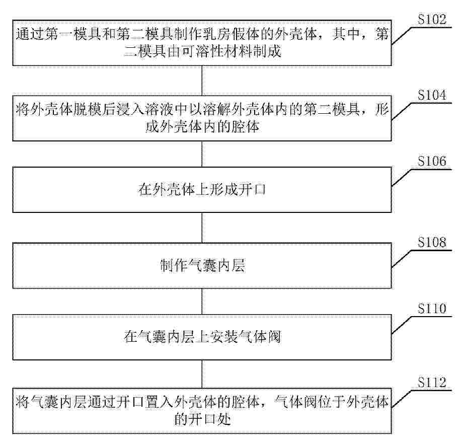
Resumen de: US20260115017A1
The present disclosure provides a breast prosthesis and a method for manufacturing the same, which relate to the field of prosthesis manufacturing technologies. The breast prosthesis includes an outer shell and an airbag inner layer. A cavity is formed inside the outer shell, and the outer shell is provided with an opening in communication with the cavity. The airbag inner layer is provided with a gas valve.
Resumen de: CN121910496A
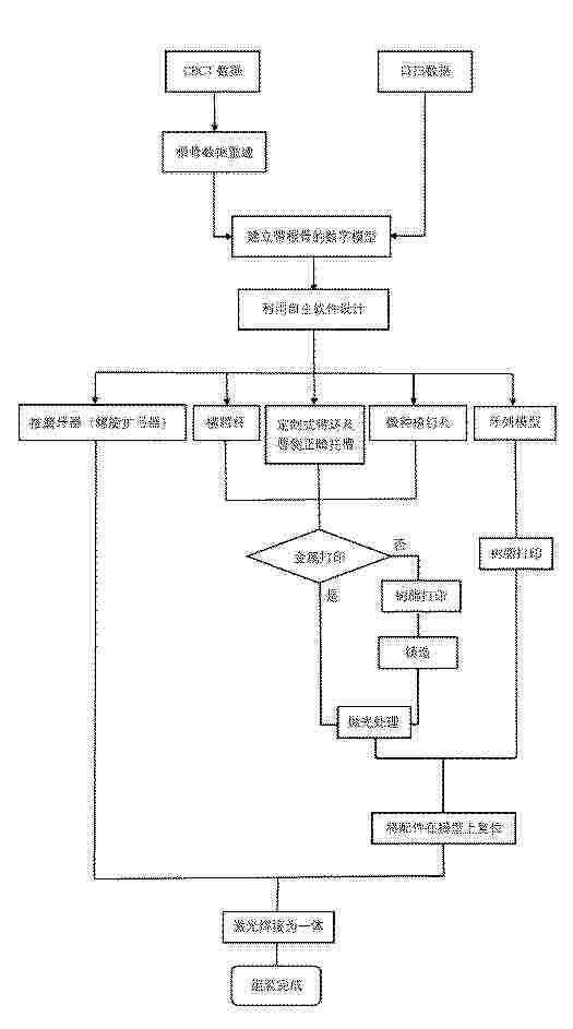
本发明提供了一种数字化定制的推磨牙矫治器及其制作方法,涉及医疗器械领域。该方法包括:获取患者口腔三维数据建立数字牙列模型;基于模型设计包括定制式带环和横腭杆的一体式结构,并在横腭杆上设计微种植钉孔;采用增材制造工艺制作该一体式结构;最后将一体式结构与一螺旋扩弓器连接。本申请提供的矫治器包括:与磨牙固定的定制式带环、横跨上腭的横腭杆、以及设置在横腭杆上用于植入微种植钉的微种植钉孔,其中带环与横腭杆为一体式结构,并与一螺旋扩弓器连接。本申请解决了现有推磨牙矫治器磨牙远中移动效率不高、支抗不足、舒适度差及个性化精度低的问题,通过数字化流程和骨性支抗,实现了高效精确的矫治,同时提升了佩戴舒适度。
Resumen de: CN121910946A
本申请提供了一种具有取向排列的生物材料基水凝胶及其制备方法,所述生物材料基水凝胶通过包含下述步骤的方法制备得到:将修饰的生物材料溶于培养基中,并加入交联剂和引发剂得到混合物;将混合物进行3D打印,并进行交联得到所述生物材料基水凝胶,其中3D打印的喷嘴直径为200‑350μm,所述的生物材料基水凝胶具有优异的取向排列,用于损伤修复场景时无二次损伤,高水合条件匹配体内组织的理化特性,所述的方法支持一体化成型,无需支撑浴、助剂或纤维加入,且成胶可在1min之内完成,具有高细胞活性;无需额外准备纤维或支持浴,溶解迅速,便于多场景使用,并且实现载多细胞打印构建取向性组织,操作简便,可满足多动物类型、不同尺寸移植需求。
Resumen de: CN121913779A
本发明提供一种氧化锆陶瓷牙周夹板的制备方法,包括以下步骤:制备具有氧化锆粉与醚类有机溶剂的陶瓷浆料;根据患者口内扫描数据,设计贴合患者牙齿舌侧形态的牙周夹板三维数字模型;将陶瓷浆料置入喷墨3D打印设备,将陶瓷浆料与支撑材料同步喷射,逐层打印出牙周夹板的生胚,并在逐层打印过程中,于牙周夹板内部生成若干微腔;对生胚进行高温烧结,得到氧化锆陶瓷牙周夹板成品,将微胶囊分散于溶剂中,通过真空浸渍使微胶囊溶液渗入微腔内部,然后干燥去除溶剂,以使微胶囊置入微腔中;本发明提高牙周夹板的生物相容性,适配不同患者的具体牙周轮廓,固定松动牙,保持牙列固定,并可实现抗菌剂的持续稳定释放,预防牙周感染。
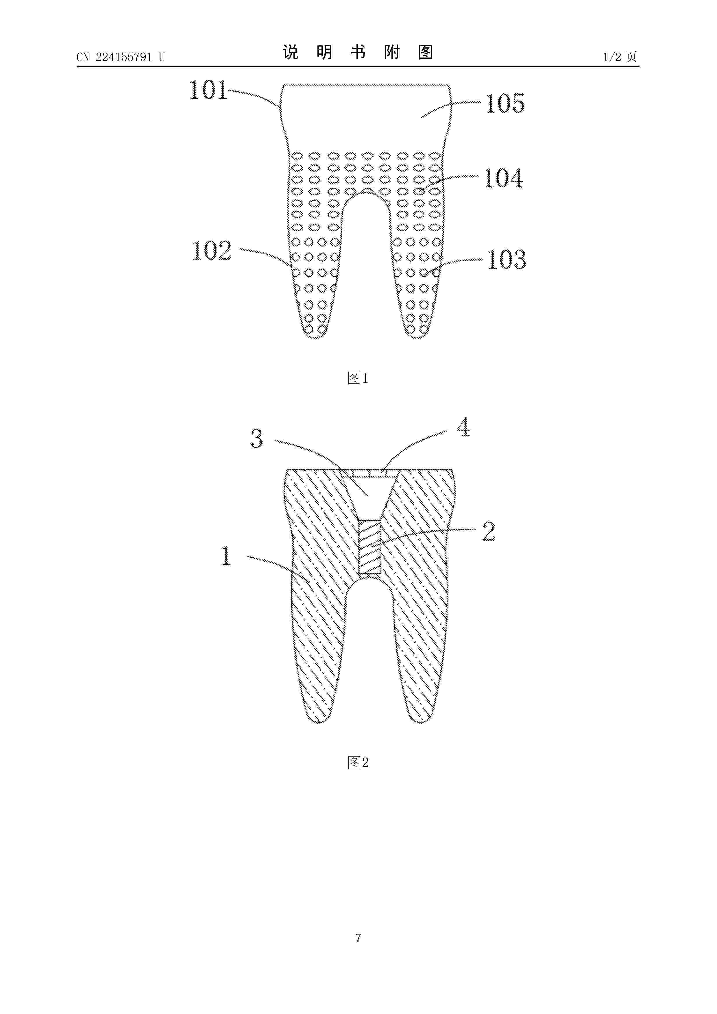
Resumen de: CN224155791U
本实用新型公开了一种个性化根型种植体;包括种植本体,所述种植本体的上端开设有中央螺丝孔,所述中央螺丝孔的上部开设有莫氏锥度孔,所述莫氏锥度孔的上端开设有内六角基座孔;本实用新型采用Ti‑Ta合金的种植体,接近人体骨组织,有效降低应力屏蔽效应;同时,钽元素的引入提高了材料的耐腐蚀性和骨结合能力;采用3D打印技术实现种植体完全匹配拔牙窝形态,骨内部分表面构建仿生骨小梁多孔结构的骨孔和凸块,使得种植体便于在牙窝中进行安装;并且在种植体上端设计中央螺丝孔和莫氏锥度孔,便于对假牙进行安装连接,以及在上端设有内六角基座孔,便于提高假牙的连接稳定性,防止假牙旋转脱落。
Nº publicación: WO2026084480A1 23/04/2026
Solicitante:
ALDAVER INC [KR]
\uC8FC\uC2DD\uD68C\uC0AC \uC54C\uB370\uBC14
Resumen de: WO2026084480A1
The present disclosure relates to: a 3D printing ink composition comprising alginate, a biocompatible hydrogel, acrylamide-based monomers, a photocrosslinkable polymer, a light absorber, and a solvent; and a 3D printing molded article formed by curing the composition.
 BOPI
BOPI
 Sede Electrónica