Si deseas distinguir tus productos, servicios o ambos de los de otra empresa, es posible que necesites una marca o nombre comercial. Descubre qué son, en qué consiste su procedimiento de registro y qué implica.

Información sobre los plazos de presentación de solicitudes de transformación de marcas de la Unión Europea en marca nacional española. Más información


Si tienes un nuevo dispositivo, producto o procedimiento que resuelva un problema técnico o tenga una ventaja práctica, existen distintas formas de protegerlo en España y en otros países. Descubre cómo hacerlo.

¿Tu innovación reside en la estética, la ornamentación o la apariencia de tu producto? Protégela mediante un diseño industrial. Descubre qué derechos confiere el registro y cómo realizar la tramitación.

Las indicaciones geográficas protegen el nombre de un producto originario de una zona geográfica, a la cual le debe una determinada calidad, reputación u otra característica. Descubre qué son, en qué consiste su procedimiento de registro y qué beneficios conceden.

Las patentes publicadas en todo el mundo son una valiosa fuente de información científica, técnica y comercial.

Si eres emprendedor/a o una empresa y quieres potenciar y mejorar la rentabilidad de tu negocio protegiendo de forma adecuada los activos intangibles de tu organización, en este espacio encontrarás lo necesario.


 175
resultados
175
resultados
 Última actualización
22/10/2026 [09:51:00]
Última actualización
22/10/2026 [09:51:00]


 Resultados 25 a 50 de 175

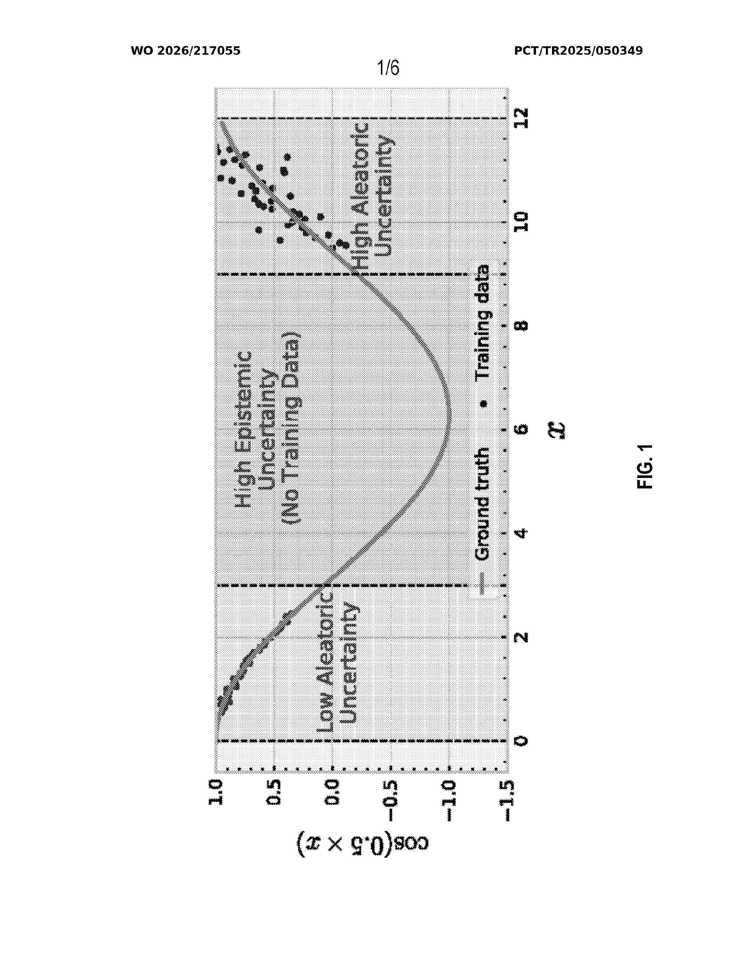
Resumen de: WO2026217055A1
A computer-implemented method (1000) for machine unlearning is presented The method comprises obtaining (1002) a request (2) indicating one or more X. X represents one or more input samples to be unlearned. The method further comprises for each X: obtaining (1004) U based on X, obtaining (1006) L based on X, updating (1008) F based on W'. W' is obtained so as to increase U, based on W, U, L, LR. F represents a machine learning model trained with at least X. W represents one or more model parameters of F. U represents one or more quantified uncertainty values for X. L represent one or more quantified loss values for X. LR represents a learning rate of F. The method (1000) further comprises initiating (1010) a deploying of F.

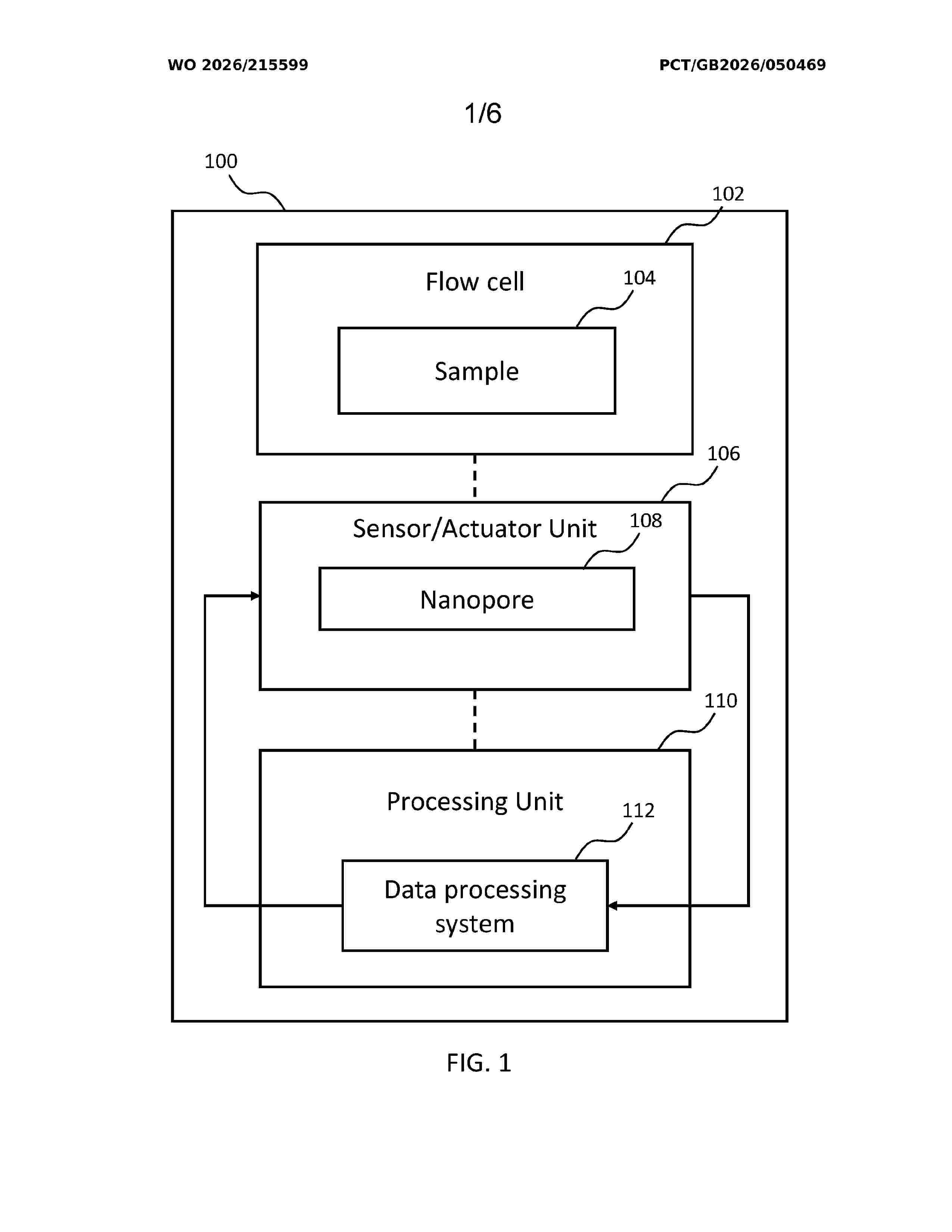
Resumen de: WO2026215599A1
A computer-implemented method of processing a measurement signal comprising measurements of a sample by a sensor unit comprising a nanopore includes analysing the measurement signal using a machine learning model to determine that a portion of the measurement signal is associated with a first state of the nanopore, the first state being one of a plurality of states of the nanopore. The method includes determining whether to further analyse the portion of the measurement signal in dependence on the determination that the portion of the measurement signal is associated with the first state of the nanopore.
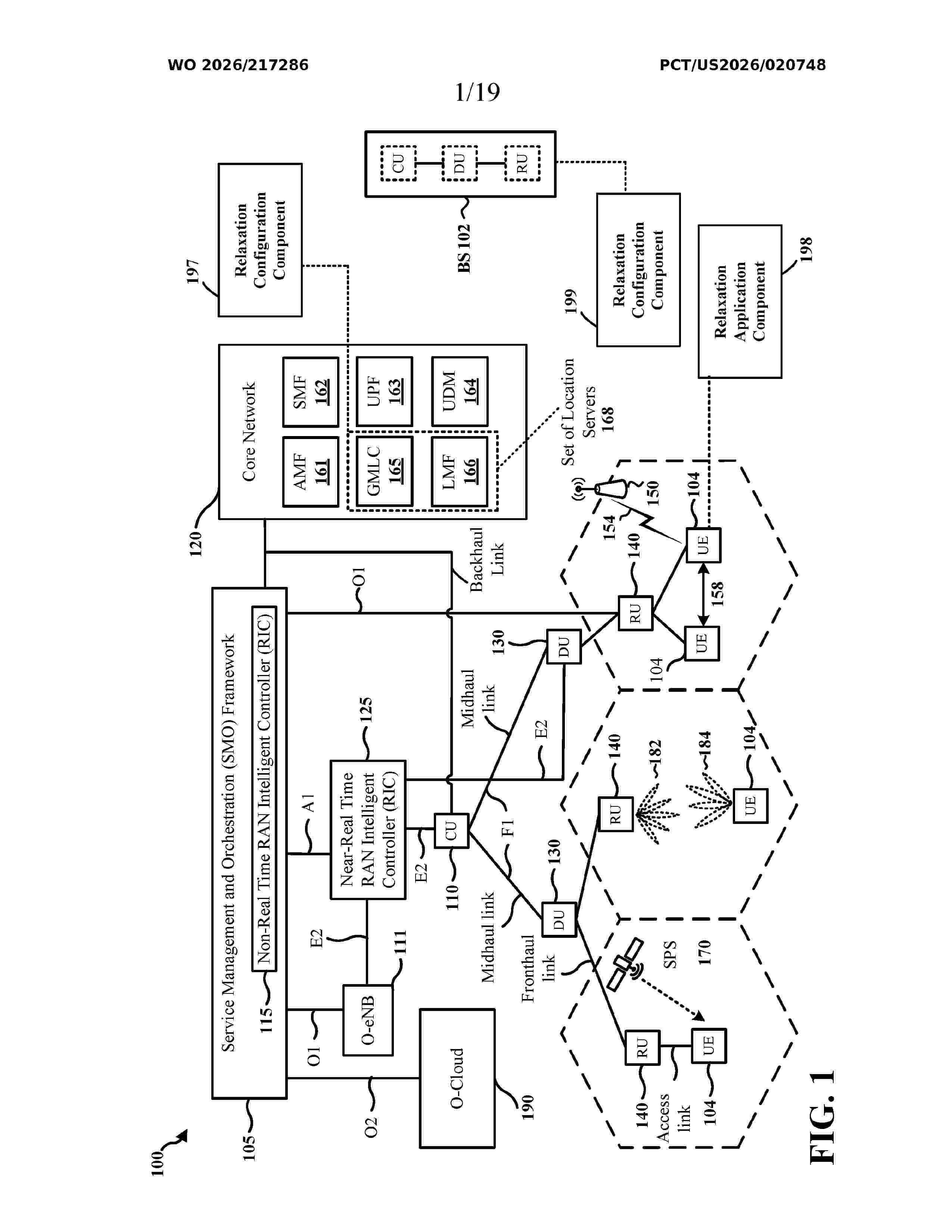
Resumen de: WO2026217286A1
Aspects presented herein may enable a user equipment (UE) to apply a set of relaxations for artificial intelligence (AI) or machine learning (ML) (AI/ML) related operations, thereby improving the resource managements for the AI/ML related operations. For example, in one aspect of the present disclosure, a UE receives an indication to perform at least one AI/ML operation. The UE obtains a set of relaxation criteria related to the at least one AI/ML operation, where the set of relaxation criteria includes at least one of: (1) a low mobility criterion, (2) a not-at-cell-edge criterion, (3) a stationary criterion, or (4) an AI/ML reduced capability device criterion. The UE performs at least one relaxation operation for the at least one AI/ML operation if the UE satisfies at least one relaxation criterion or all relaxation criteria in the set of relaxation criteria.
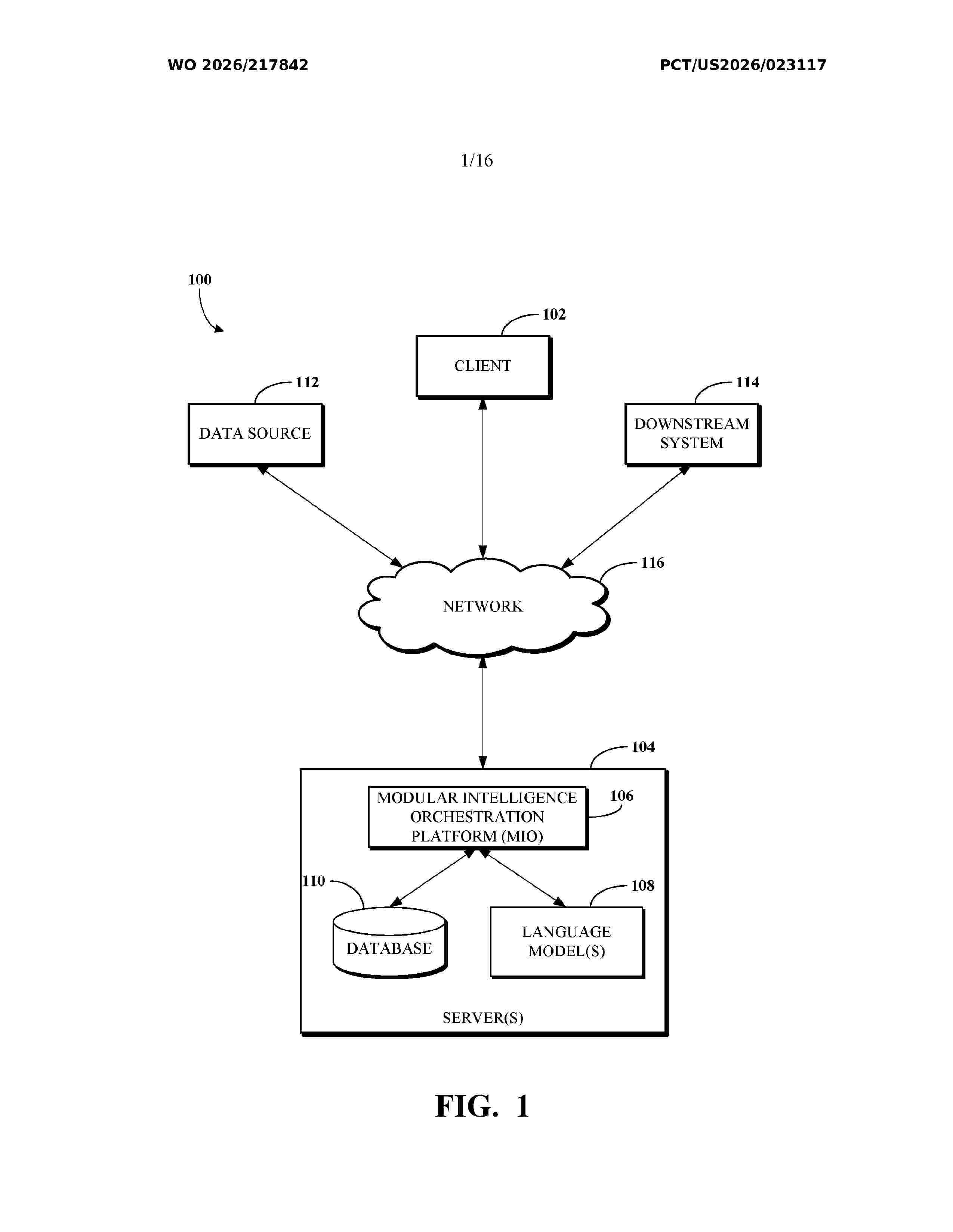
Resumen de: WO2026217842A1
A natural language input from a user specifying a challenge or objective is received. The input is processed using a retrieval-augmented generation (RAG) engine and context-augmented guidance (CAG) engine to identify one or more relevant machine learning modules from a predefined library. A modular execution pipeline that includes the identified modules is dynamically configured by resolving interdependencies using dependency graph analysis that models modules as nodes and inter-module data dependencies as edges. Goal-aware cleansing of user-provided data is applied. The goal-aware cleansing is based on data requirements of at least one of the identified modules. The modular pipeline is executed on the cleansed data to generate actionable insights. The actionable insights are then transmitted to a client device.
Resumen de: WO2026216662A1
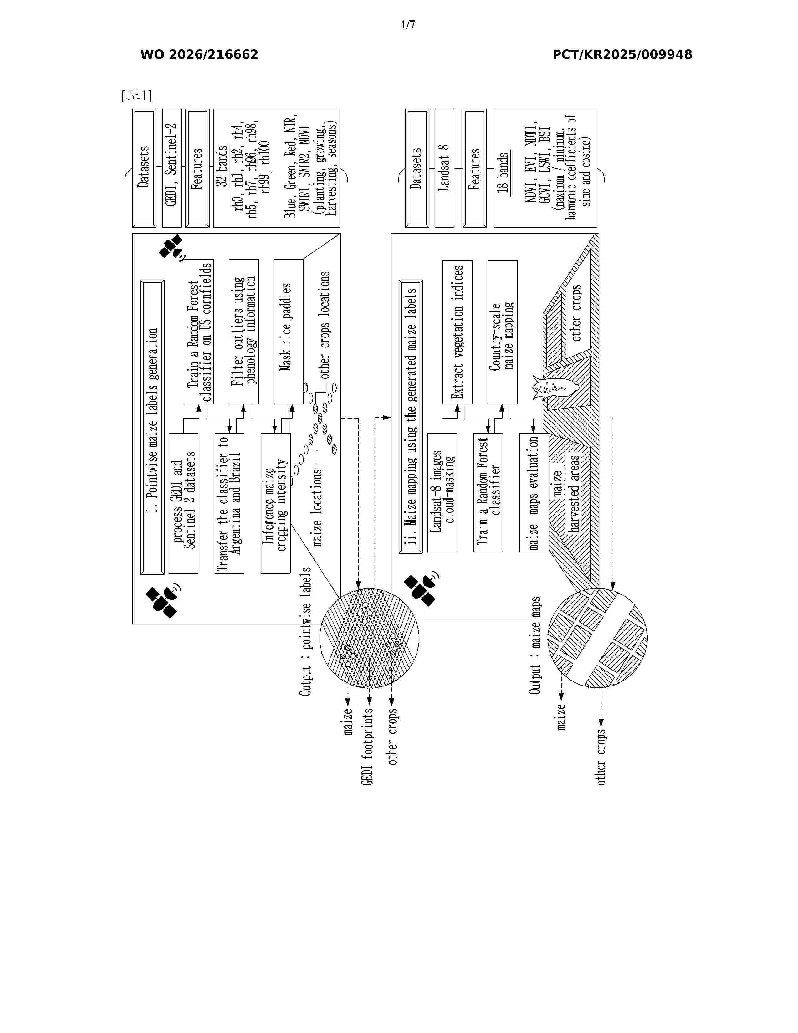
The present invention relates to a method and system for maize mapping using multi-source satellite imagery and machine learning. The method for maize mapping, according to an embodiment of the present invention, is performed by the system for maize mapping and comprises: a first step of acquiring LiDAR data and optical data captured by a satellite; a second step of training a first random forest classifier by using data of a first area including a height and optical characteristics of maize; a third step of inputting LiDAR data and optical data captured by a satellite of a second area into the trained first random forest classifier to infer maize cultivation areas; a fourth step of filtering outliers in the inferred maize cultivation areas by using phenological growth cycle information of maize; a fifth step of inferring a maize cropping intensity in the filtered maize cultivation areas; and a sixth step of outputting maize location points including point data by excluding paddy field areas from the maize cultivation areas in which the maize cropping intensity is inferred.
Resumen de: WO2026217316A1
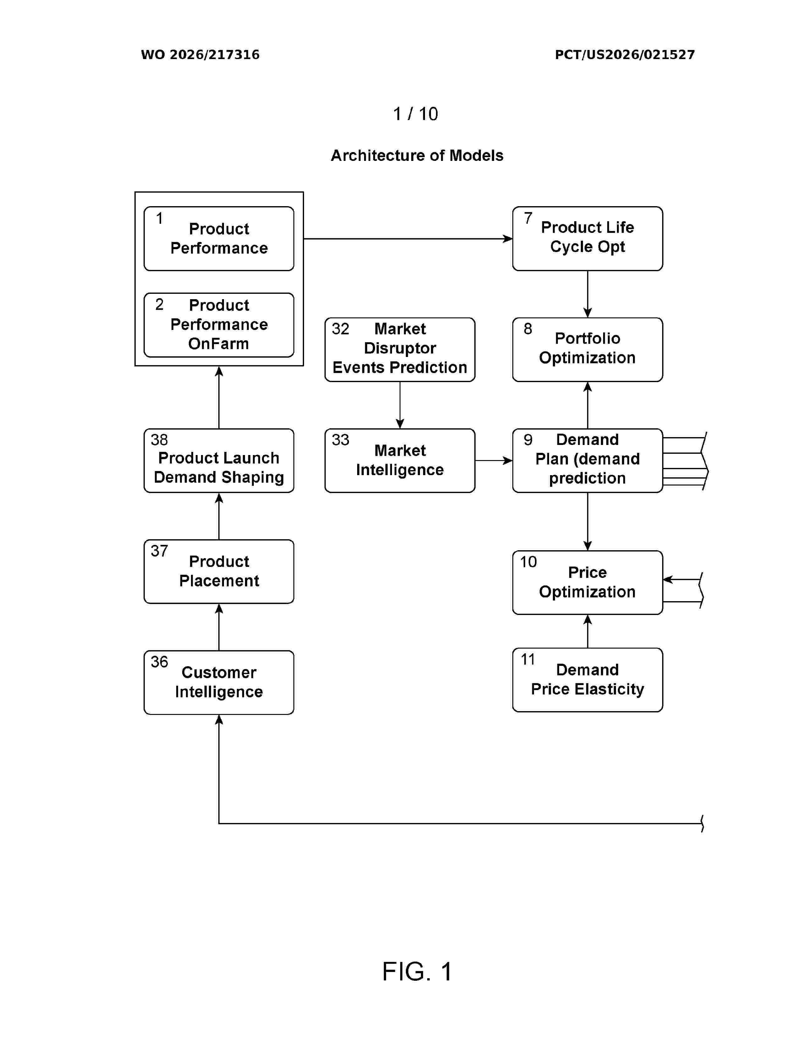
Systems, methods, and apparatuses for a system for seed production and logistics are described. Seed production and logistics system data comprising seed variety demand, crop status, environmental conditions, seed conditioning and inventory status associated with a seed production and logistics process may be received, and a data representing a digital twin of the end to end seed production process may be aggregated and harmonized. A machine learning model may process the current state of the digital twin data to generate future state predictions and to recommend seed productions and logistics decisions that will result in improved seed quality and production efficiency.
Resumen de: TW202528173A
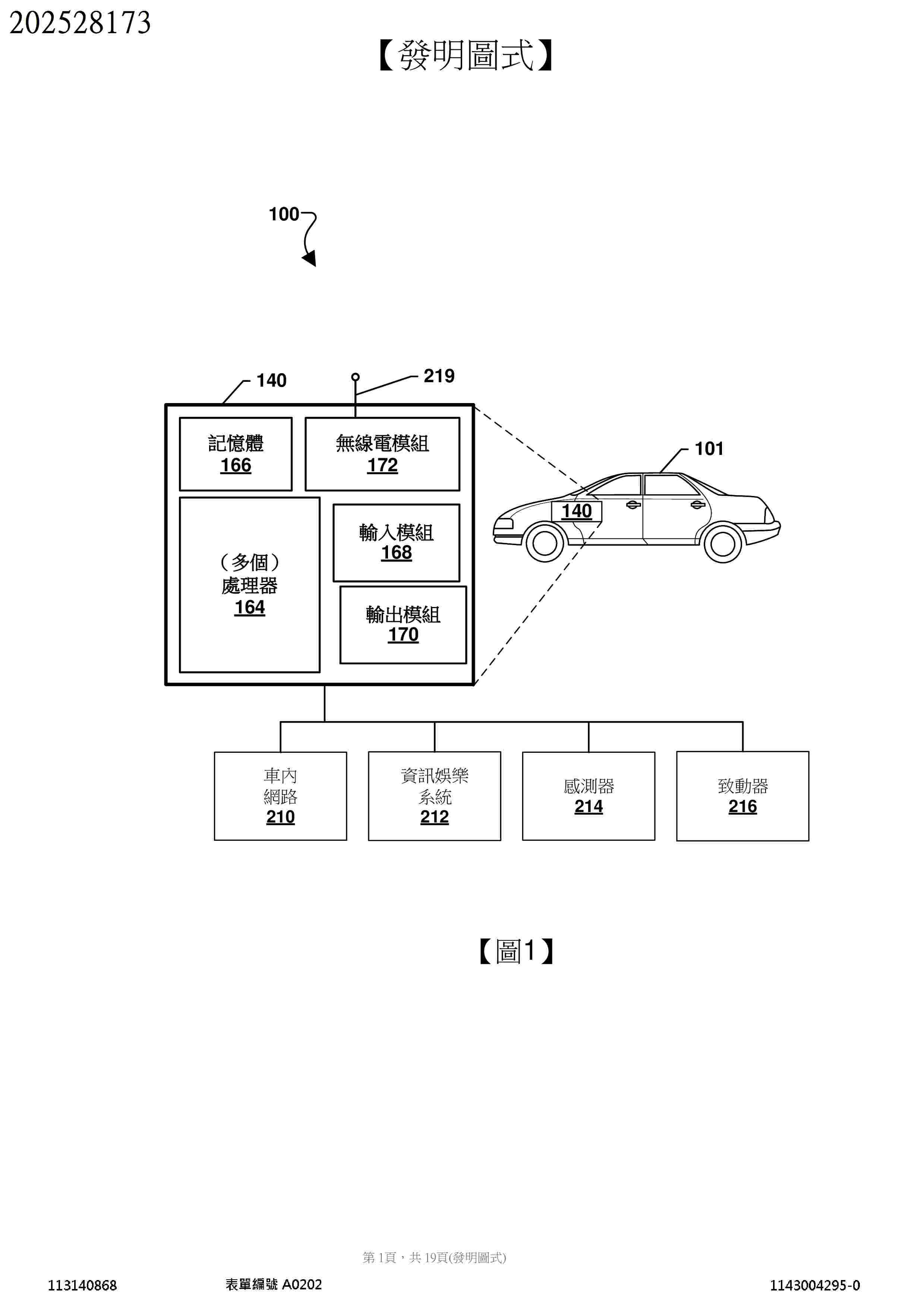
Various embodiments include methods and vehicles that may include managing driving policies in a vehicle advanced driver assist system (ADAS) that include obtaining vehicle sensor data, determining a current vehicle context based on the vehicle sensor data, selecting a modified vehicle driving policy from a plurality of saved modified vehicle driving policies based on the determined current vehicle context, and controlling vehicle behavior by the ADAS based upon the selected modified vehicle driving policy. Methods may also include receiving user voice inputs from a vehicle microphone, using a generative AI to infer relevance of the user voice inputs to vehicle driving policies or actions of the ADAS, adjusting a vehicle driving policy of the ADAS based on the inferred relevance of the user voice inputs, and commanding vehicle behavior based upon the adjusted vehicle driving policy.
Resumen de: EP4822508A1
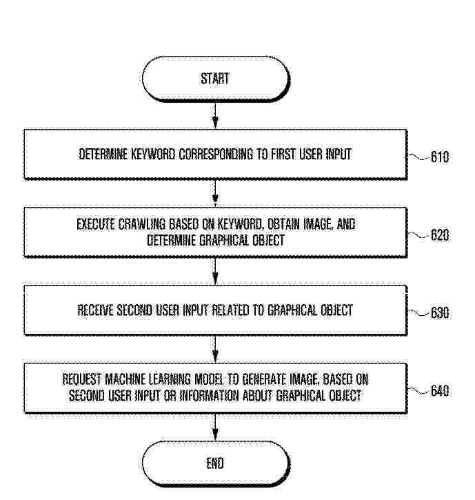
0001 This electronic device may comprise: a memory for storing instructions; a display; and a processor. The instructions, when executed by the processor, may cause the electronic device to: determine a keyword corresponding to a first user input on the basis of reception of the first user input; obtain at least one image through crawling on the basis of the determined keyword; determine at least one graphical object corresponding to the at least one image; receive a second user input related to the at least one graphical object; and generate an artificial intelligence image using a machine learning model on the basis of any one of the second user input or information about the at least one graphical object.
Resumen de: US20260310246A1
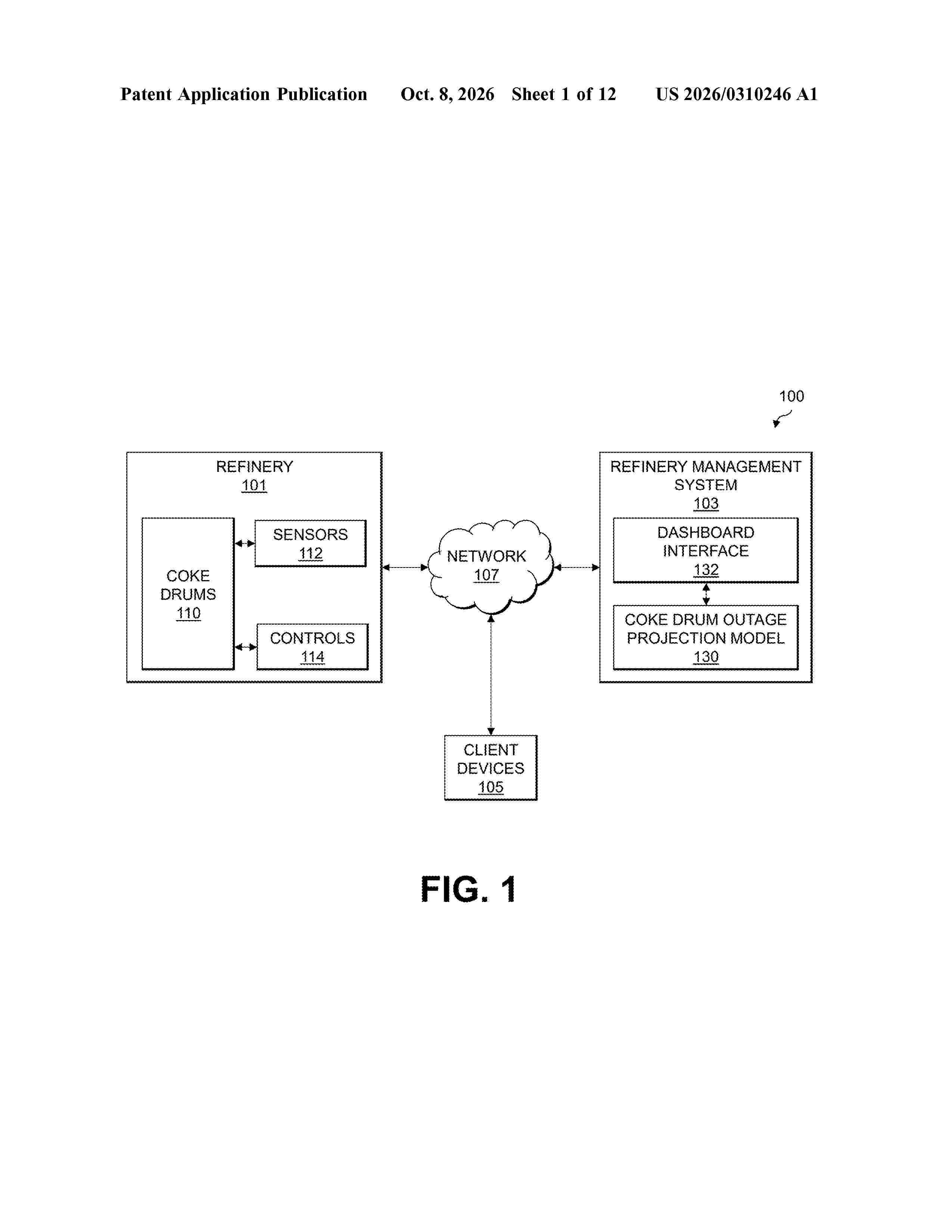
0000 An apparatus includes at least one processing device configured to obtain sensor data from sensors associated with one or more coke drums in a refinery. The at least one processing device is also configured to generate an outage projection for an end of a feed cycle of at least one of the coke drums by processing, utilizing a machine learning model, information derived from the sensor data characterizing (i) an amount of feedstock which has been fed to the at least one coke drum during the feed cycle, (ii) a time at which a level indicator for the at least one coke drum first exceeds a designated threshold value, and (iii) a density of the feedstock fed to the at least one coke drum during the feed cycle. The at least one processing device is further configured to provide, at a user interface, the generated outage projection.
Resumen de: WO2026210775A1
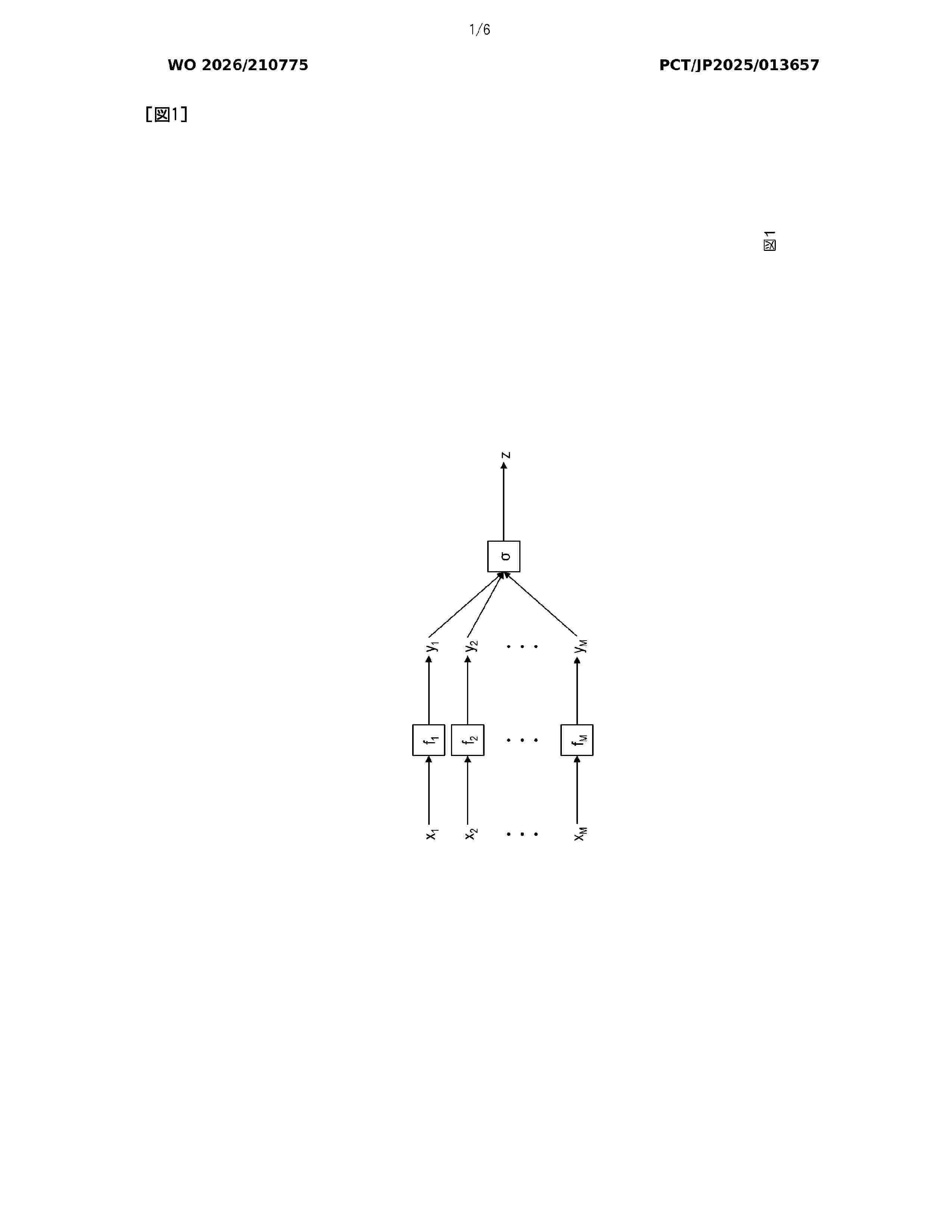
Provided is an erroneous answer risk determination device capable of accurately determining an erroneous answer risk of a machine learning model. The erroneous answer risk determination device includes an erroneous answer risk determination unit that determines an erroneous answer risk on the basis of a result of comparing a first set and a second set, the first set being a set of errors in an estimation function, which is an estimation result of an unknown function, allowed on the basis of correctness of an output domain with respect to M input domains in a first composite function obtained by composing the estimation function and a first known function, and the second set being a set of errors in the estimation function allowed on the basis of correctness of an output domain with respect to M input domains in a second composite function obtained by composing the estimation function and a second known function different from the first known function.
Resumen de: US20260308751A1
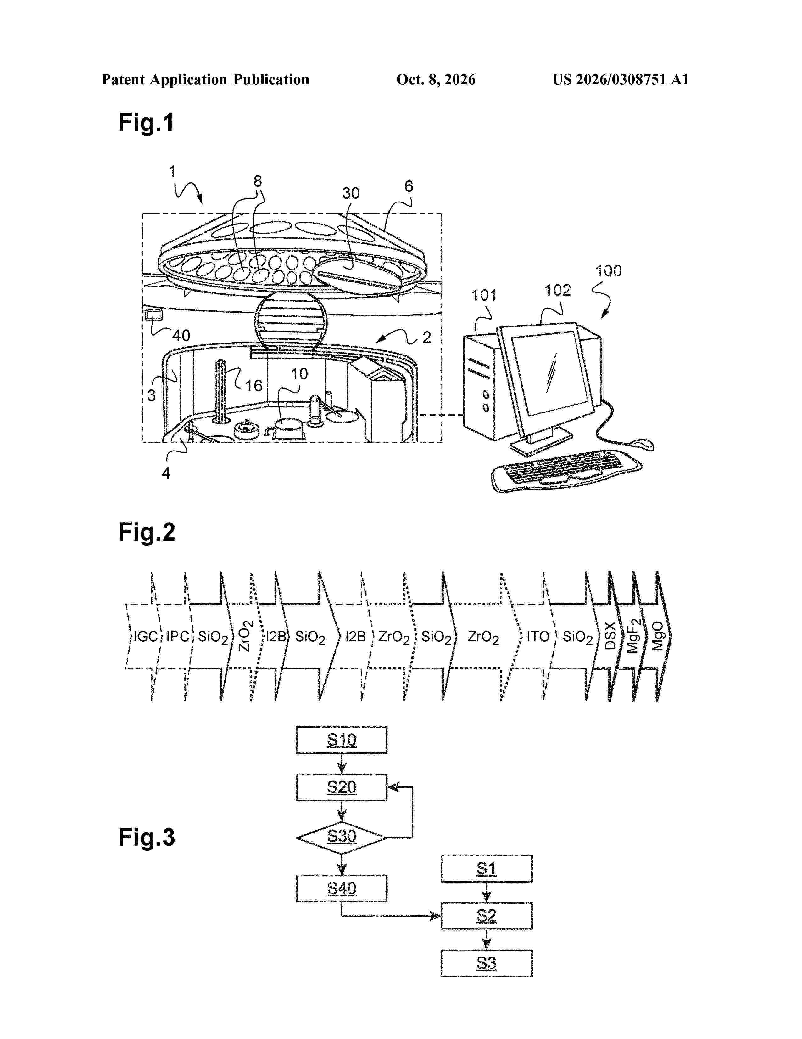
The invention relates to a computer implemented method for predicting a level of quality of a vacuum into a vacuum chamber (2) of a coating machine (1) suitable for coating optic elements (8). According to the invention, this method comprises steps of: —acquiring at least one technical feature of the coating machine during coating processes of optic elements, and —predicting said level of quality from the at least one measured technical feature by means of a processing unit that registers a machine learning model, said at least one technical feature being an input of said machine learning model and said level of quality being an output of said machine learning model.
Resumen de: US20260311570A1
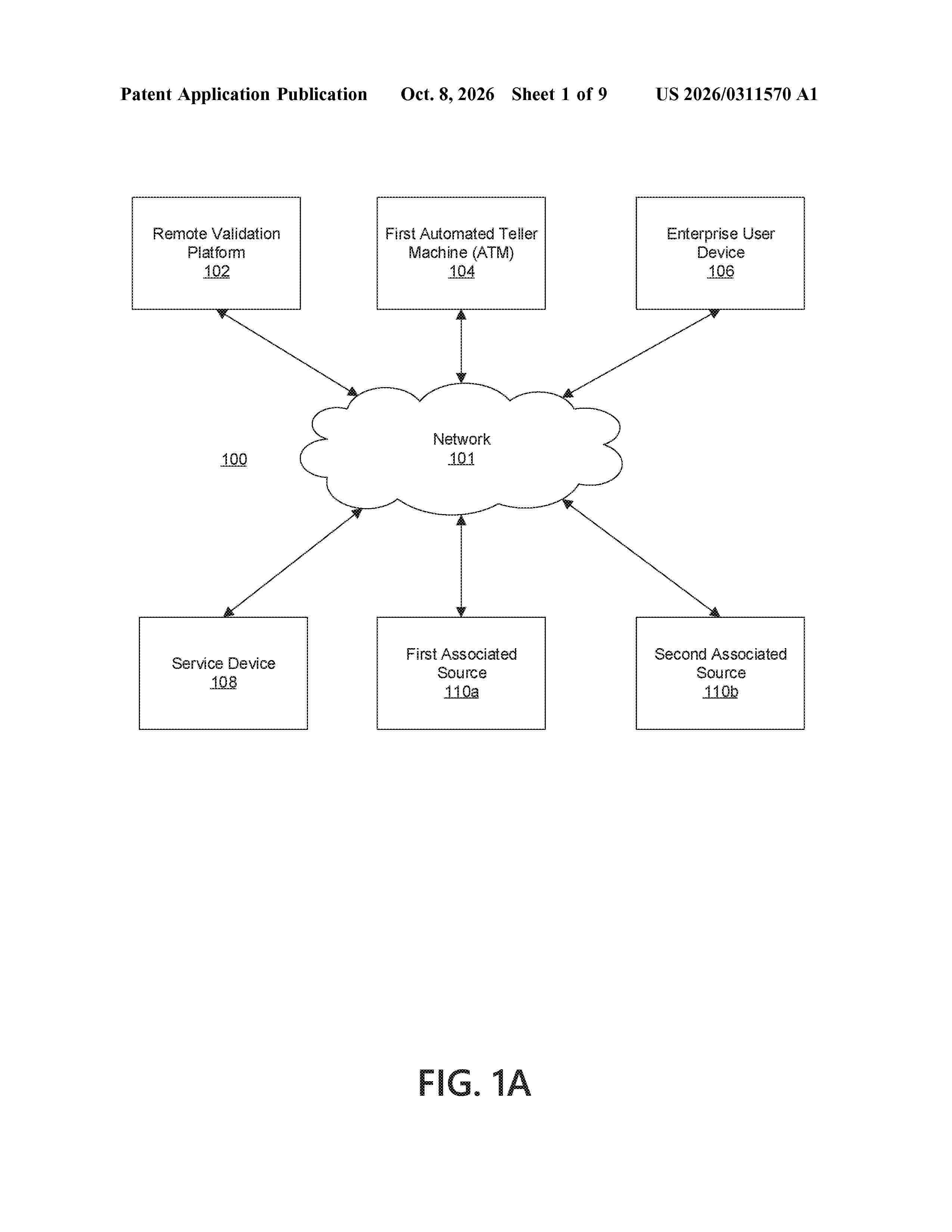
0000 Aspects of the disclosure relate to a remote validation platform for ATMs. The platform may train a reconfiguration model to output sets of configuration instructions. The platform may receive a first information stream from a first ATM. The platform may receive one or more additional information streams from sources associated with the first ATM. The platform may generate, using the reconfiguration model, a first set of configuration instructions based on the first information stream and the one or more additional information streams. The platform may cause reconfiguration of the first ATM based on the first set of configuration instructions. The platform may refine the reconfiguration model based on the first set of configuration instructions.
Resumen de: US20260310999A1
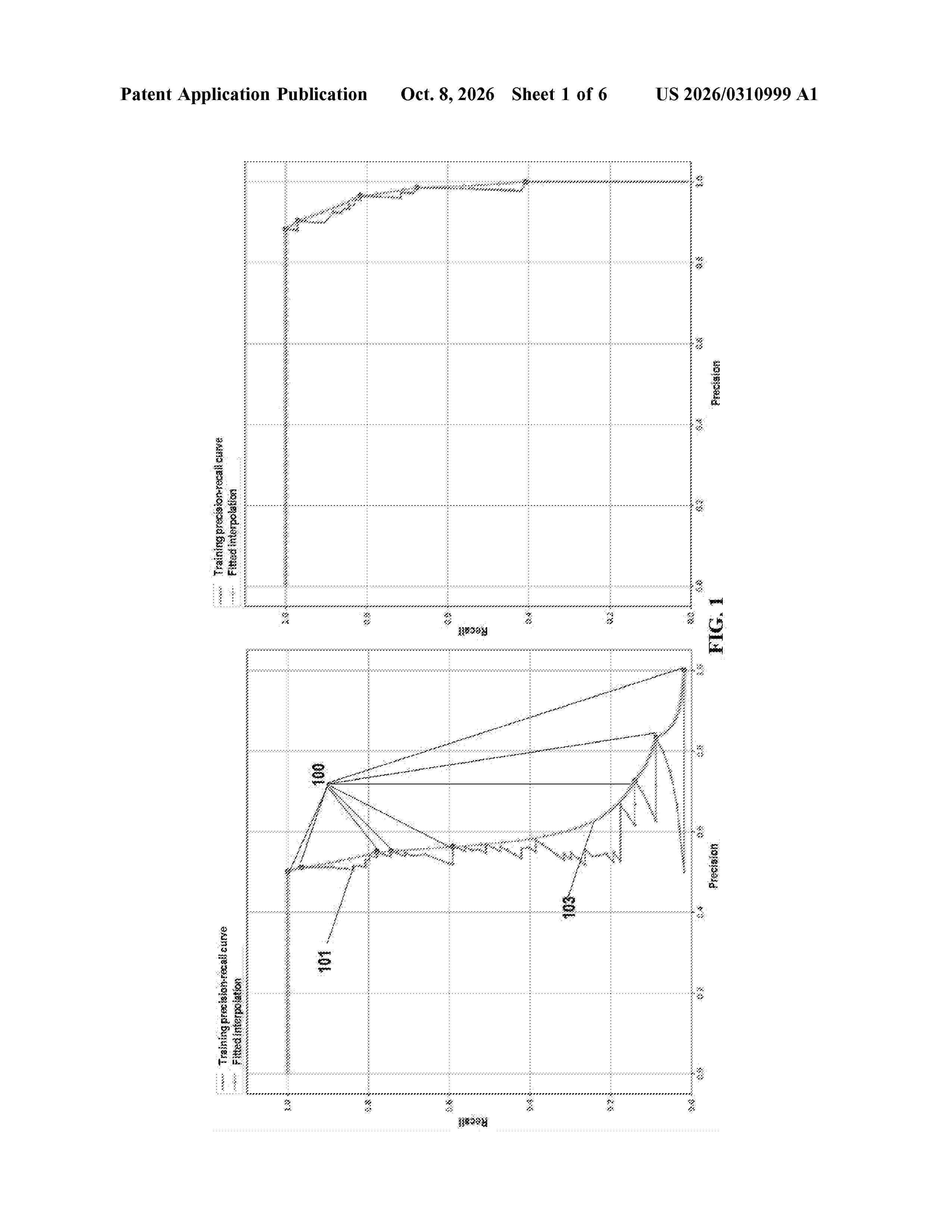
0000 The present disclosure provides methods and systems for generating a confidence label for a prediction produced by a predictive model. The method comprises: (a) generating training datasets for training a confidence model, the training datasets are generated using data collected from a cross validation process for evaluating the predictive model; (b) training the confidence model using the training datasets to learn a relationship between a score assigned by the predictive model to a prediction and a correctness measure of the prediction; and (d) feeding an input to the trained confidence model to output a confidence label. The input comprises a target precision or a target recall for a new prediction produced by the predictive model and a score assigned to the new prediction, and the confidence label indicates whether the new prediction is high confidence or low confidence.
Resumen de: US20260310899A1
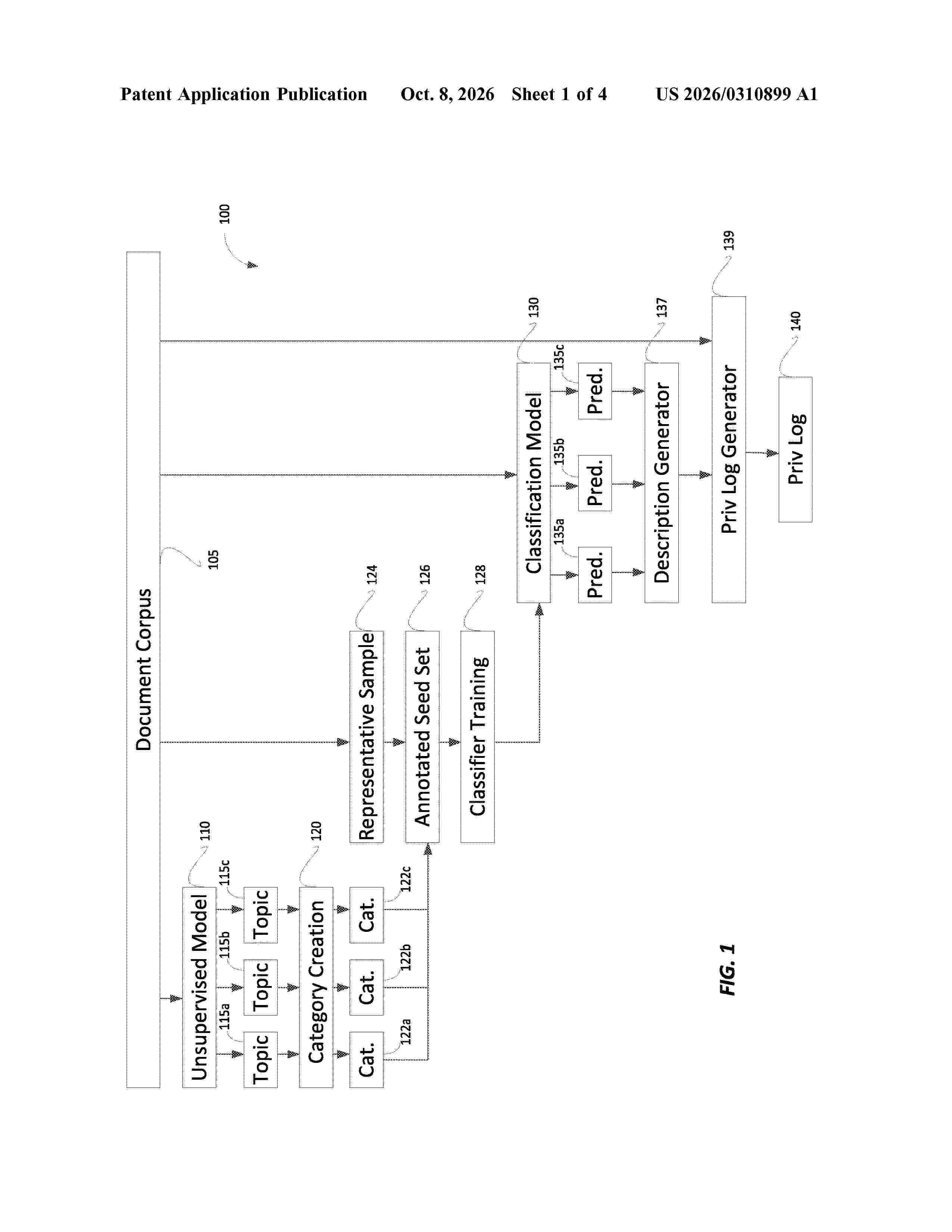
Systems, methods, and computer readable media for intelligent generation of privilege logs are provided. These techniques may include applying an unsupervised machine learning model to a corpus of documents to identify a plurality of topics associated with the corpus of documents, associating a plurality of categories with respective subsets of the plurality of topics, executing a first classifier training model to train a first classifier for a category, and a second classifier training model to train a second classifier for the category, determining a performance metric for the first classifier for the category and a performance metric for the second classifier for the category, selecting one of the first classifier or the second classifier for the category based on the performance metrics, applying the selected classifier to documents in the corpus of documents, and generating a privilege log based upon the classifiers applied to documents in the corpus of documents.
Resumen de: US20260311056A1
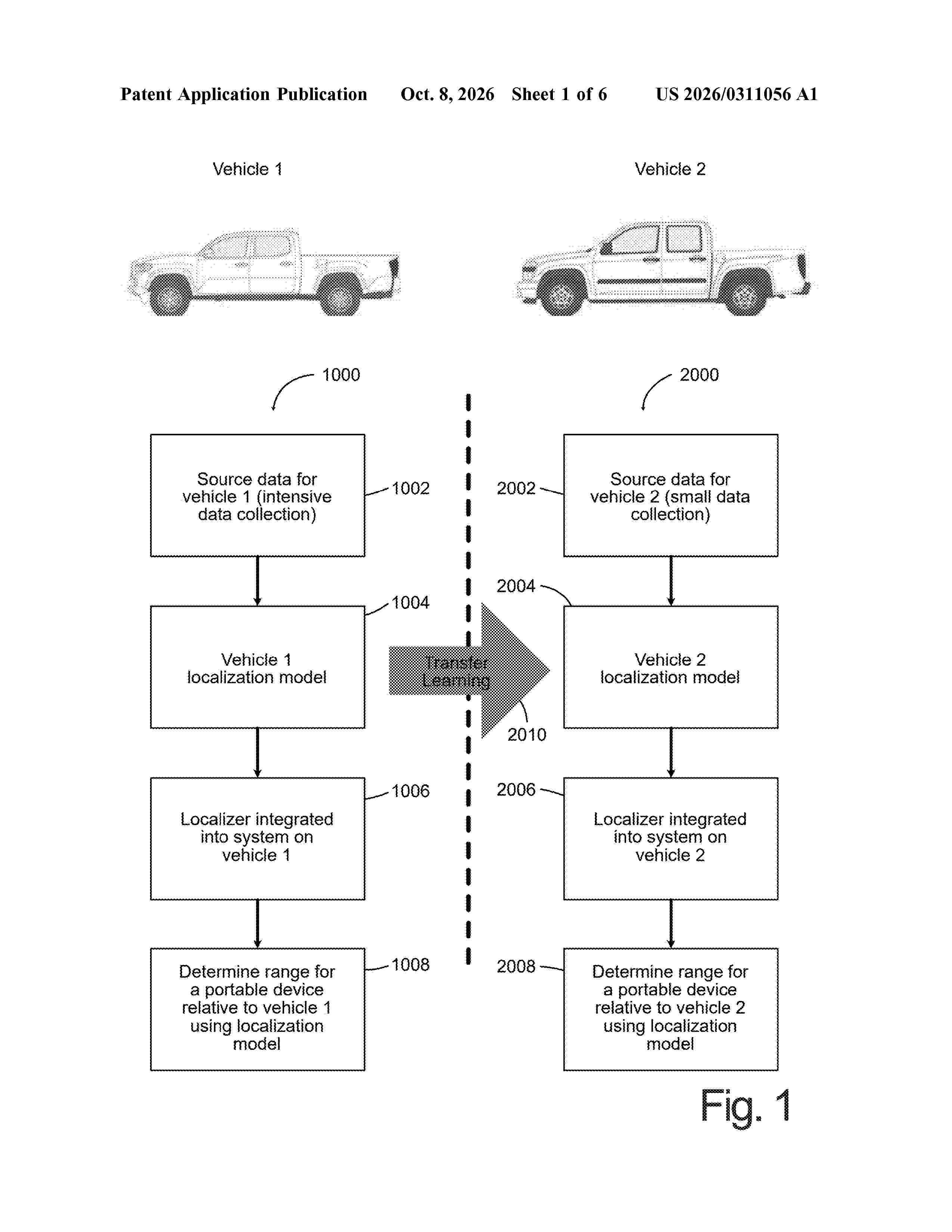
A system and method for determining location information of a portable device relative to an object is provided. In one embodiment, aspects of the system to determine location with respect to the portable device may be transferred to another system for training that system to determine location with respect to the portable device or another type of portable device.
Resumen de: AU2026234064A1
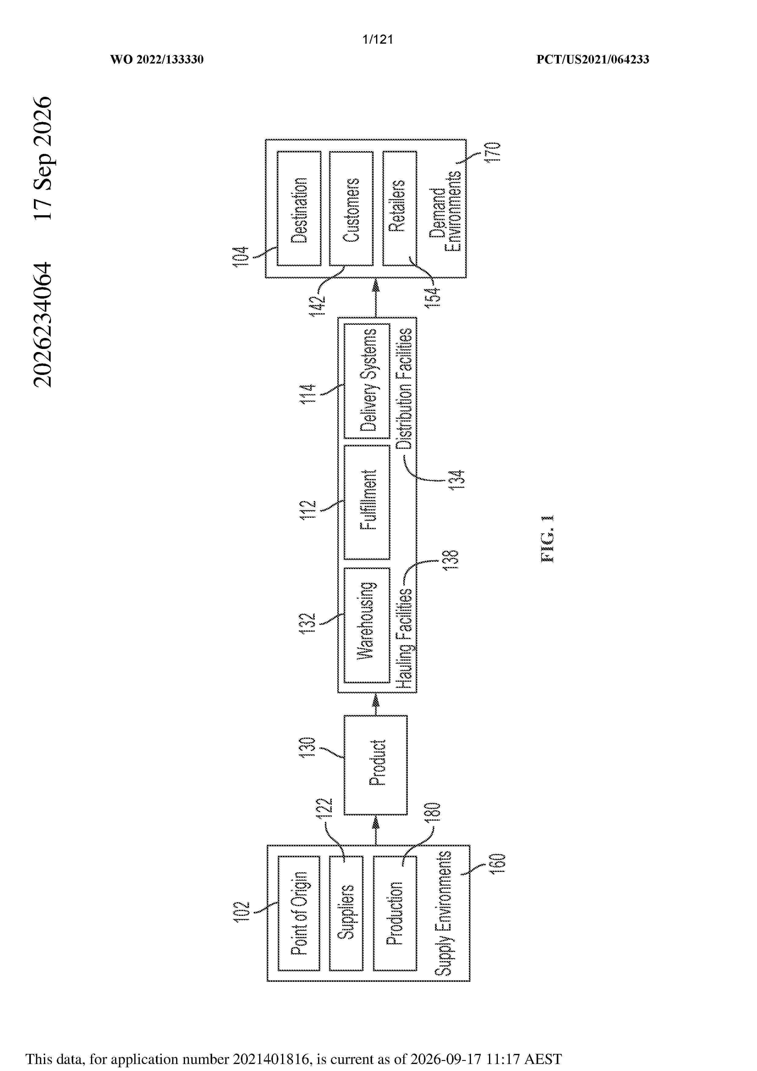
Abstract A robot fleet management platform, includes a set of datastores that store a governance library that defines a set of governance standards that include at least one set of security standards, legal standards, ethical standards, regulatory standards, quality standards, or engineering standards that are applied to decisions made by one or more respective intelligence services; and includes: a set of one or more processors that execute a set of computer-readable instructions, wherein the set of one or more processors collectively execute: a governance-enabling intelligence layer that receives and responds to intelligence requests received from respective intelligence service clients, wherein the intelligence layer includes: a set of artificial intelligence services that includes at least one of a machine learning service, a rules-based intelligence service, a digital twin service, a robot process automation service, or a machine vision service; and an intelligence layer controller that coordinates performance of respective intelligence services on behalf of the respective intelligence service clients and performance of a set of analyses corresponding to the respective intelligence services based in part on the set of governance standards, wherein the intelligence layer returns decisions determined collectively by the artificial intelligence service in response to the intelligence requests, such that the decisions are determined based on a set of intelligence service dat
Resumen de: US20260311066A1
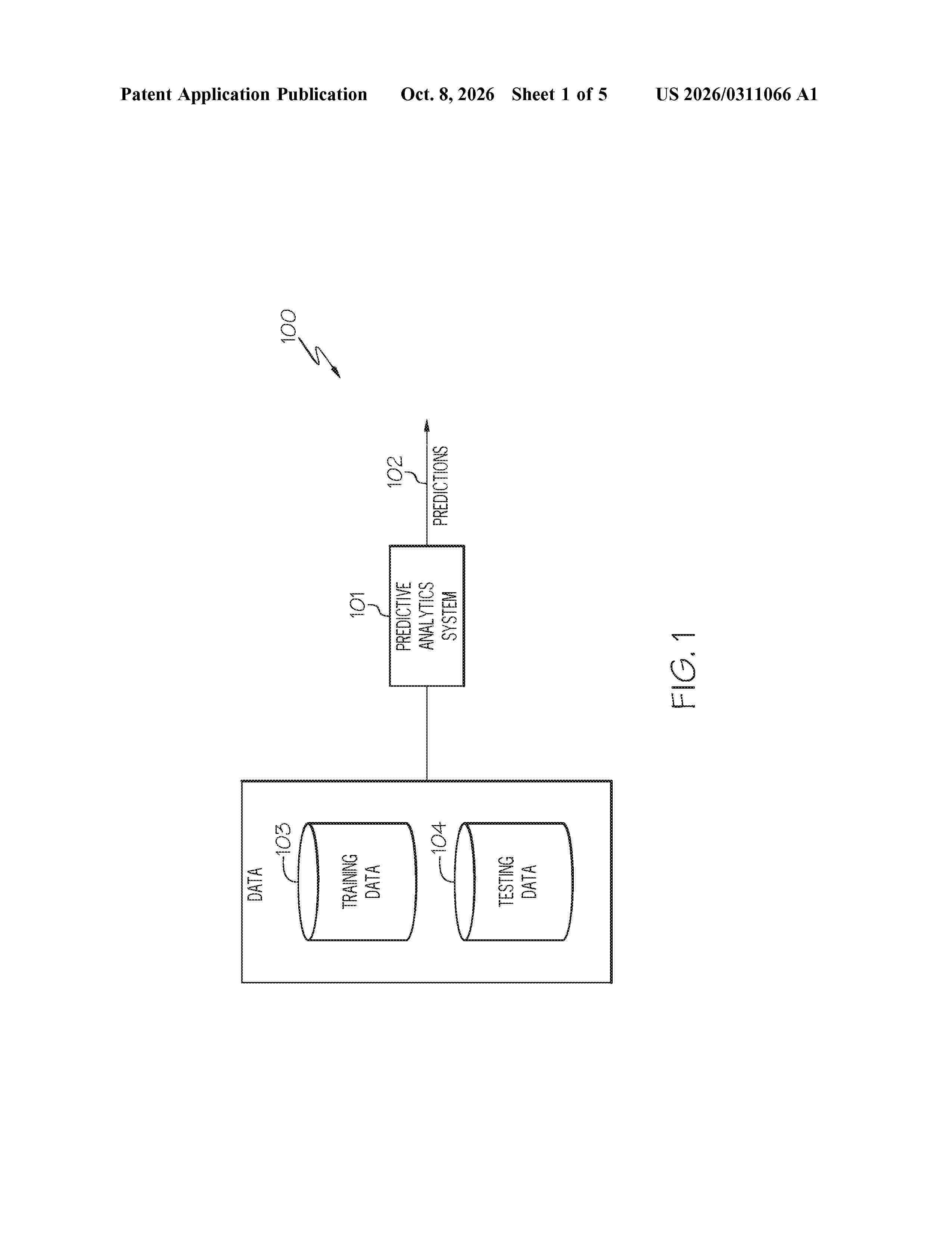
0000 A computer-implemented method, system and computer program product for improving prediction accuracy in machine learning techniques. A teacher model is constructed, where the teacher model generates a weight for each data case. The current student model is then trained using training data and the weights generated by the teacher model. After training the current student model, the current student model generates state features, which are used by the teacher model to generate new weights. A candidate student model is then trained using training data and these new weights. A reward is generated by comparing the current student model with the candidate student model using training and testing data, which is used to update the teacher model if a stopping rule has not been satisfied. Upon a stopping rule being satisfied, the weights generated by the teacher model are deemed to be the “optimal” weights which are returned to the user.
Resumen de: US20260310752A1
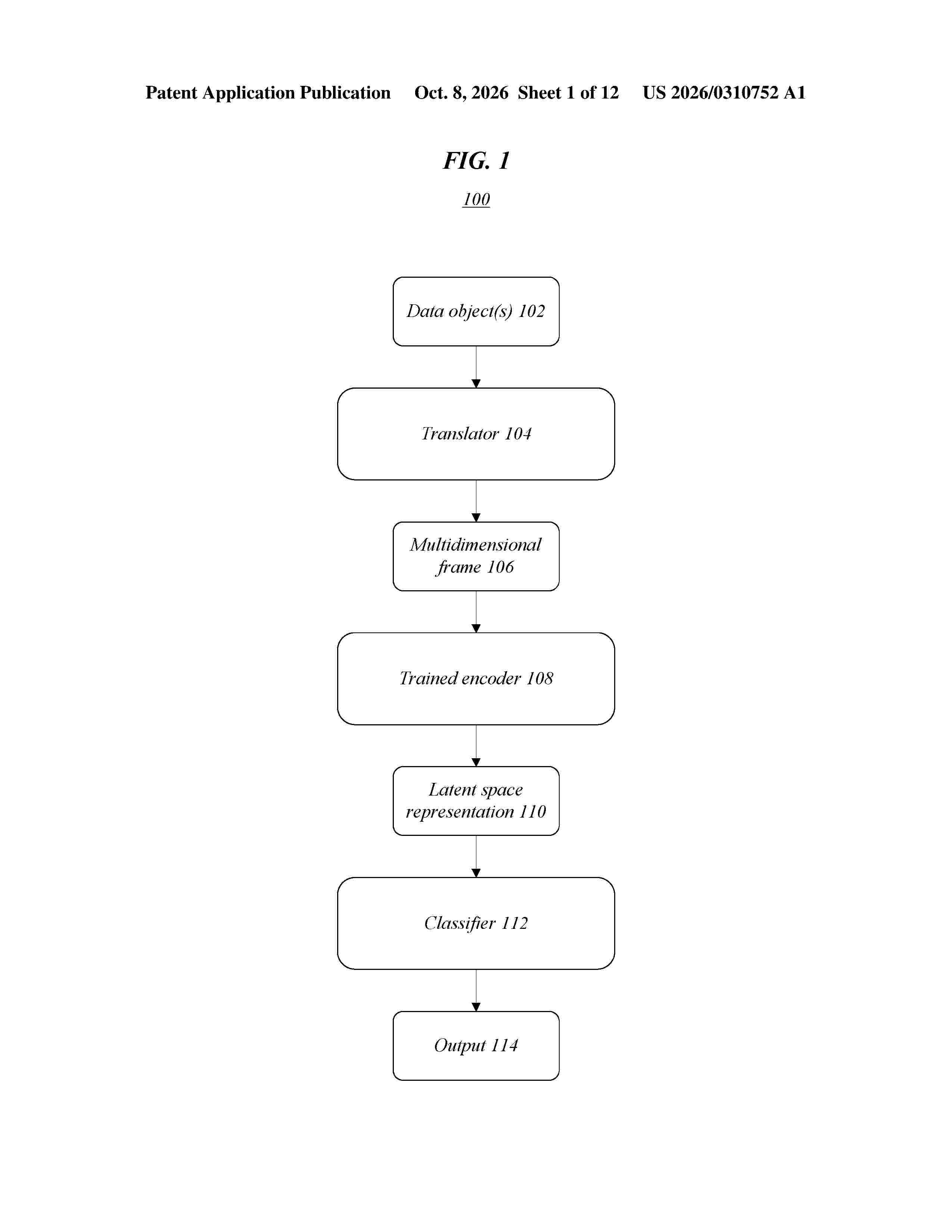
0000 Various embodiments are generally directed to techniques for embedding a data object into a multidimensional frame, such as for training an autoencoder to generate latent space representations of the data object based on the multidimensional frame, for instance. Additionally, in one or more embodiments latent space representations of data objects may be classified, such as with a machine learning algorithm. Some embodiments are particularly directed to embedding a data object comprising a plurality of object entries into a three-dimensional (3D) frame.
Resumen de: US20260311067A1
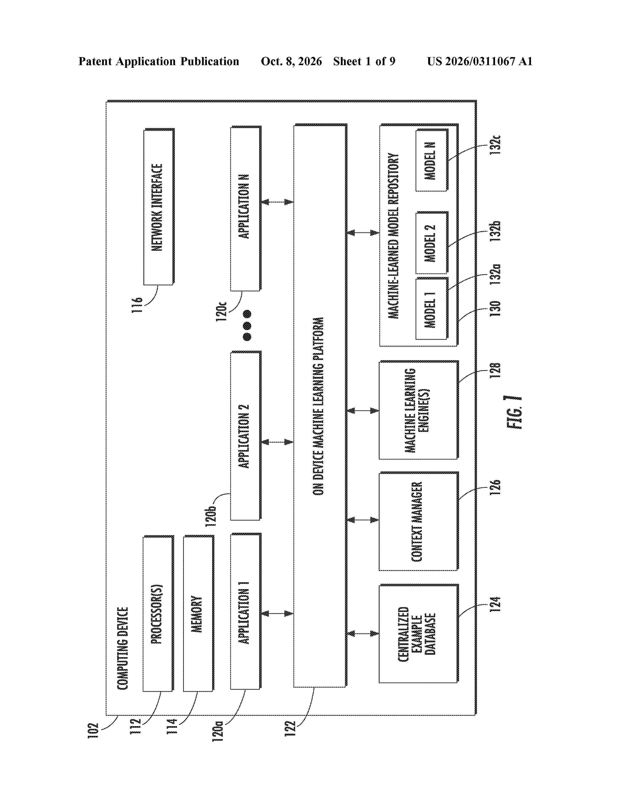
0000 The present disclosure provides systems and methods for on-device machine learning. In particular, the present disclosure is directed to an on-device machine learning platform and associated techniques that enable on-device prediction, training, example collection, and/or other machine learning tasks or functionality. The on-device machine learning platform can include a context provider that securely injects context features into collected training examples and/or client-provided input data used to generate predictions/inferences. Thus, the on-device machine learning platform can enable centralized training example collection, model training, and usage of machine-learned models as a service to applications or other clients.
Resumen de: US20260311061A1
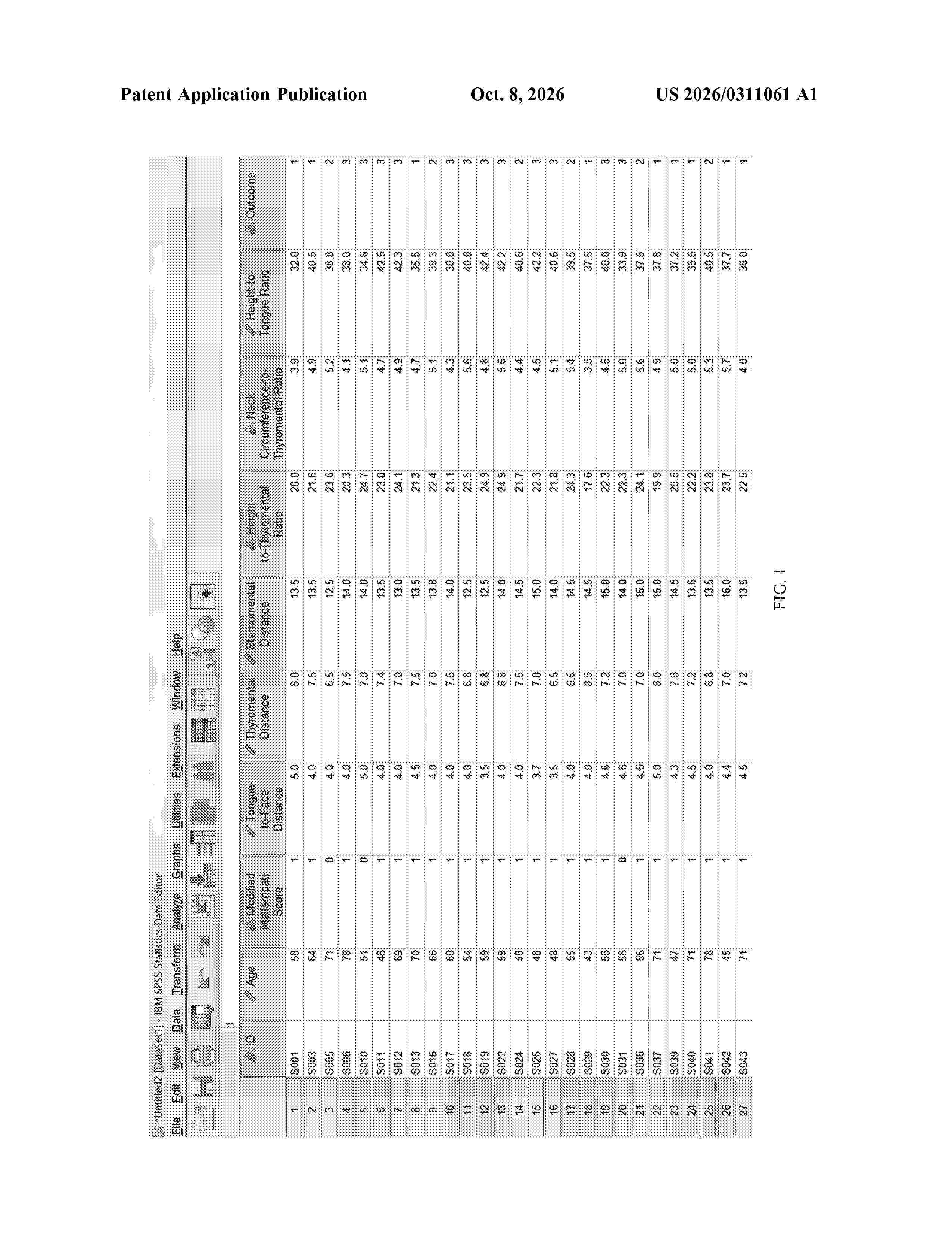
0000 Disclosed is a method for constructing a machine learning model for recommending an airway opening posture angle. The method includes collecting independent variable data affecting a glottis exposure effect when opening a patient airway, conducting an airway opening experiment, taking a posture angle corresponding to an optimal glottis exposure image as an optimal posture angle, performing single-factor analysis on the independent variables, and screening out statistically significant independent variable data as candidate prediction variables. The method further includes preselecting machine learning models, training and verifying the models, evaluating using AUC values, and selecting a model with a highest AUC value as a prediction model for recommending the optimal airway opening posture angle. The obtained prediction model accurately recommends the optimal airway opening posture angle to expose a glottis of the patient for medical personnel, reducing or avoiding a risk of glottis exposure difficulty in bronchoscopy diagnosis and treatment.
Resumen de: US20260310634A1
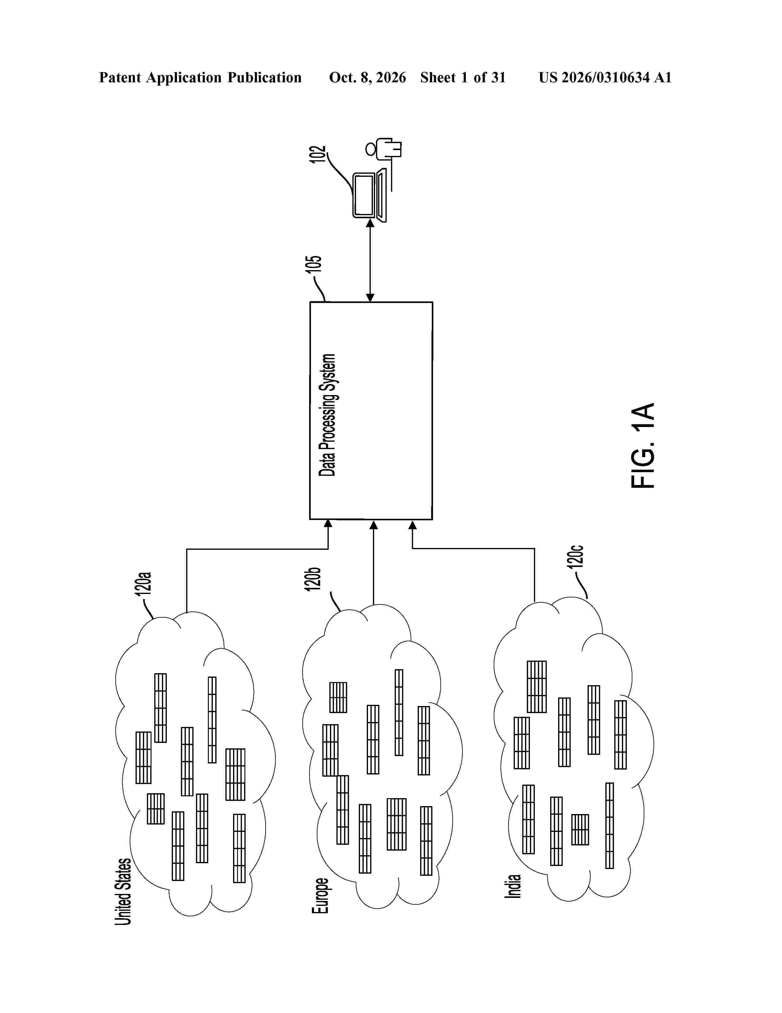
0000 Techniques for discovering primary, unique, and/or foreign keys for relational datasets are described. The techniques include profiling the relational datasets to obtain respective data profiles; identifying one or more primary key candidates for a first relational dataset using a first data profile of the first relational dataset and a first trained machine learning model; identifying one or more foreign key proposals for a second relational dataset using the one or more primary key candidates by performing a subset analysis of the second relational dataset with respect to the first relational dataset; identifying one or more foreign key candidates for the second relational dataset using the first data profile, a second data profile of the second relational dataset, and a second trained machine learning model different from the first trained machine learning model; and outputting the at primary key candidate(s) and the foreign key candidate(s).
Resumen de: WO2026209468A1
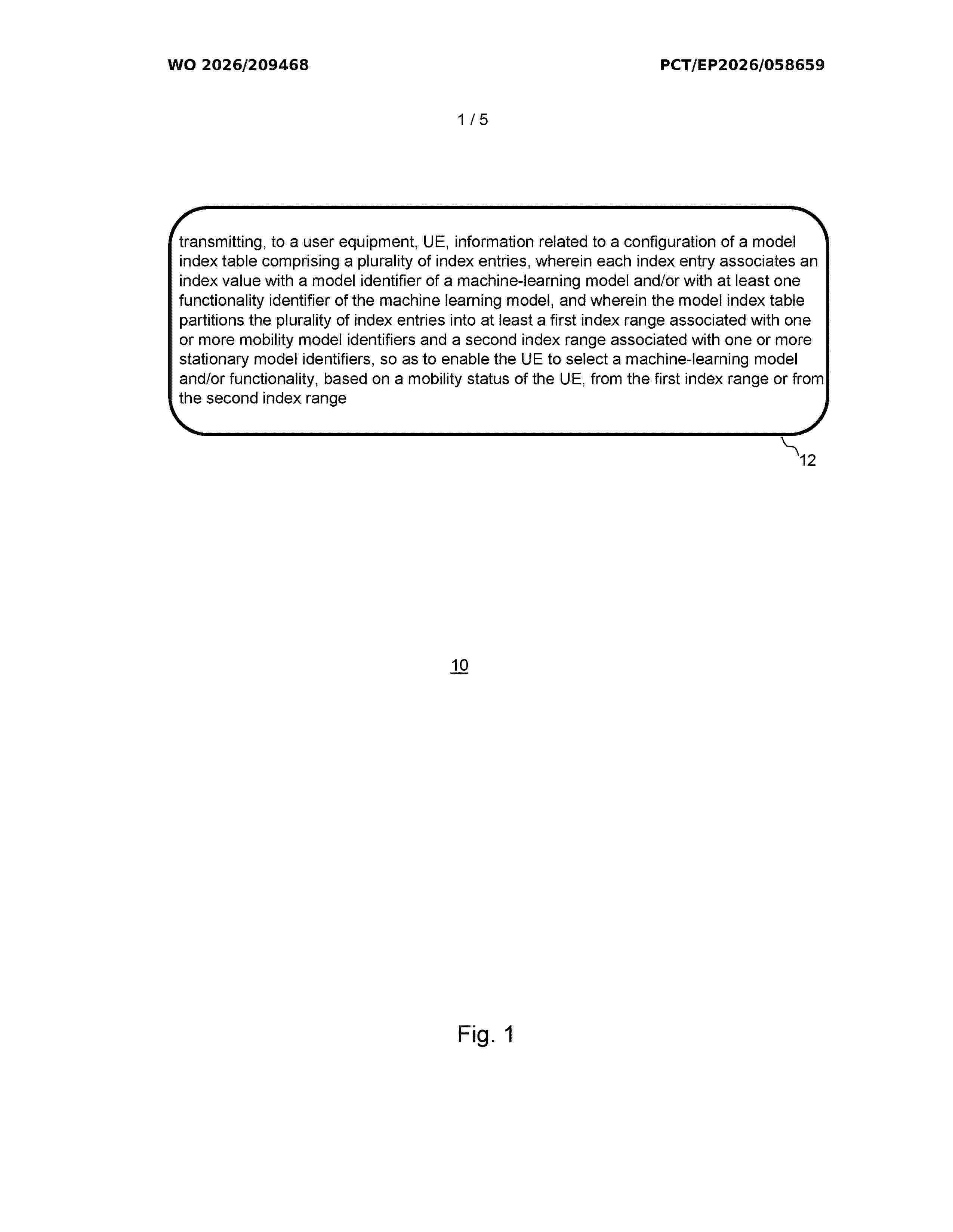
Provided are apparatuses, methods, and computer programs for a network node and for user equipment. The method (10) for the network node (300) of a mobile communication system (500) comprises transmitting (12), to a user equipment (400), UE, information related to a configuration of a model index table comprising a plurality of index entries. Each index entry associates an index value with a model identifier of a machine-learning model and/or with at least one functionality identifier of the machine-learning model. The model index table partitions the plurality of index entries into at least a first index range associated with one or more mobility model identifiers and a second index range associated with one or more stationary model identifiers, so as to enable the UE to select a machine-learning model and/or functionality, based on a mobility status of the UE, from the first index range or from the second index range.
Resumen de: US20260311057A1
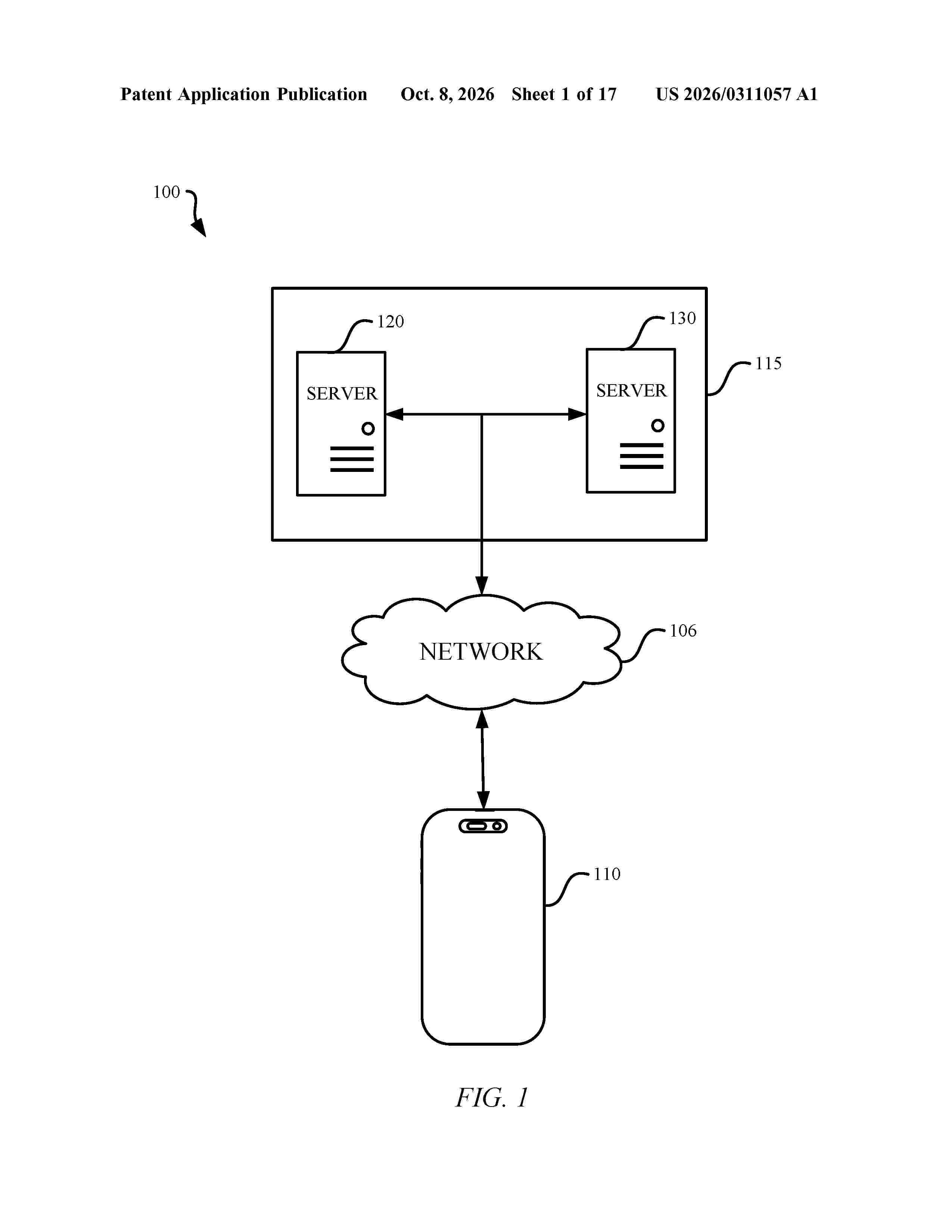
The subject technology provides for distributed training and inferencing of a machine learning model across multiple processing resources. During a forward pass, each processing resource generates a portion of an update for its corresponding portion of a feature set. While performing this computation, each resource can approximate, compress, and transmit its portion of the features to other resources, and can receive an approximated and compressed portion generated elsewhere. Each resource then decompresses the received representation and updates its local feature portion without waiting for full-precision data from other nodes. By allowing approximation, compression, communication, and computation to occur in parallel, the system may reduce communication overhead that typically causes idle compute periods. This structure may improve sustained utilization and responsiveness when models operate across multiple distributed compute nodes.
Resumen de: EP4818902A1
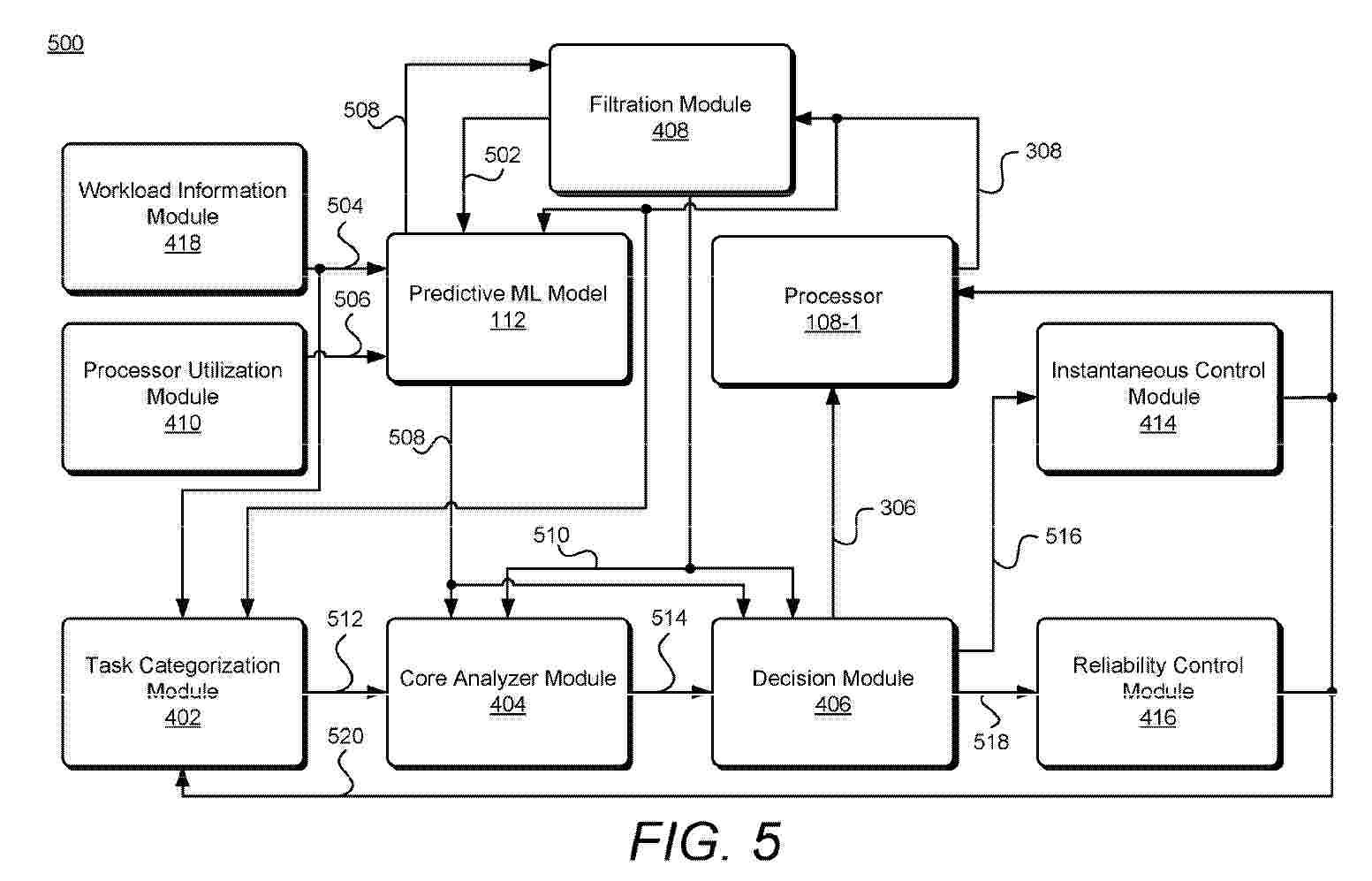
0001 Techniques and apparatuses are described for implementing a machine-learning (ML) based prediction and dynamic core allocation architecture. In example aspects, a plurality of parameters related to one or more operating conditions (e.g., temperature of a core) of one or more processors are generated. The plurality of parameters are used as an input for an ML-based model, which outputs a prediction of future parameters. The prediction is used as input for one or more process modules. The one or more process modules parse the prediction to provide a decision output, which is used to modify one or more facets of the one or more processors (a frequency, a voltage, core assignments for tasks, etc.). In this way, a system-on-chip can dynamically manage multiple operations concems for the one or more processors, such as balancing performance and temperature concerns to realize a tailored user experience.
Nº publicación: EP4818985A1 07/10/2026
Solicitante:
MANGOHAMMOCK LTD [GB]
Mangohammock Limited
Resumen de: EP4818985A1
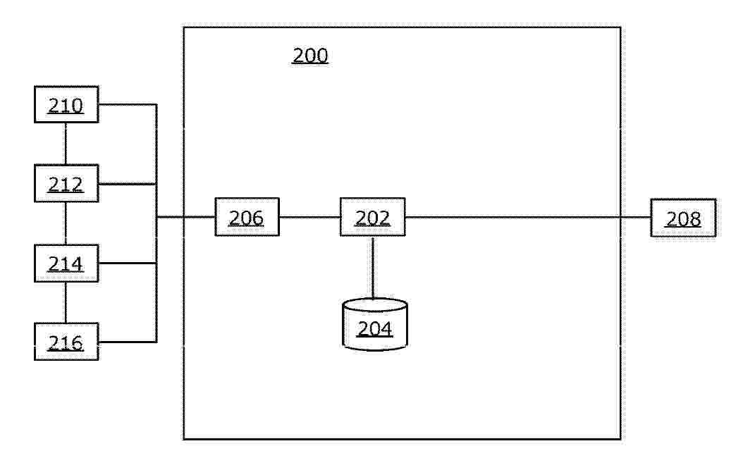
Disclosed is a computer-implemented method for an automated valuation of non-fungible asset is performed by computing node(s) (202), communicably coupled to user device(s) (208) and data repository(s) (204), wherein the computing nodes, the data repository and the user device are connected via a computing network, comprises: receiving input data comprises at least one parameter that is a unique identifier of the non-fungible asset; capturing published data, wherein the published data comprises a second set of parameters; preprocessing the captured data for normalization of the published data to generate an output data that is structured; executing a pre-trained valuation model (212) to identify a proxy for parameters that are missing from the captured data; and applying a predictive machine learning model by executing a predictive valuation model (214) to generate valuation result for the non-fungible asset with a pre-set accuracy and display the valuation result at a screen.
 BOPI
BOPI
 Sede Electrónica