Si deseas distinguir tus productos, servicios o ambos de los de otra empresa, es posible que necesites una marca o nombre comercial. Descubre qué son, en qué consiste su procedimiento de registro y qué implica.

Información sobre los plazos de presentación de solicitudes de transformación de marcas de la Unión Europea en marca nacional española. Más información


Si tienes un nuevo dispositivo, producto o procedimiento que resuelva un problema técnico o tenga una ventaja práctica, existen distintas formas de protegerlo en España y en otros países. Descubre cómo hacerlo.

¿Tu innovación reside en la estética, la ornamentación o la apariencia de tu producto? Protégela mediante un diseño industrial. Descubre qué derechos confiere el registro y cómo realizar la tramitación.

Las indicaciones geográficas protegen el nombre de un producto originario de una zona geográfica, a la cual le debe una determinada calidad, reputación u otra característica. Descubre qué son, en qué consiste su procedimiento de registro y qué beneficios conceden.

Las patentes publicadas en todo el mundo son una valiosa fuente de información científica, técnica y comercial.

Si eres emprendedor/a o una empresa y quieres potenciar y mejorar la rentabilidad de tu negocio protegiendo de forma adecuada los activos intangibles de tu organización, en este espacio encontrarás lo necesario.


 172
resultados
172
resultados
 Última actualización
27/07/2026 [08:08:00]
Última actualización
27/07/2026 [08:08:00]


 Resultados 50 a 75 de 172

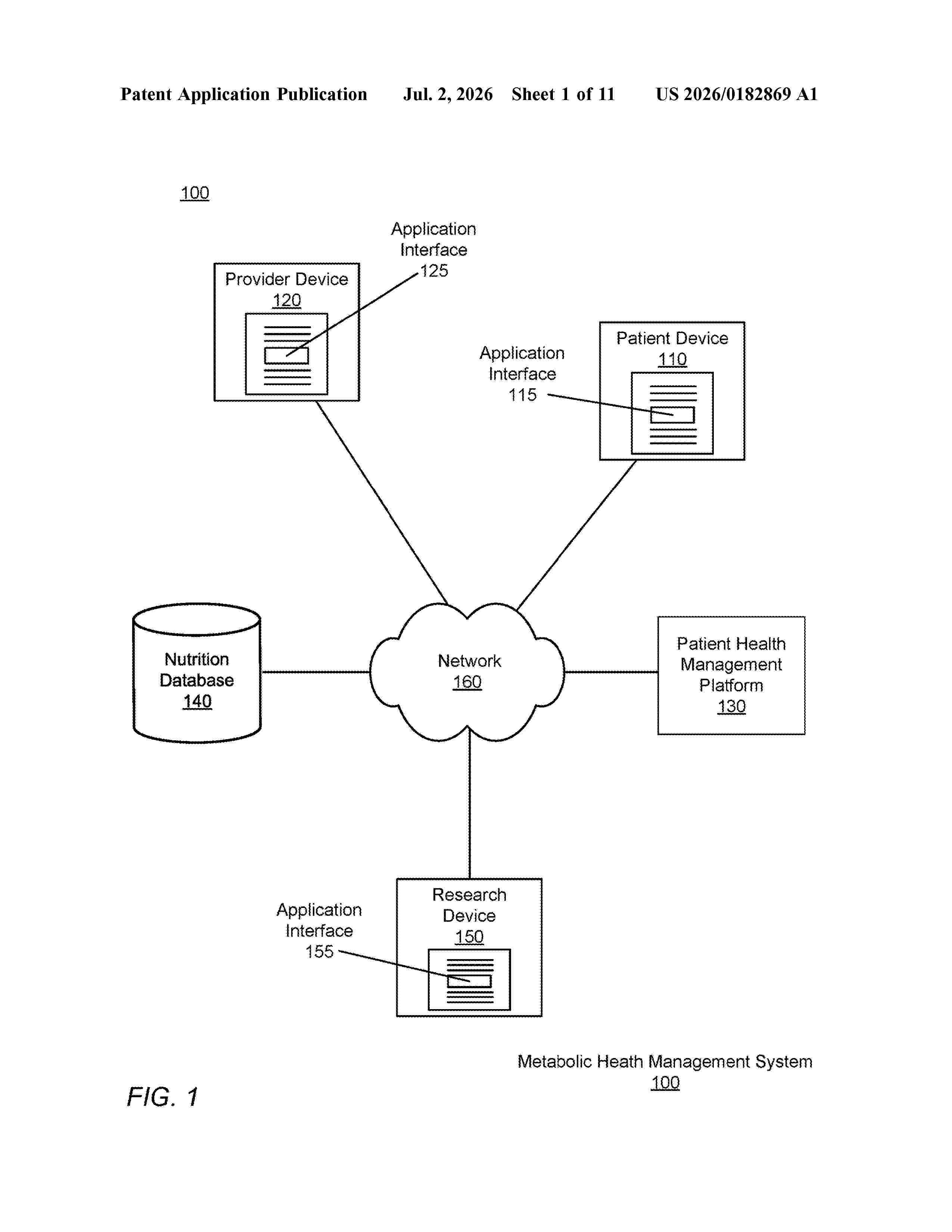
Resumen de: US20260182869A1
0000 A patient health management platform implements a machine-learned metabolic model to generate a prediction of a patient's glucose level. The platform implements a short-term prediction model to generate a daily prediction of the patient's glucose level based on nutrition data reported by the patient and sensor data and lab test data collected for the patient. The platform implements a long-term prediction model generate a prediction of the patient's glucose level during an extended time period based on sensor data and lab test data collected for the patient. Using the short-term prediction model, the long-term prediction model, or both, the patient health management platform generates predictions of the patient's glucose level and updates a digital twin of the patient's metabolic profile.

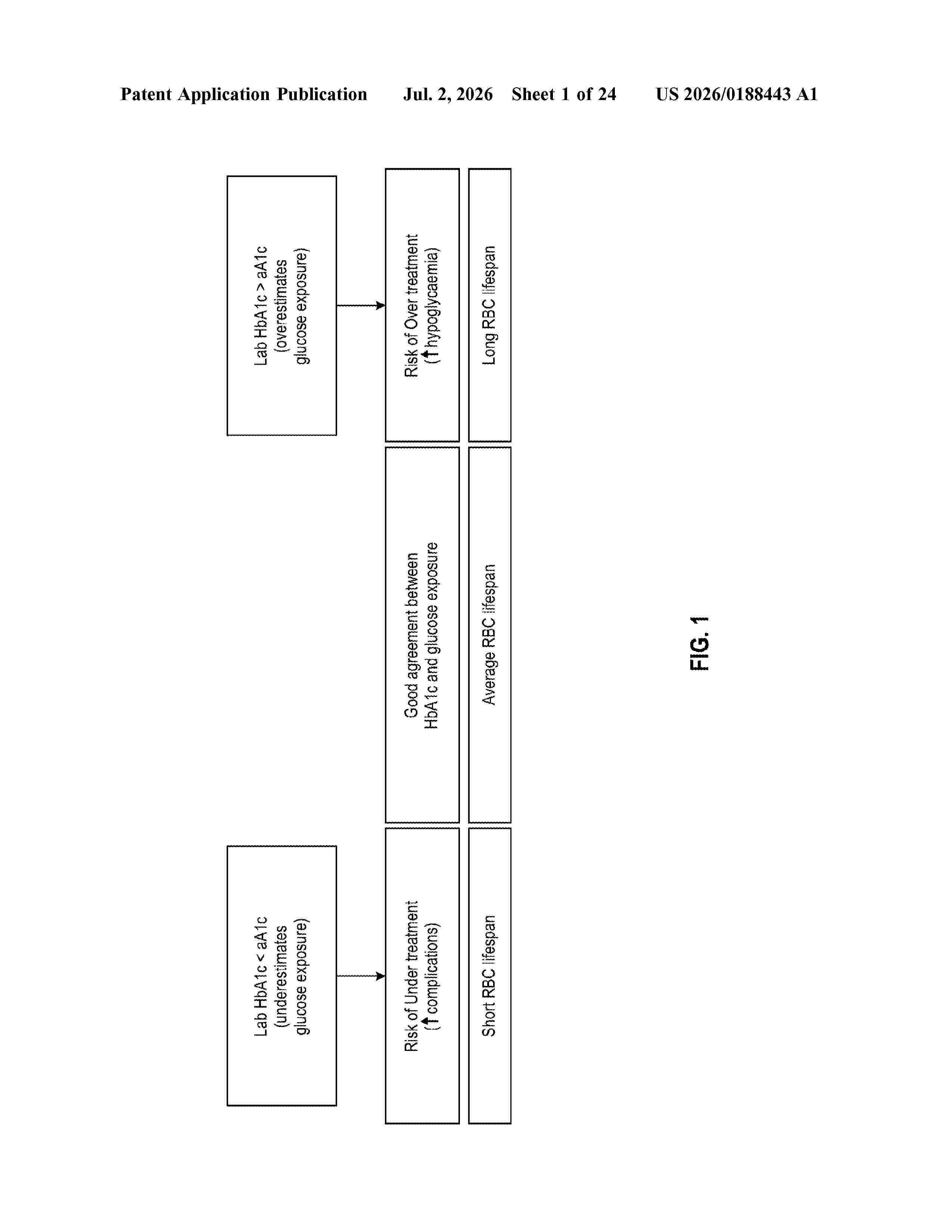
Resumen de: US20260188443A1
0000 A method of providing personalized treatment for a diabetes patient including a remote device which is configured to receive a first data indicative of an analyte level of a subject during a first time period, retrieve a first glycated hemoglobin level for the subject associated with the first time period, calculate a first personal apparent glycation ratio for the first time period using the received first data and the retrieved first glycated hemoglobin level, compare the calculated first personal apparent glycation ratio to a representative apparent glycation ratio, generate a recommendation based on the comparison, and display a graphical interface comprising the calculated first personal apparent glycation ratio, the representative apparent glycation ratio, and the comparison.
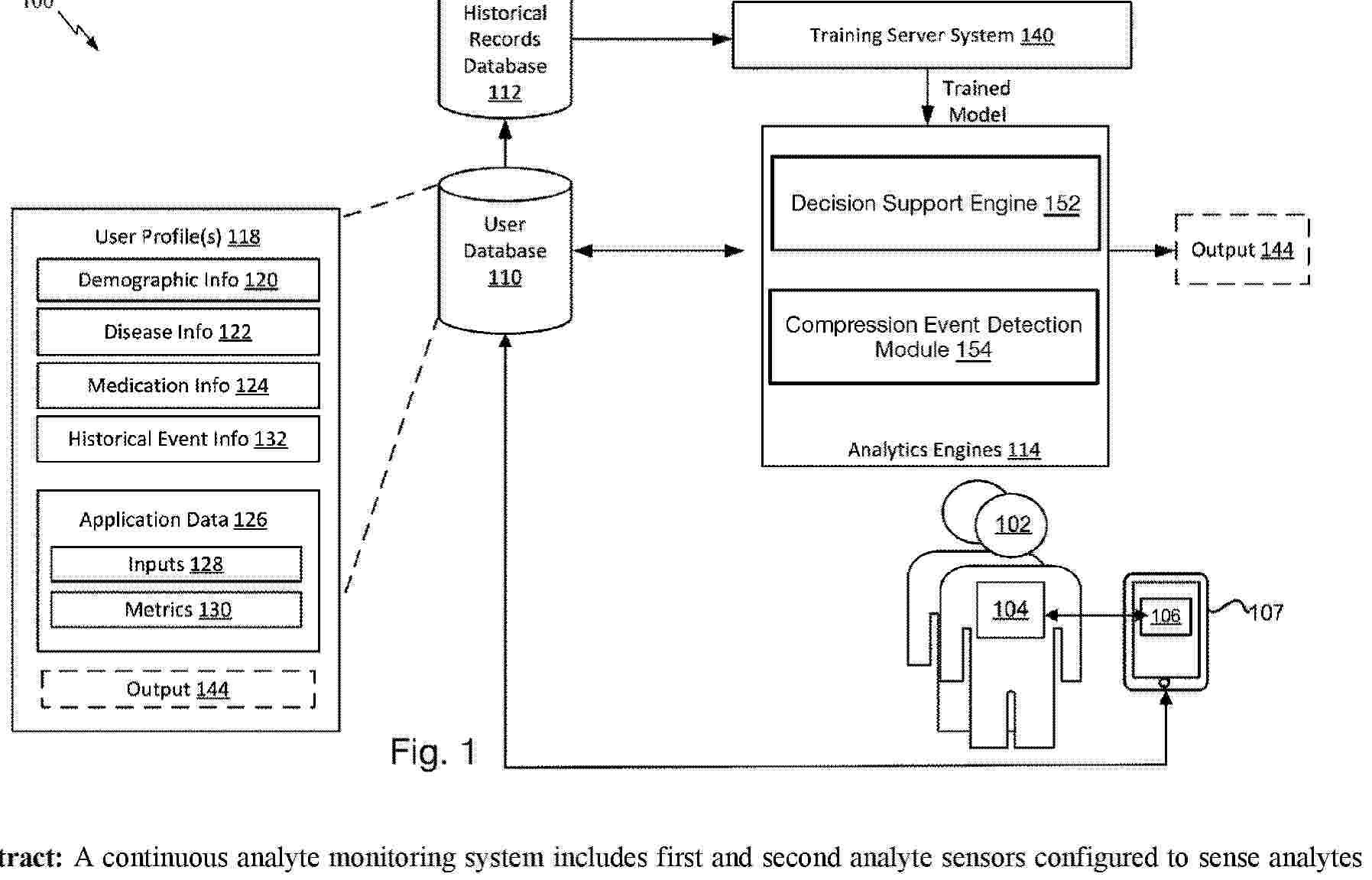
Resumen de: AU2024389410A1
A continuous analyte monitoring system includes first and second analyte sensors configured to sense analytes such as lactate and glucose in the tissue of a user. A controller Is coupled to the analyte sensors and configured evaluate first samples of outputs of the first analyte sensor and second samples of outputs of the second analyte sensor with respect to one another to determine whether the first samples and the second samples indicate compression of the tissue. If the first samples and the second samples indicate compression of the tissue, compensate for the compression of the tissue with respect to the first samples. The controller may evaluate the machine learning models using a machine learning model or a filter.
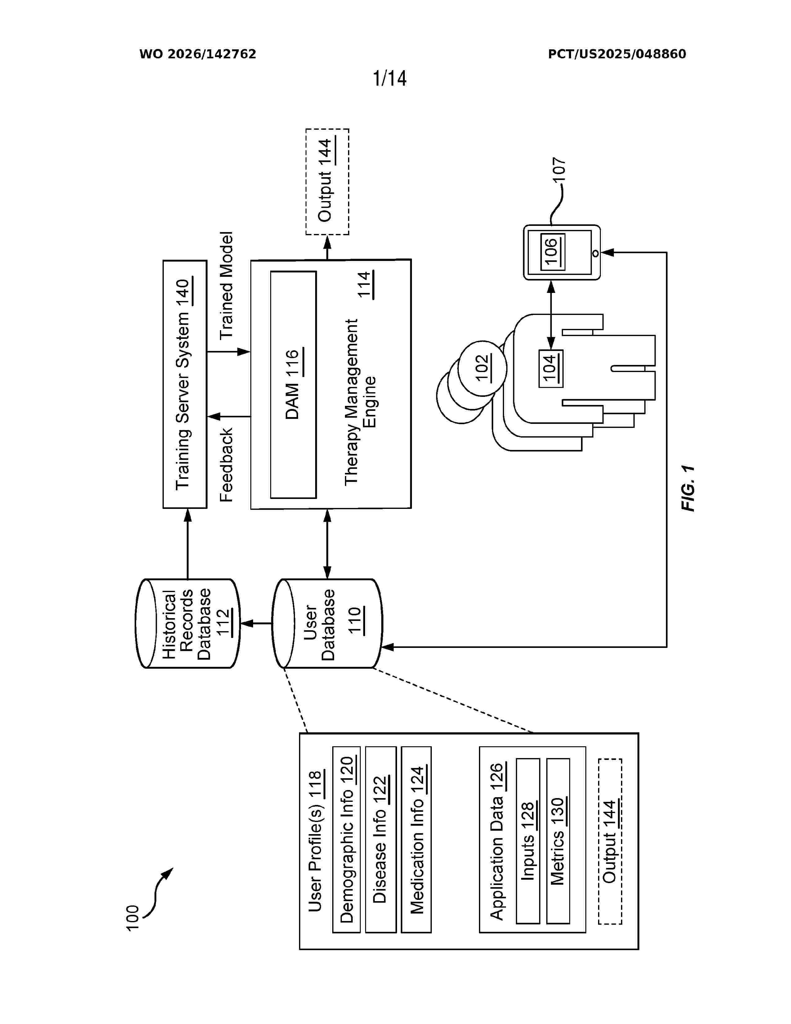
Resumen de: WO2026142762A1
In an embodiment, a system for mitigating diabetic ketoacidosis (DKA) risk for a patient in real-time includes a continuous glucose monitoring (CGM) system configured to generate one or more glucose measurements associated with a cunent glucose level of a patient over a period, one or more memories including executable instructions, and one or more processors in data communication with the one or more memories. The one or more processors are configured to execute the executable instructions to receive glucose measurements for the patient from the CGM system, receive insulin data for the patient, evaluate criteria associated with DKA using first information related to the glucose measurements and second information related to the insulin data, detect a risk of DKA for the patient responsive to a combination of the first information and the second information satisfying the criteria associated with DKA, and alert the patient based on the detected risk.
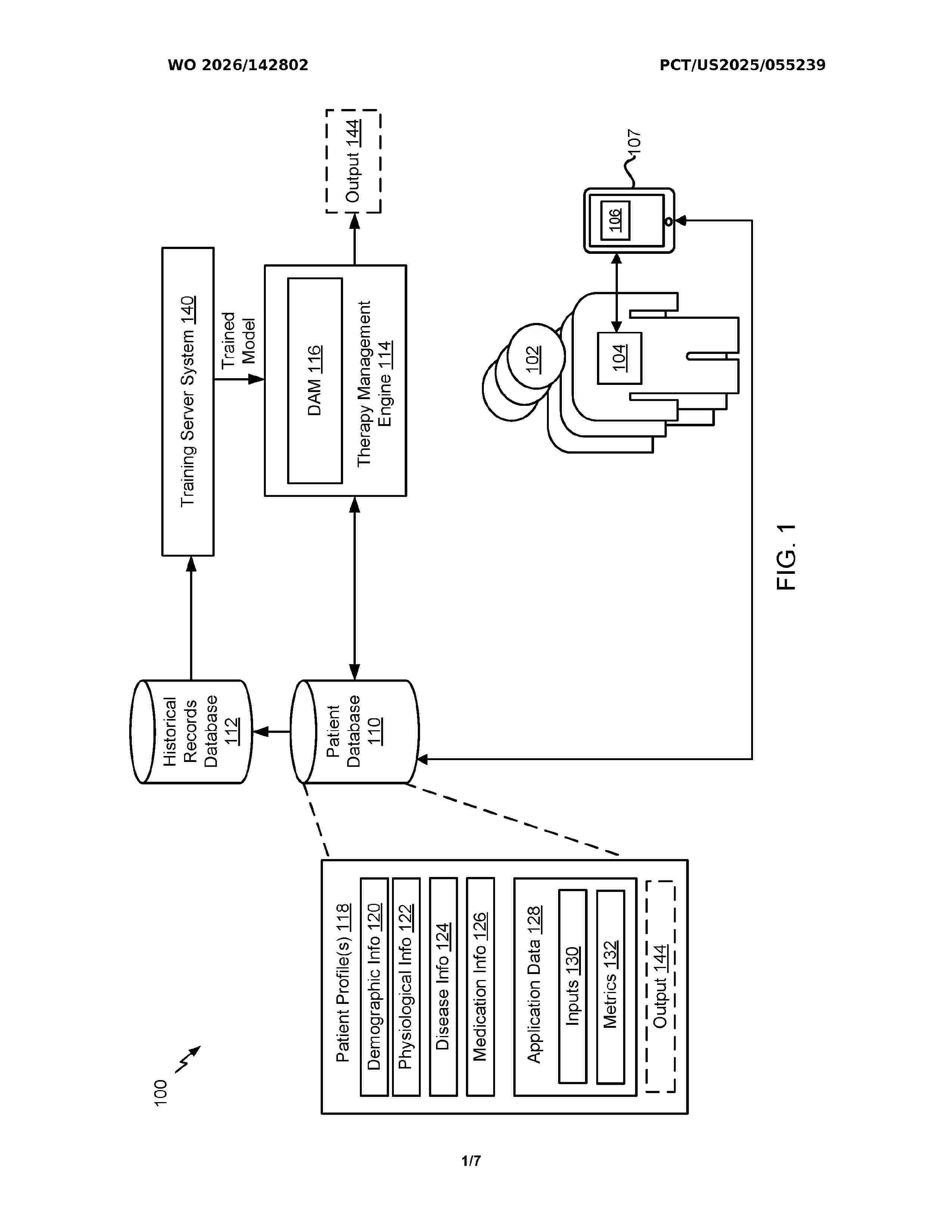
Resumen de: WO2026142802A1
Certain aspects of the present disclosure provide a therapy management system configured to monitor at least glucose concentration levels of a patient during a first time period using one or more glucose sensors to obtain glucose data, and determine a diabetic state classification of the patient based on the glucose data. The diabetic state classification indicates a diabetic state of the patient and comprises one of: a healthy classification, a pre-diabetic classification, a mild type 2 diabetes (T2D) classification, a severe T2D classification, or a gestational diabetes classification. The therapy management system is further configured to provide personalized feedback to the patient based on the determined diabetic state classification, wherein the personalized feedback comprises a recommendation for at least one of an exercise session, a dietary change, a lifestyle modification, a medication adjustment, or a continuous glucose monitoring wear schedule.
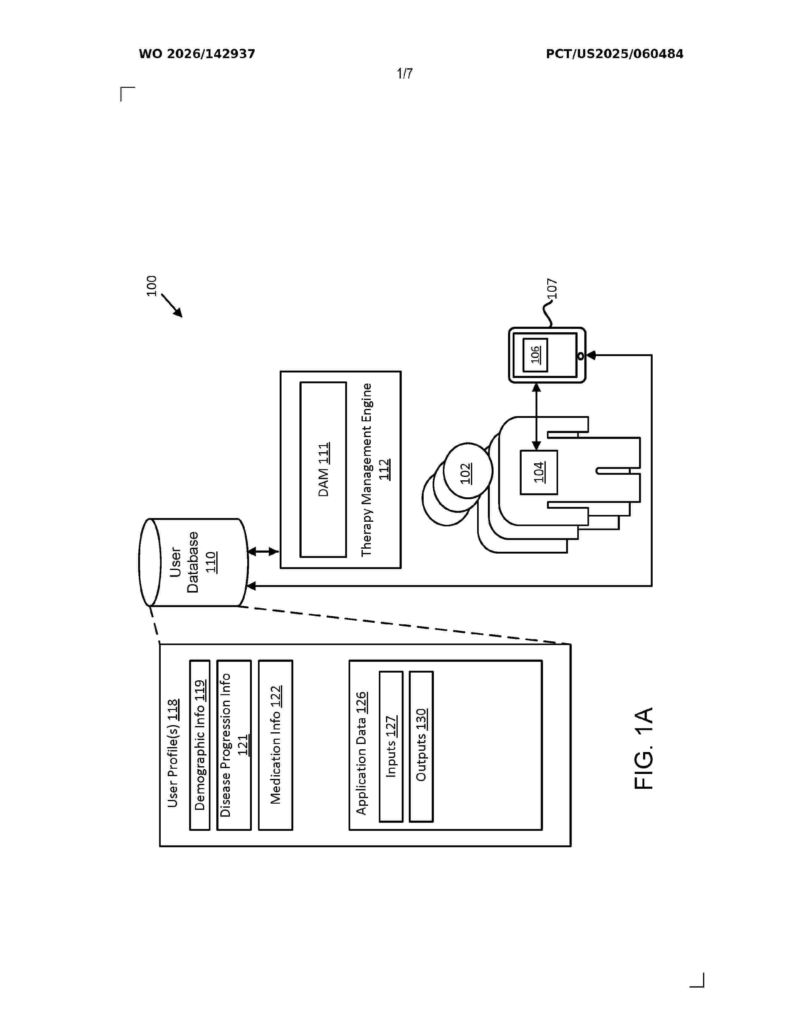
Resumen de: WO2026142937A1
In some embodiments, a method of automatically adjusting insulin therapy for a patient includes receiving, at a device associated with the patient, one or more glucose measurements generated by a continuous glucose monitoring (CGM) system of the patient for a period. The method also includes determining, at the device, basal insulin data and bolus insulin data of the patient for the period. The method also includes determining, at the device, a relationship between the basal insulin data and the bolus insulin data. The method also includes generating, at the device, an insulin therapy instruction for the patient based on the one or more glucose measurements and the determined relationship between the basal insulin data and the bolus insulin data, where the insulin therapy instruction includes a total bolus amount.
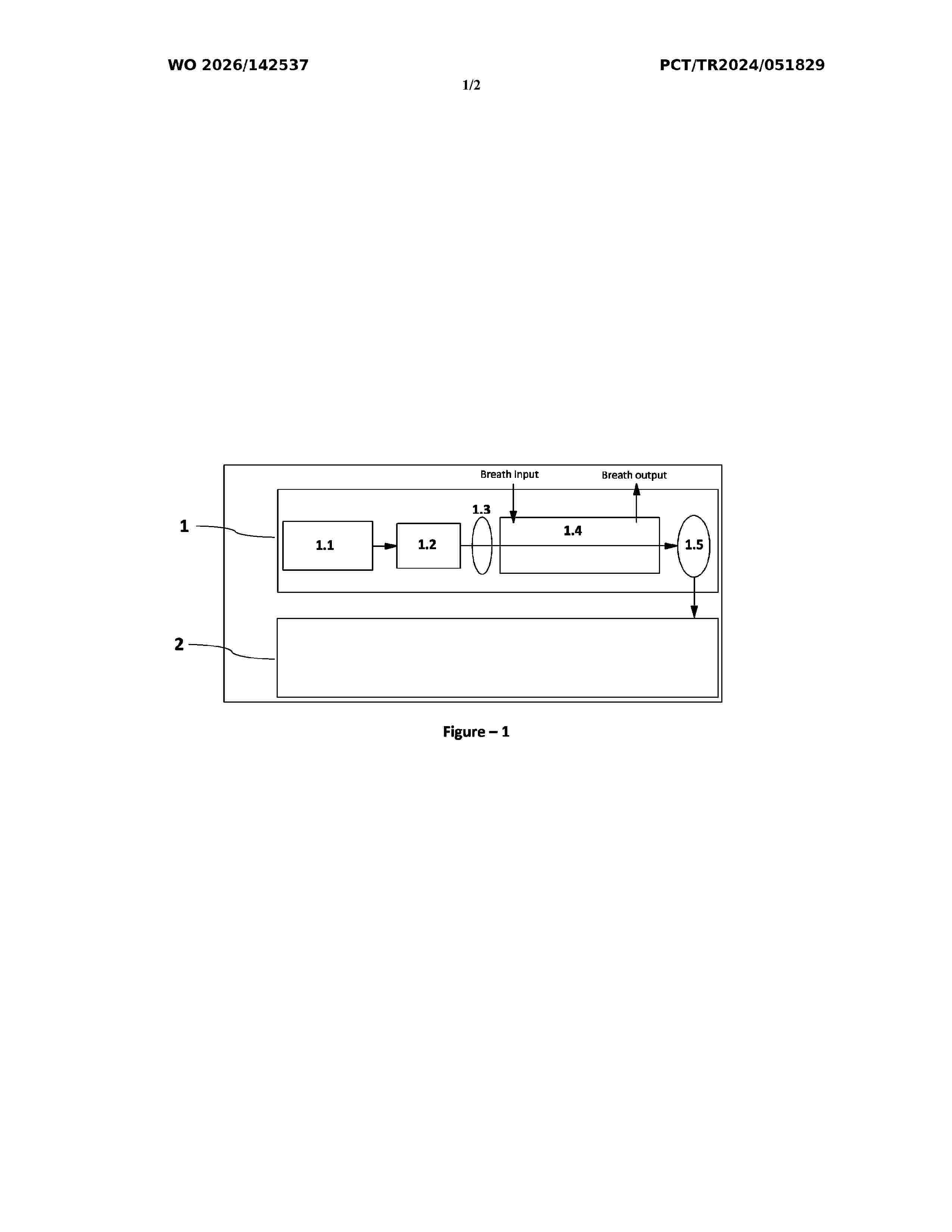
Resumen de: WO2026142537A1
Present invention relates to a needle-free glucose measurement system which enable measurement of blood glucose level using concentration value of N2O molecule available in breath air, which comprises an electronic assembly (1.1) which stabilizes the temperature and flow of wavelength-adjustable laser light source (1.2) and controls wavelength, a laser-based sensor (1) which determines concentration values of N2O molecule by measuring the adsorption amount of detector (1.5) wherein beams coming from laser light source (1.2) are adsorbed by N2O molecules in the breath air in the adsorption cell (1.4), and a processor (2) which determine glucose values in the blood using the concentration data coming from laser-based sensor (1).
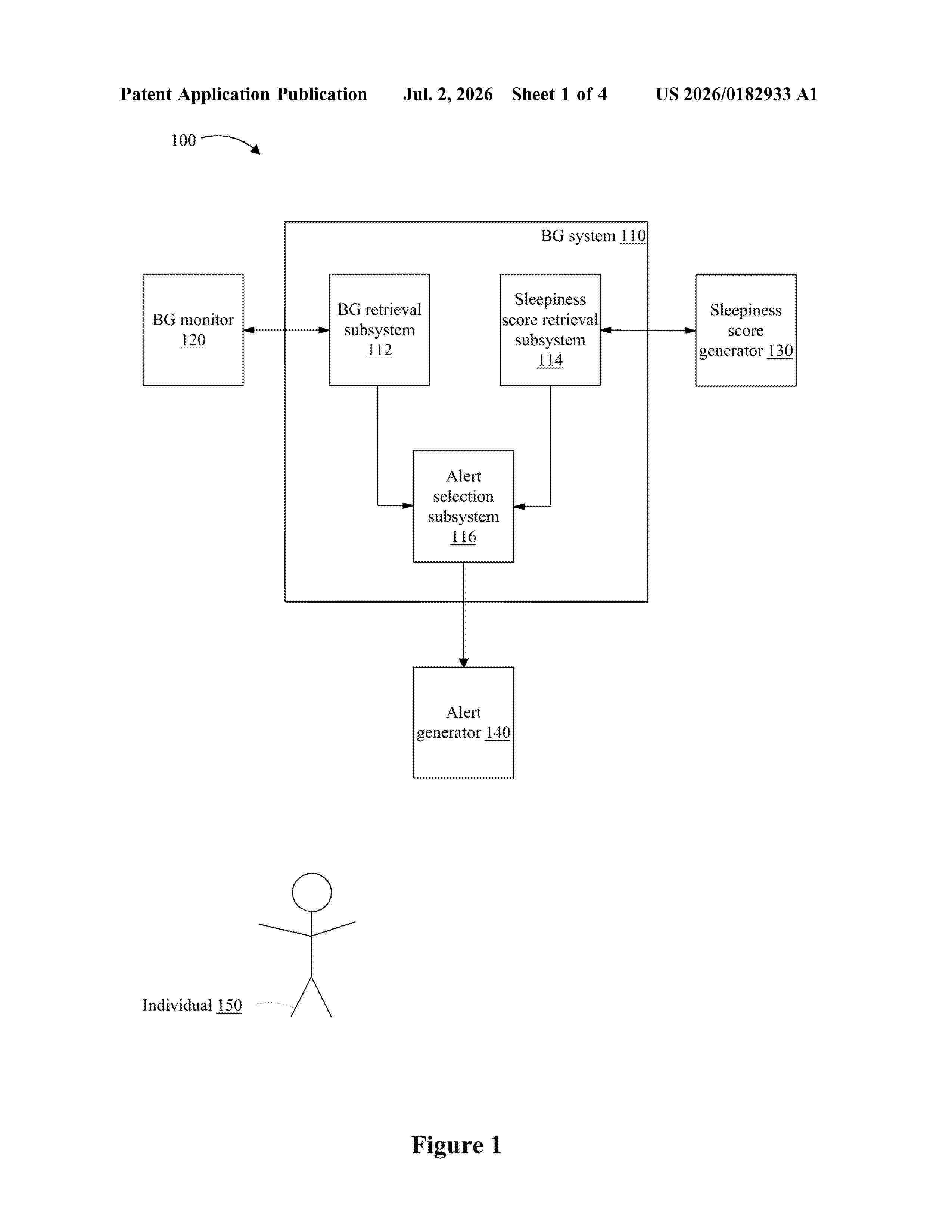
Resumen de: US20260182933A1
In accordance with some implementations, a method includes obtaining blood glucose (BG) data indicative of BG levels of an individual. The method includes obtaining a first sleepiness score associated with the individual. The method includes determining, based at least in part on the BG data, that a low blood sugar alert condition is satisfied. The method includes, in response to determining that the low blood sugar alert condition is satisfied, directing an alert subsystem to generate a first alert output based on the first sleepiness score.
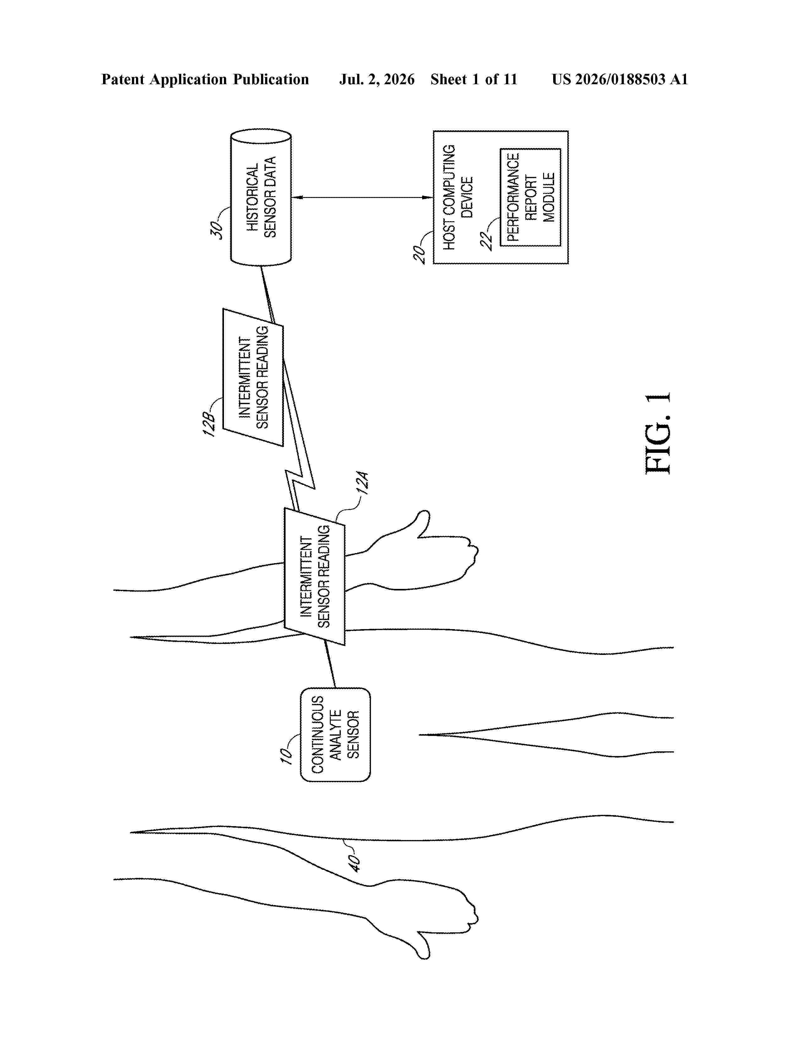
Resumen de: US20260188503A1
A method of providing performance reports to persons associated with the monitoring of an analyte level, such as blood glucose concentrations in a host, is provided. The performance reports can indicate a measure of an analyte variability of the host over a first analysis time period compared to a measure of analyte variability of the host over one or more other analysis time periods. The analyte level of the host can be received in each of a plurality of sensor readings from a continuous analyte sensor, such as multiple sensor readings per hour, so that the analyte variability that is used in determining the performance report of the host can be based on not just a few metered readings taken by the host, but a plurality of intermittent readings that are taken by a continuous analyte sensor, for example. Various types of performance reports can be generated, such as reports that comprise charts, graphs, tables, and/or other visual indicia of a host's performance during one analysis period versus another analysis period.
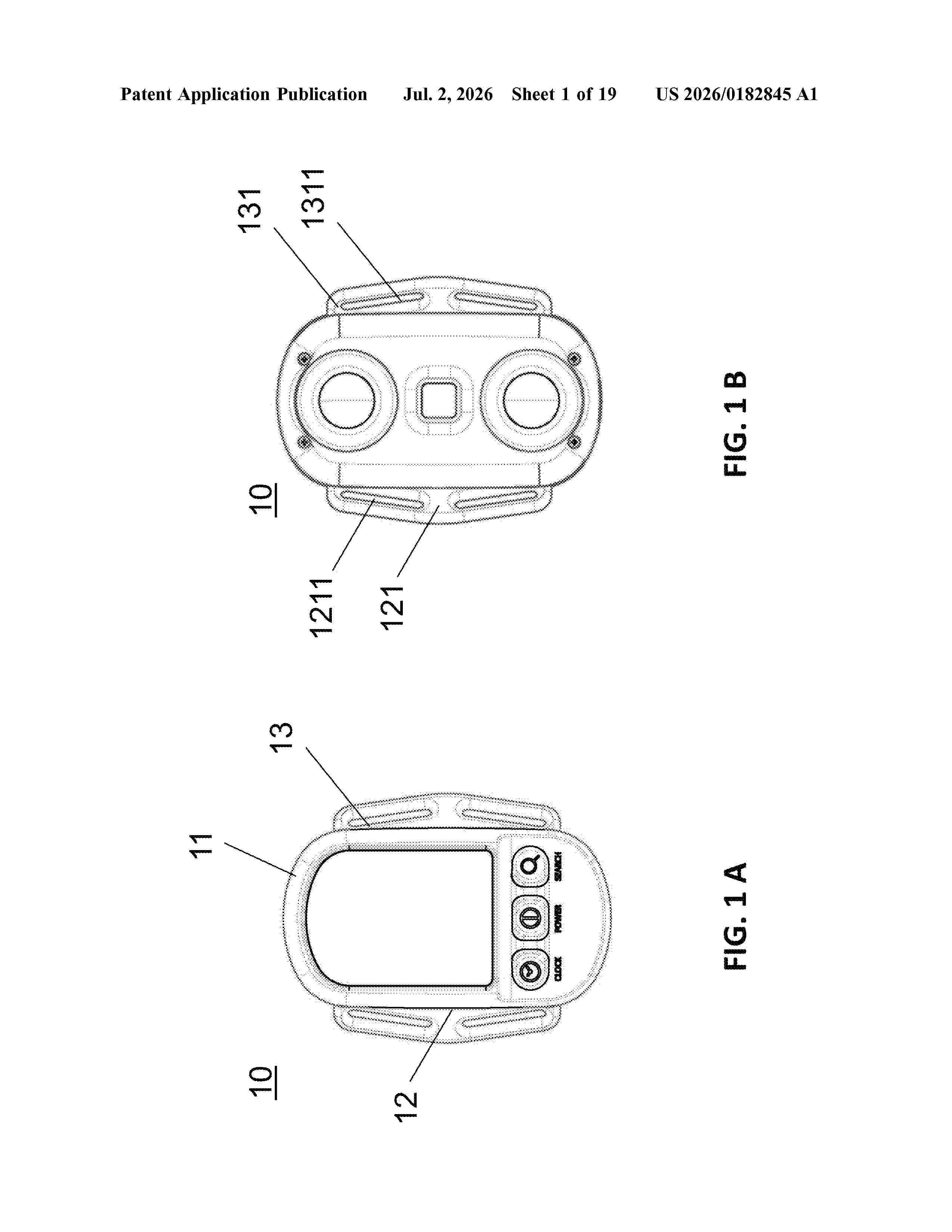
Resumen de: US20260182845A1
0000 A wearable device for measuring blood glucose and blood pressure for a user includes a main body including a first side and a second side formed on the opposite side of the first side; a first supporting frame protruding from the first side and a second supporting frame protruding from the second side; a belt portion having a first belt end detachably coupled to the first supporting frame, a second belt end detachably coupled to the second supporting frame, and at least one aligning mark, wherein the belt portion is positioned around a wrist of the user; wherein the main body includes at least one pressure sensor array assembly overlaid on a radial artery of the user; wherein the pressure sensor array assembly includes a plurality of apertures arranged one after another to form a cross-shaped pattern and at least four sensor elements arranged in a symmetrical configuration.
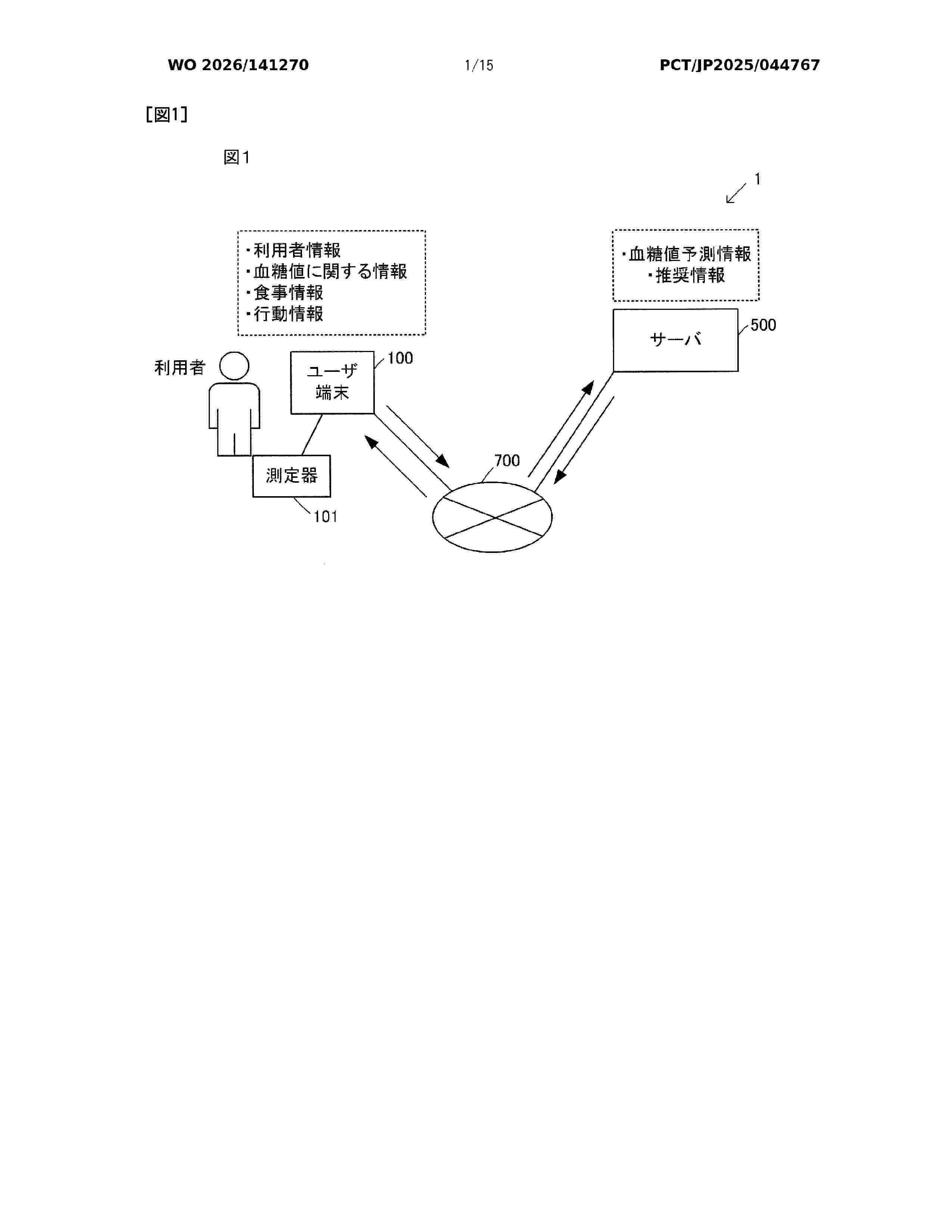
Resumen de: WO2026141270A1
Provided is an information processing system that provides users more useful information for improving blood sugar levels. The information processing system acquires meal information about meals consumed by a user, and outputs recommendation information on the basis of the meal information, user information, blood glucose level information, and behavioral information. Additionally, the information processing system outputs predicted blood glucose levels on the basis of meal information, user information, blood glucose level information, and behavioral information, the predicted blood glucose levels being for the case in which the user consumes a meal corresponding to the meal information. The information processing system also outputs blood glucose level prediction information on the basis of the recommended information. Moreover, the information processing system provides the user with blood glucose level prediction information based on meal information, recommendation information, and blood glucose level prediction information based on the recommendation information.
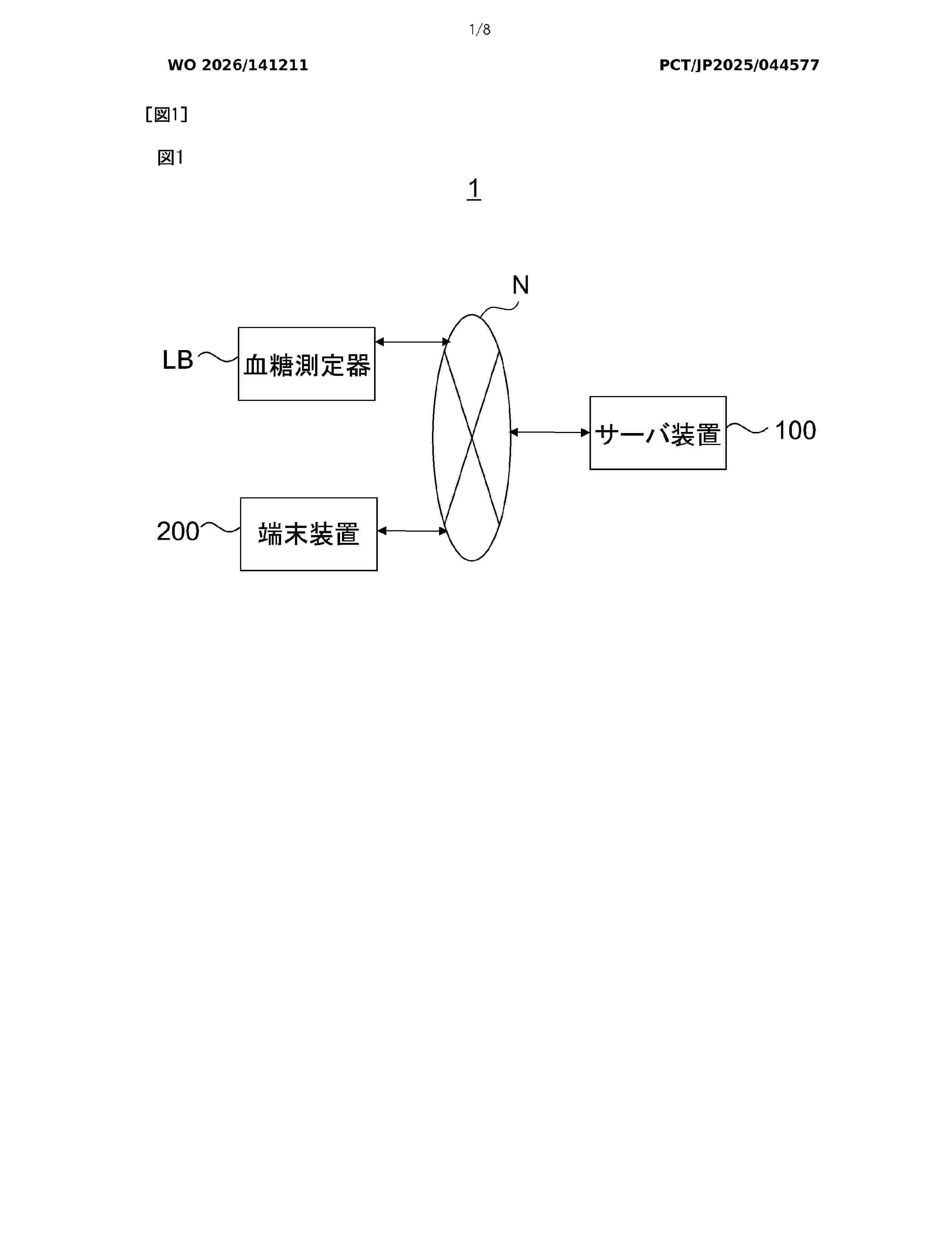
Resumen de: WO2026141211A1
The present invention accurately predicts and notifies the degree of daily discomfort after food intake. The present invention comprises: an acquisition unit that acquires blood glucose-related data related to the blood glucose level of a subject; a prediction unit that, on the basis of the blood glucose-related data, predicts the degree of daily discomfort after a prescribed amount of time from food intake by the subject; and an output unit that outputs the result of prediction by the prediction unit.
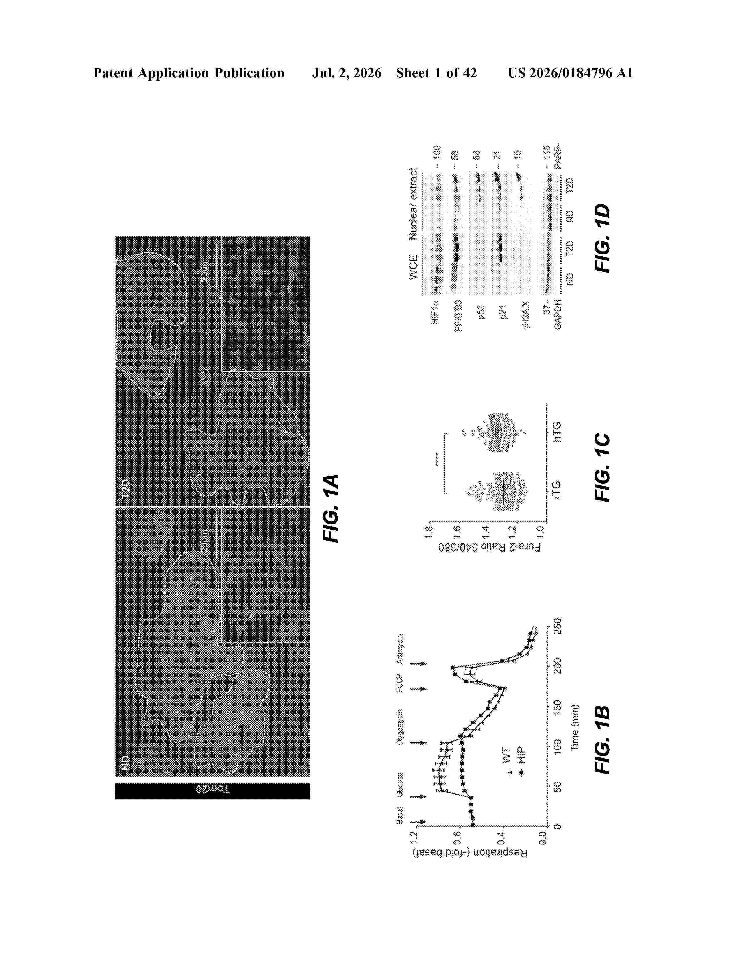
Resumen de: US20260184796A1
0000 Aspects of the present disclosure are directed to methods and compositions for facilitating β-cell regeneration by inhibition of the HIF1α-PFKFB3 pathway. Certain aspects describe methods for treatment and prevention of prediabetes and diabetes, including type 2 diabetes. Also disclosed are methods and compositions for enhancing β-cell regeneration.
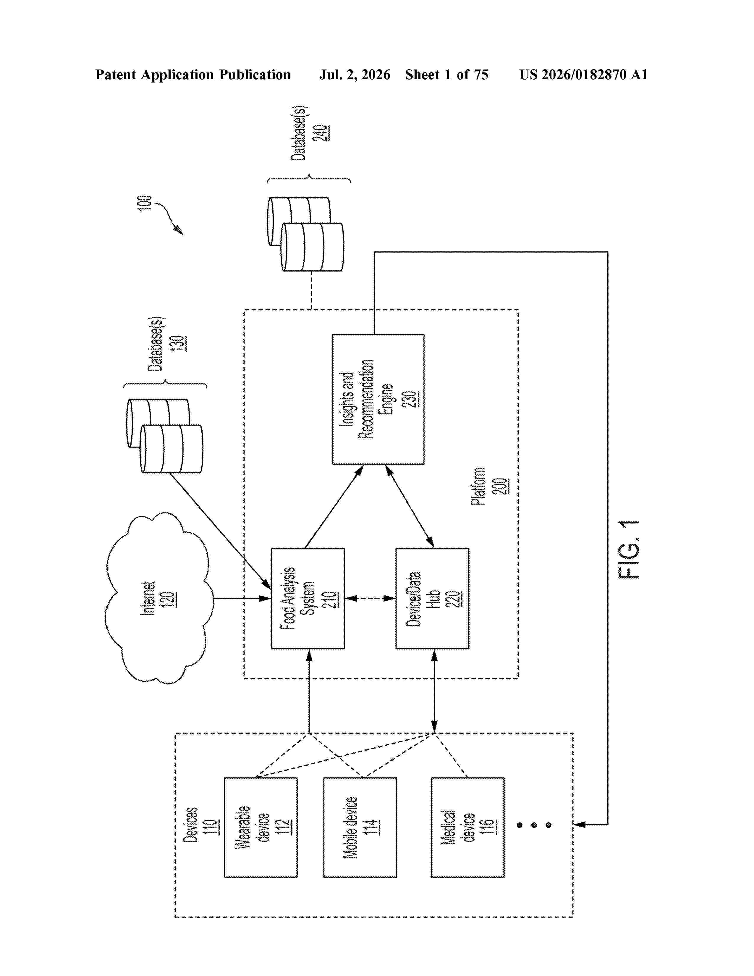
Resumen de: US20260182870A1
0000 Techniques disclosed herein relate to food analysis and glucose level management. In some examples, the techniques involve determining a consumption of a food item by a user, receiving data indicative of a glucose level of the user, determining an effect of the food item on the glucose level of the user based on a change of the glucose level of the user, and storing the effect of the food item on the glucose level of the user that includes data associated with one or more effects of the food item on the glucose level of the user.
Resumen de: US20260183397A1
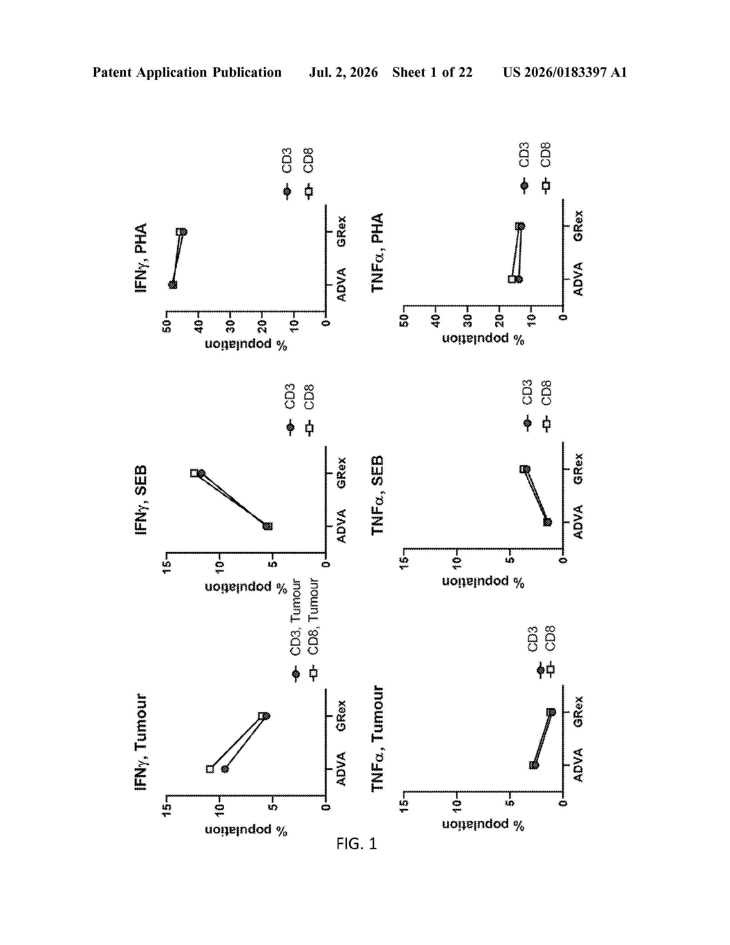
0000 The present invention relates to a method for expansion of a population of lymphocytes in a controlled single culture vessel, the method comprising a step of (a) culturing a tissue or blood sample from a subject, which sample is known or suspected to contain lymphocytes; or (b) culturing lymphocytes, which lymphocytes are isolated from a tissue or blood sample from a subject; wherein the lymphocytes are expanded in a culture medium in which at least one of the following parameters is monitored and adjusted to a predefined value or range: pH, dissolved oxygen (DO) concentration, glucose concentration, lactate concentration and/or temperature; and wherein the method comprises a step of adjusting the culture volume to the expansion rate of the lymphocytes. Furthermore, the invention relates to a population of lymphocytes comprising at least 90% CD3+ T cells and less than 5% B cells, wherein at least 70% of said T cell portion are viable, optionally, and less than 10% are triple positive for CD45RA, CD57 and KLRG1.
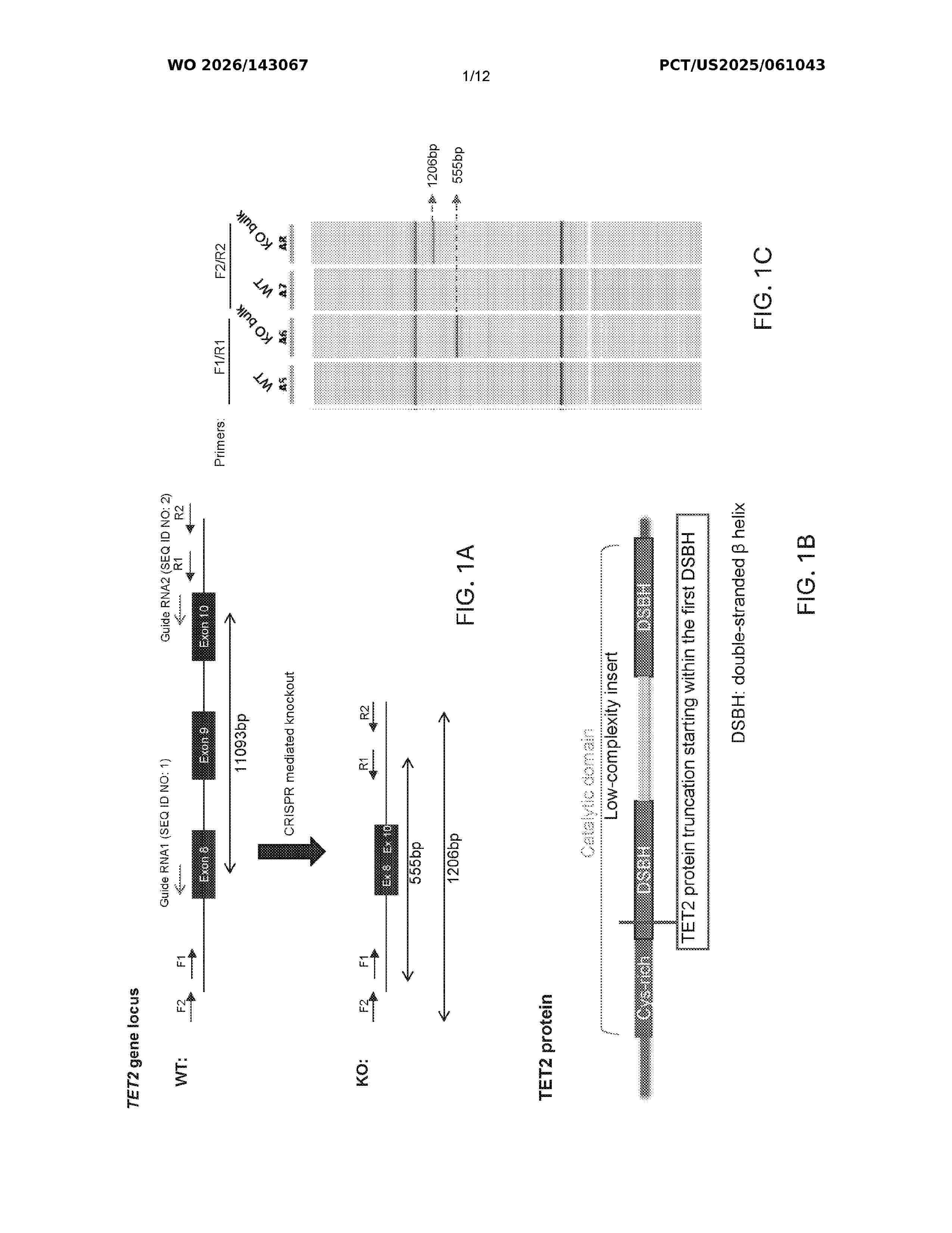
Resumen de: WO2026143067A1
The present disclosure provides compositions of stem cell-derived pancreatic islet cells engineered for improved engraftment and methods of use thereof for treating a subject having an insulin deficiency.
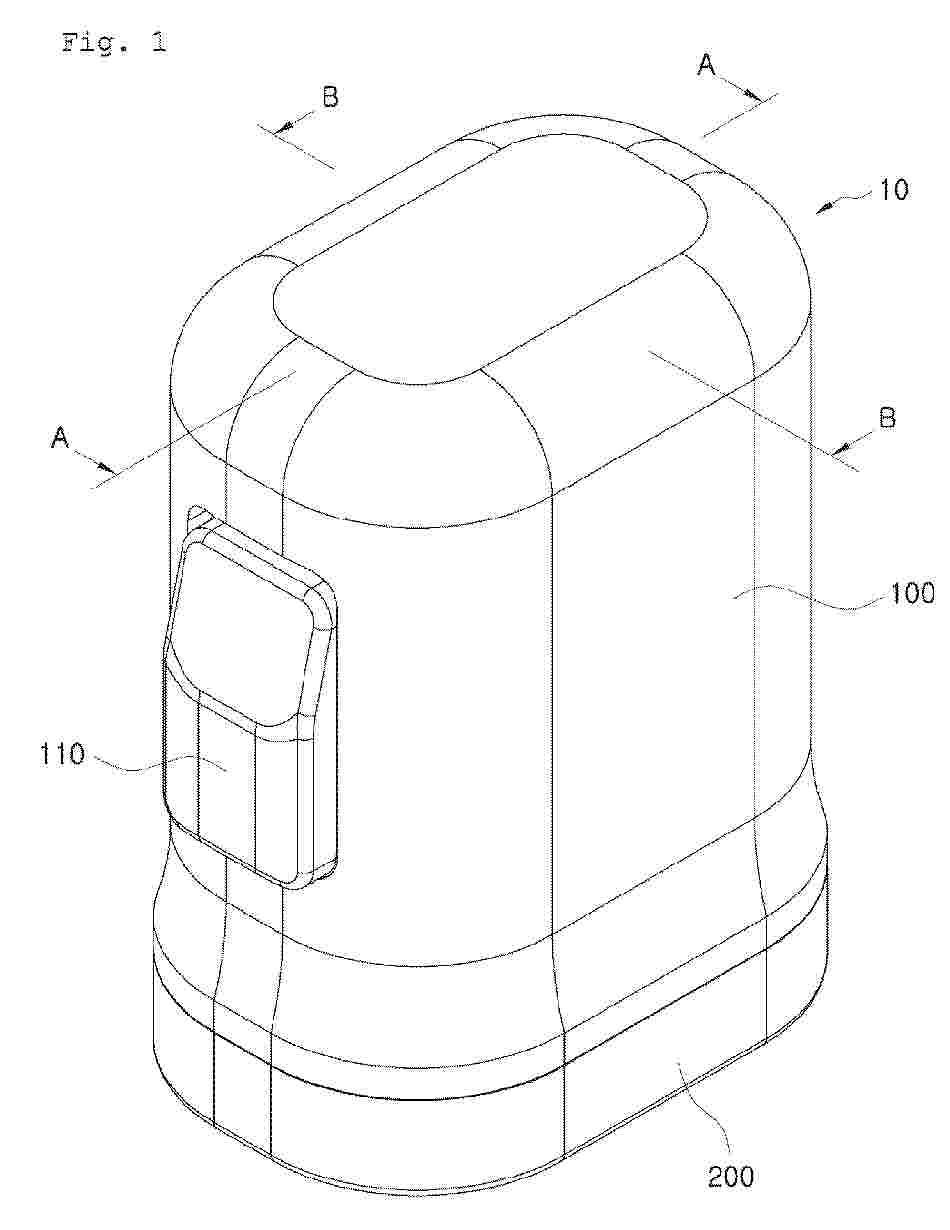
Resumen de: EP4767938A2
0001 The present disclosure relates to a continuous blood glucose measurement device and an applicator, wherein the continuous blood glucose measurement device is produced in a state in which a body attachment unit has been assembled inside an applicator, so as to minimize a user's additional work of attaching the body attachment unit to the body, thereby enabling the body attachment unit to be attached to the body only by simply activating the applicator. Particularly, the present invention provides a continuous blood glucose measurement device and an applicator, wherein the applicator comprises a plunger body disposed at a first locating in an inner space of a main case and configured to downwardly move to a second location according to a manipulation of a pressure button. The pressure button is configured to be mode-changeable from a first mode, in which pressurizing movement by pressurizing manipulation is prevented, to a second mode, in which the pressurizing movement by the pressurizing manipulation is allowed.
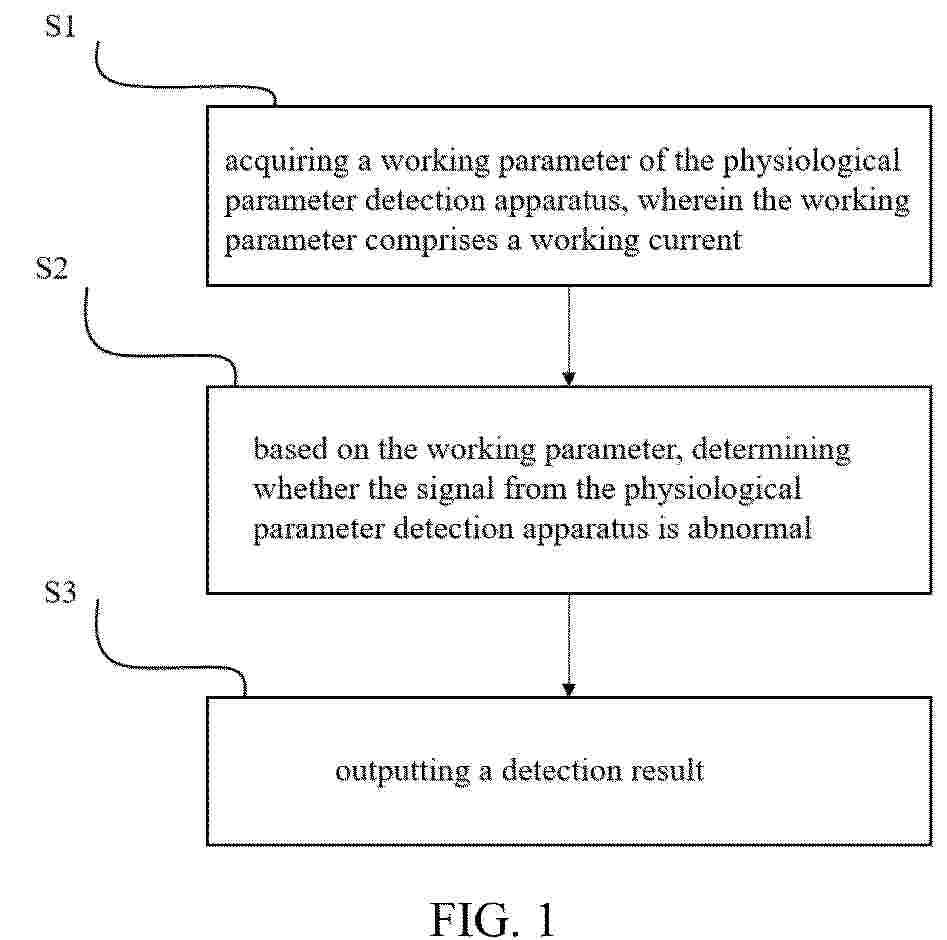
Resumen de: EP4769427A1
This application discloses a method and device for detecting an anomaly of a signal from a physiological parameter detection apparatus, particularly a continuous glucose monitor, CGM, comprising: acquiring a working parameter of the physiological parameter detection apparatus, wherein the working parameter comprises a working current; determining whether the signal from the physiological parameter detection apparatus is abnormal based on the working parameter: and outputting a detection result. Through the foregoing solution, an anomaly of a signal from the physiological parameter detection apparatus can be effectively detected, thereby ensuring signal accuracy.
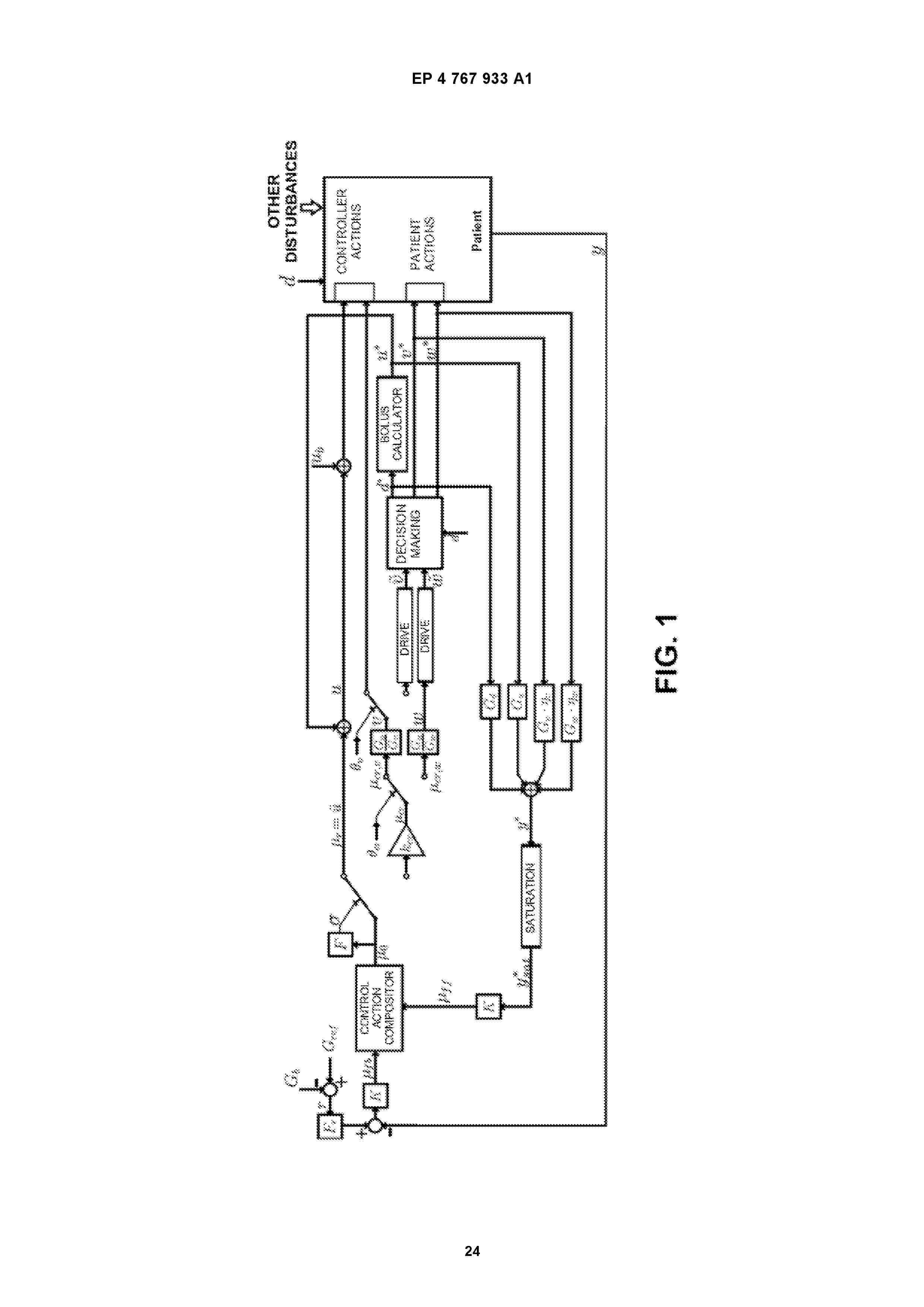
Resumen de: EP4767933A1
0001 A method for controlling glucose in a flexibly structured dual-hormone artificial pancreas that manages optional meal and/or exercise notifications using coordinated control actions, which comprises: measuring a plasma glucose signal; calculating an incremental plasma glucose measurement (y); defining a model for incremental plasma glucose; defining a carbohydrate intake as dependent on a carbohydrate content estimated by the patient; defining an expected postprandial incremental plasma glucose y*(s) depending on the insulin bolus administered; defining a corrected incremental plasma glucose y(s), a corrected insulin infusion u(s), and a corrected carbohydrate d(s) intake; defining a virtual control action µ(s), divided into regulatory actions µr and counter-regulatory actions µcr and calculating control actions using a 2-DOF feedback controller with a nominal value pre-filter Fr(s).
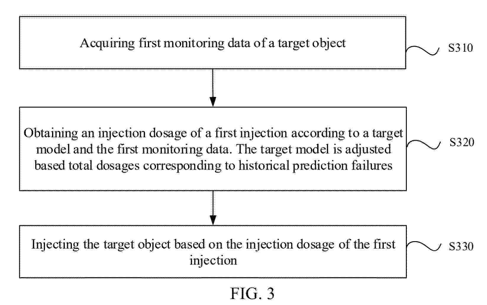
Resumen de: EP4769422A1
An insulin pump control method and apparatus, an artificial pancreatic closed-loop system, and a storage medium are provided. The method includes: acquiring first monitoring data of a target object, obtaining an injection dosage of a first injection according to a target model and the first monitoring data, and injecting the target object based on the injection dosage of the first injection. The target model is adjusted based on total dosages corresponding to historical prediction failures.
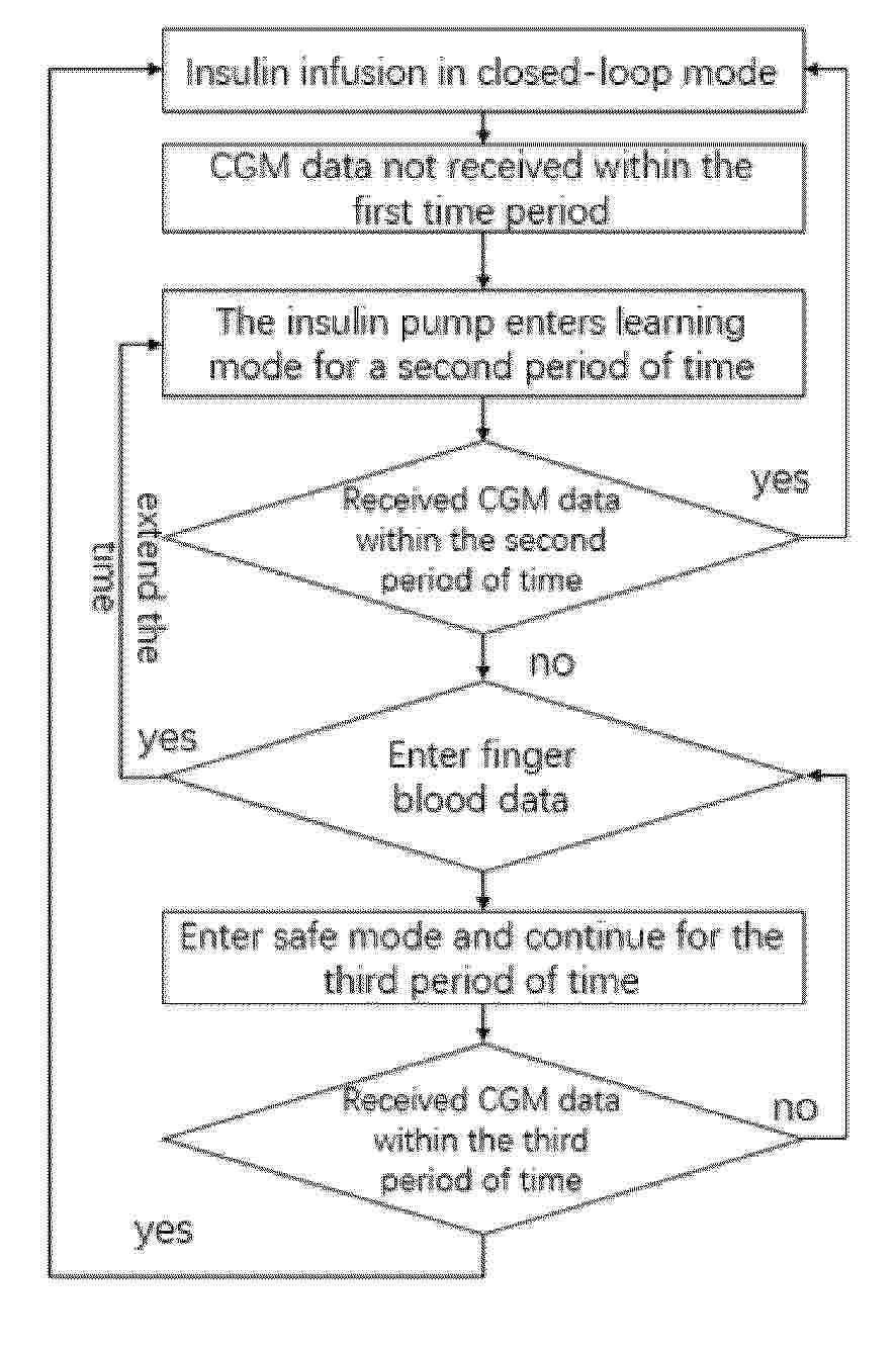
Resumen de: EP4769421A1
0001 The invention discloses a closed-loop artificial pancreas system for safe infusion, during a long period of real-time blood glucose value loss, the closed-loop artificial pancreas system can enter a learning mode or a safe mode. Based on historical blood glucose values and finger blood glucose values, it continues to calculate the required insulin infusion amount for the patient and infuses an appropriate amount of insulin to the patient. It will not infuse an inappropriate amount of insulin or even interrupt insulin infusion due to the lack of real-time blood glucose value, avoiding life-threatening situations for the patient and benefiting the patient's treatment.
Resumen de: EP4767931A1
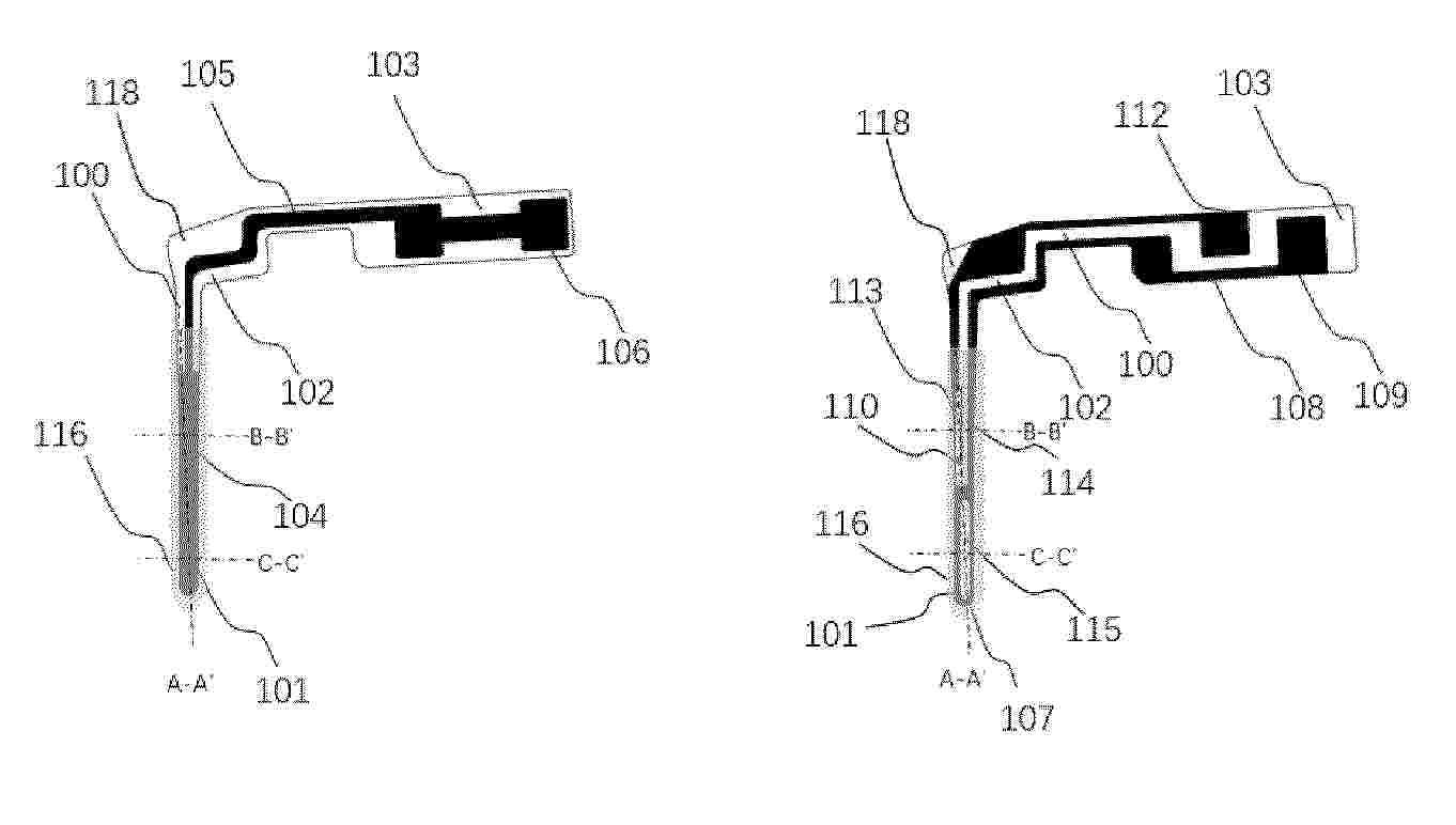
The present application provides an analyte sensor based on blood glucose monitoring. The analyte sensor comprises a substrate, a working electrode, a reference electrode, a counter electrode, and a sensing layer; the sensing layer is provided on the working electrode; the working electrode, reference electrode, and counter electrode are provided at surfaces of the substrate, and the two electrodes from the working electrode; the working electrode, reference electrode, and counter electrode located at the same surface of the substrate are spatially separated by a channel. The analyte sensor can be implanted into a human body for monitoring, and has the characteristics of good flexibility, small size, low manufacturing cost, simple process flow, and long-term monitoring.
Resumen de: US20260174961A1
A system for monitoring a patient includes one or more processors and a sensor device implemented in circuitry. The system is configured to measure, using the sensor device, a blood glucose status of the patient, and determine, using one or more processors, an initial insulin dose for the patient based on the blood glucose status of the patient. The system is further configured to optimize the initial insulin dose for the patient, based at least in part on a correction factor, to create an optimized insulin dose for the patient. The system is configured to facilitate therapy, using the one or more processors, based on the determined optimized insulin dose.
Resumen de: US20260177565A1
0000 An agent is for inhibiting glucose uptake into red blood cells and a blood collection tube suppresses glucose concentration decrease. The agent for inhibiting glucose uptake into red blood cells includes inosine as an active ingredient. The inhibition agent can be used for suppressing a decrease in glucose concentration of whole blood collected in a blood collection tube. The blood collection tube includes inosine in an internal space. The blood collection tube can be used to measure glucose concentration in blood. The blood collection tube can further include a glycolysis inhibitor.
Nº publicación: WO2026131972A1 25/06/2026
Solicitante:
INST NAT SANTE RECH MED [FR]
UNIV GRENOBLE ALPES [FR]
CENTRE NAT RECH SCIENT [FR]
INST POLYTECHNIQUE GRENOBLE [FR]
UNIVERSITE GRENOBLE ALPES
CENTRE NATIONAL DE LA RECHERCHE SCIENTIFIQUE
INSTITUT POLYTECHNIQUE DE GRENOBLE
INSTITUT NATIONAL DE LA SANT\u00C9 ET DE LA RECHERCHE M\u00C9DICALE
Resumen de: WO2026131972A1
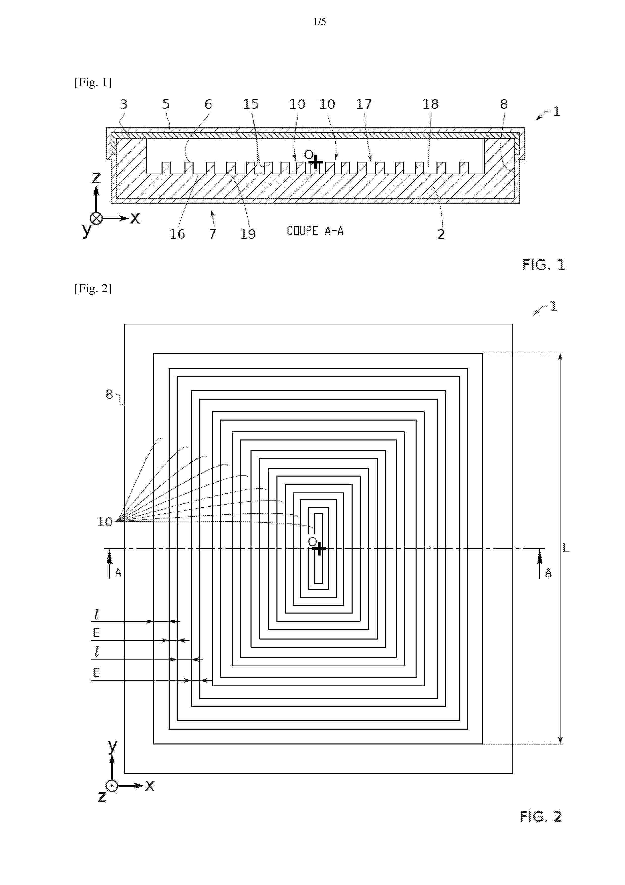
The invention relates to a device for distributing and receiving a population of pancreatic islets heterogeneous in size, the device comprising a matrix having a first face opposite a second face, characterized in that the matrix comprises concentric channels centered on a center O, the channels have an opening for receiving the pancreatic cells, the opening leading out onto the first face, the opening extending over the entire length (L) of the channel, the opening having a width (I) taken along the section Si, the device being configured such that the widths of the opening of two adjacent channels have an equal or increasing value moving away from the center O and such that the widths of the opening of a channel closest to the center O and of a channel furthest from the center O have an increasing value moving away from the center O. The invention relates to the field of implantable devices, in particular artificial pancreas devices.
 BOPI
BOPI
 Sede Electrónica