Si deseas distinguir tus productos, servicios o ambos de los de otra empresa, es posible que necesites una marca o nombre comercial. Descubre qué son, en qué consiste su procedimiento de registro y qué implica.

Información sobre los plazos de presentación de solicitudes de transformación de marcas de la Unión Europea en marca nacional española. Más información


Si tienes un nuevo dispositivo, producto o procedimiento que resuelva un problema técnico o tenga una ventaja práctica, existen distintas formas de protegerlo en España y en otros países. Descubre cómo hacerlo.

¿Tu innovación reside en la estética, la ornamentación o la apariencia de tu producto? Protégela mediante un diseño industrial. Descubre qué derechos confiere el registro y cómo realizar la tramitación.

Las indicaciones geográficas protegen el nombre de un producto originario de una zona geográfica, a la cual le debe una determinada calidad, reputación u otra característica. Descubre qué son, en qué consiste su procedimiento de registro y qué beneficios conceden.

Las patentes publicadas en todo el mundo son una valiosa fuente de información científica, técnica y comercial.

Si eres emprendedor/a o una empresa y quieres potenciar y mejorar la rentabilidad de tu negocio protegiendo de forma adecuada los activos intangibles de tu organización, en este espacio encontrarás lo necesario.


 166
resultados
166
resultados
 Última actualización
31/03/2026 [07:16:00]
Última actualización
31/03/2026 [07:16:00]


Resumen de: WO2025031978A1
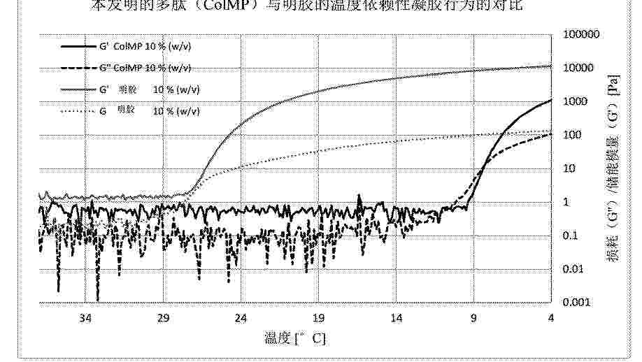
The invention relates to a polypeptide that is excellently suited for use in an ink for 3D printing. Furthermore, the present invention also relates to a polynucleotide that encodes the polypeptide according to the invention, and to a host cell that expresses the polypeptide according to the invention. Another embodiment is a method for producing an ink for 3D printing, wherein the ink contains a polypeptide. Additional embodiments relate to methods for producing a 3D scaffold and to the 3D scaffold that can be obtained by said methods, including for use in medicine.

Resumen de: CN121732829A
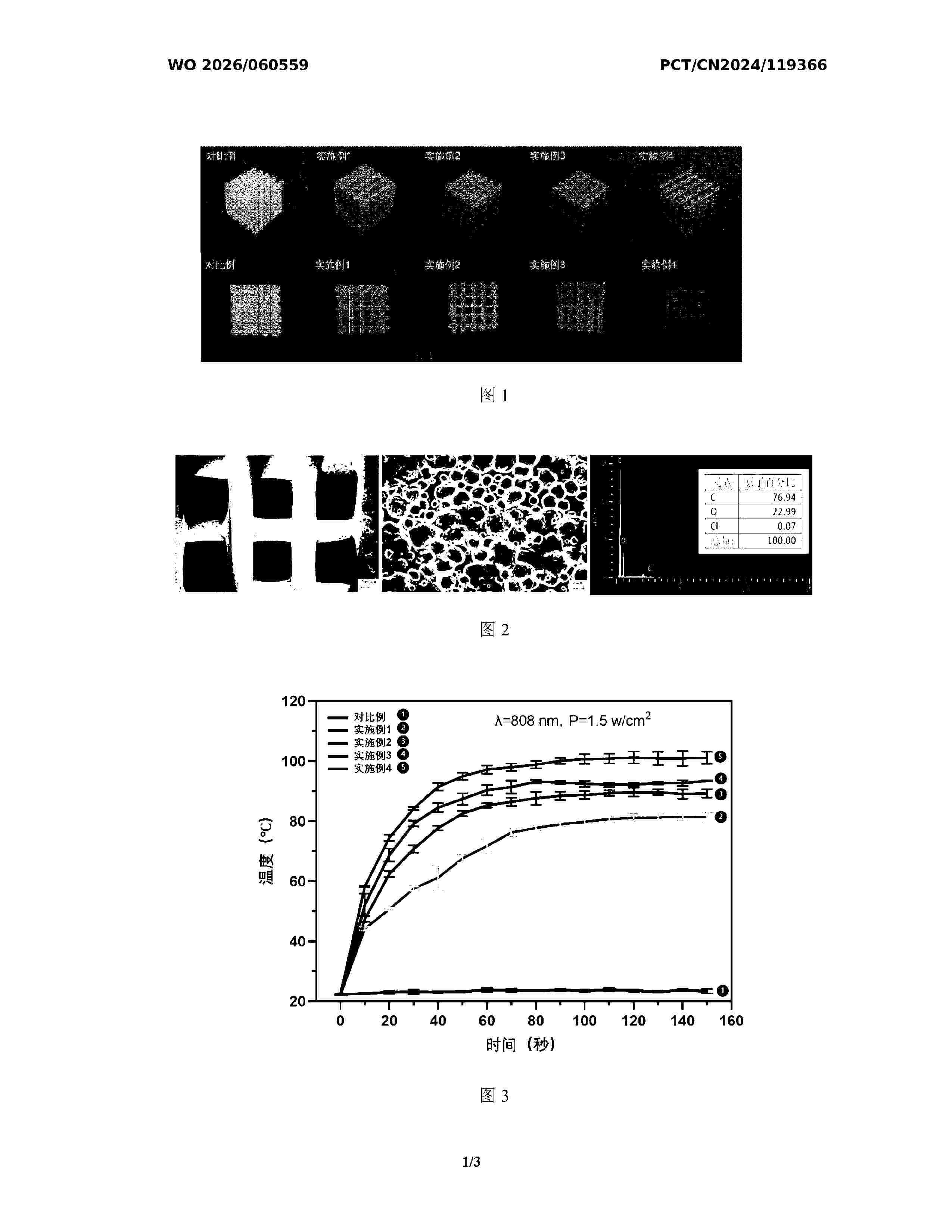
本发明公开了金属玻璃仿生人工骨植入体激光粉末床熔融增材制造方法,属于增材制造技术与生物医用材料技术领域,采用激光粉末床熔融技术,通过三维模型设计、调控激光工艺参数,在不同位置成形具有不同孔隙率的显微组织,从而实现具有梯度孔隙结构的人工骨植入体一体化成形;本发明从三维建模和工艺参数两个角度出发,可在宏观、介观、微观三个层级实现对零件内部孔隙结构的控制,显著提升金属玻璃植入体的能量吸收能力及细胞组织渗透能力,并能有效降低金属玻璃植入体的弹性模量,实现对植入体性能的综合调控,有效缓解应力屏蔽效应,增强金属玻璃植入体整体的稳定性,有助于金属玻璃仿生人工骨植入体在人体骨缺损修复临床治疗中的应用。
Resumen de: WO2026060559A1
The present invention provides a biomedical scaffold, a preparation method therefor, a use method therefor, and use thereof. The biomedical scaffold comprises the following starting material components in percentage by mass: 96.0%-99.5% of shape memory polyurethane and 0.5%-4% of active particles, wherein the active particles comprise one or more of manganese dioxide, nano magnesium oxide, ferroferric oxide, and magnesium metal. According to the present invention, by means of utilizing the easy volatility of dichloromethane, the pressure in a container is increased, thereby enabling homogeneous dissolution of shape memory polyurethane and uniform dispersion and mixing of active ingredients. By means of room-temperature volatilization and deposition, such shape memory materials are 3D printed, and the printed structures, such as pore size and porosity, are further designed to achieve good shape memory performance and mechanical properties. In addition, a plurality of scaffolds can be heated in various ways, such as near-infrared light irradiation and hot water soaking. Thus, the scaffolds can be assembled into a specific shape and exhibit strong stability and mechanical properties.
Resumen de: WO2026062643A1
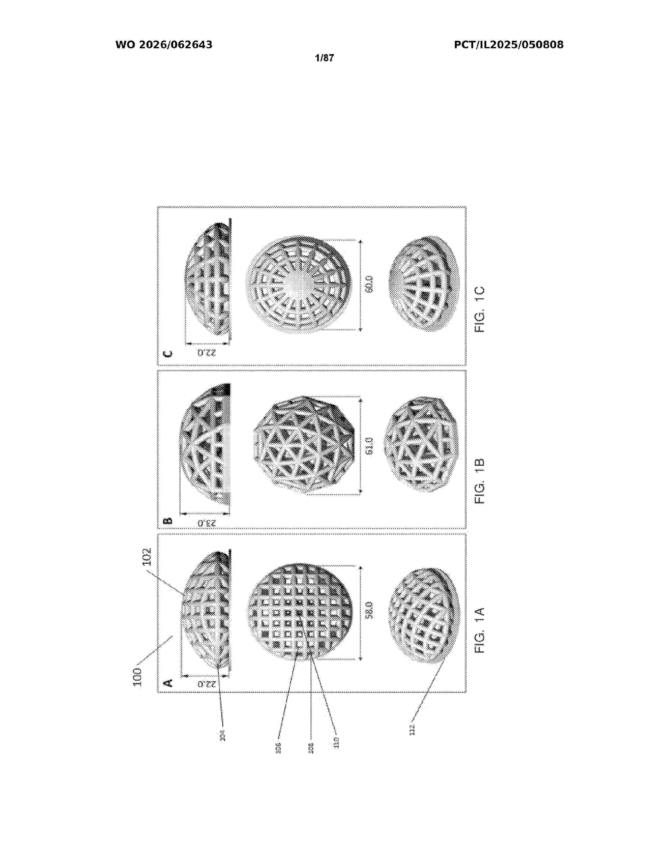
A photocurable formulation usable in additive manufacturing a three-dimensional object is disclosed. The formulation features, in at least a portion thereof, a biological or a biocompatible material, the photocurable formulation comprising a photoinitiator, a photocurable biological or biocompatible material, and a carrier, the formulation further comprising at least two photocurable polymeric materials. Implants comprising same are also disclosed.
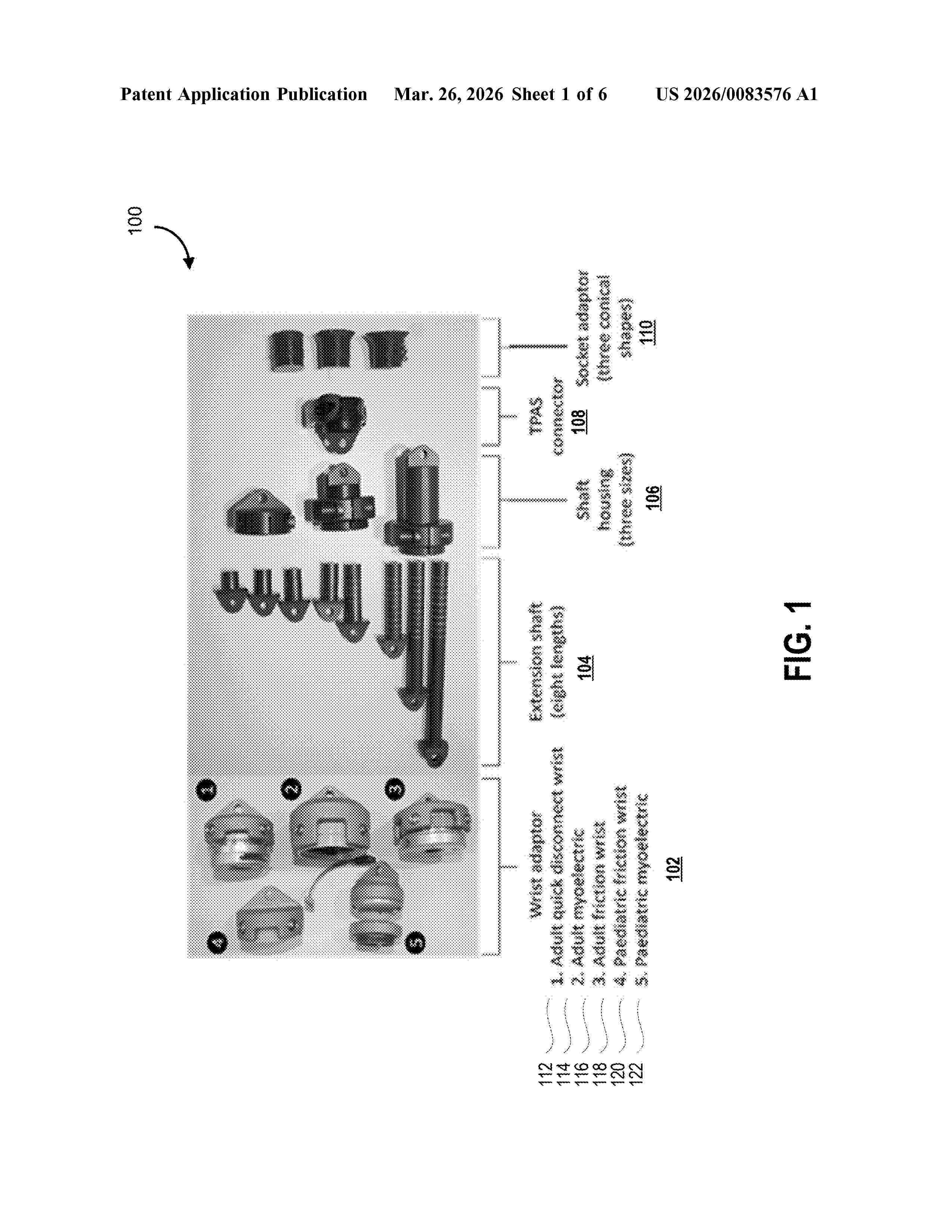
Resumen de: US20260083576A1
A transradial prosthesis assessment system apparatus and method for simulating and evaluating alignment and functionality of a definitive prosthesis system for a user's diagnostic socket is provided. The apparatus includes socket adaptors, a connector, shaft housings of different sizes, extension shafts of different lengths and wrist adaptors that can be connected through different combinations to provide length and alignment adjustability to emulate the definitive prosthesis.
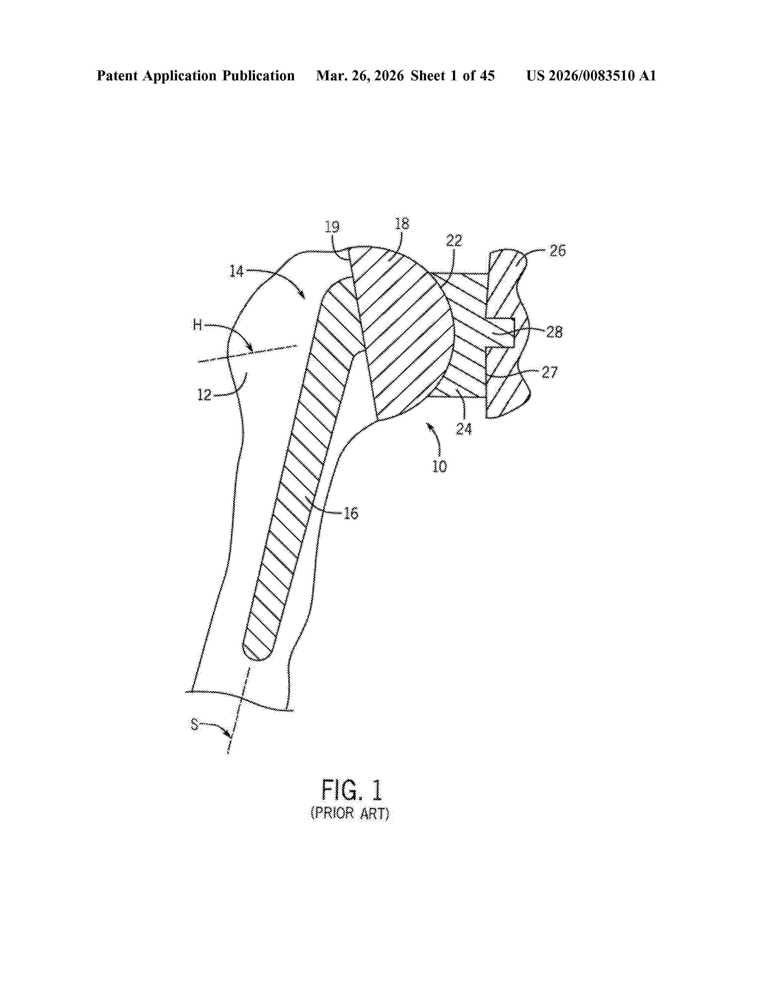
Resumen de: US20260083510A1
Methods for understanding external and internal anatomy of bones through the use of imaging data and 3D modeling to facilitate the design of anatomically correct plates, devices and implants are disclosed. In one aspect the method results in an implant or plate that includes at least one curved surface wherein a contour of the at least one curved surface corresponds to an anatomic shape of a subject. The anatomic shape of the subject being determined based on an image of the bone.
Resumen de: US20260083568A1
Systems and methods include a component of a bone implant that includes a patient specific bone contacting surface and, optionally, at least one protrusion adjacent to or extending from the patient specific bone contacting surface. The at least one protrusion is configured for engaging a bone preparation in the bone. To prepare the bone for receiving the component, one or more cutting tool templates may be used. The one or more cutting tool templates may be fabricated to have a patient specific bone contacting surface that engages the bone that is to receive the implant component. A cutting tool is engaged in one or more of one or more slots defined by the cutting tool template(s) to remove surface portions of the bone to create the bone preparation for engaging the protrusion. The patient specific bone contacting surface of the component engages the bone.
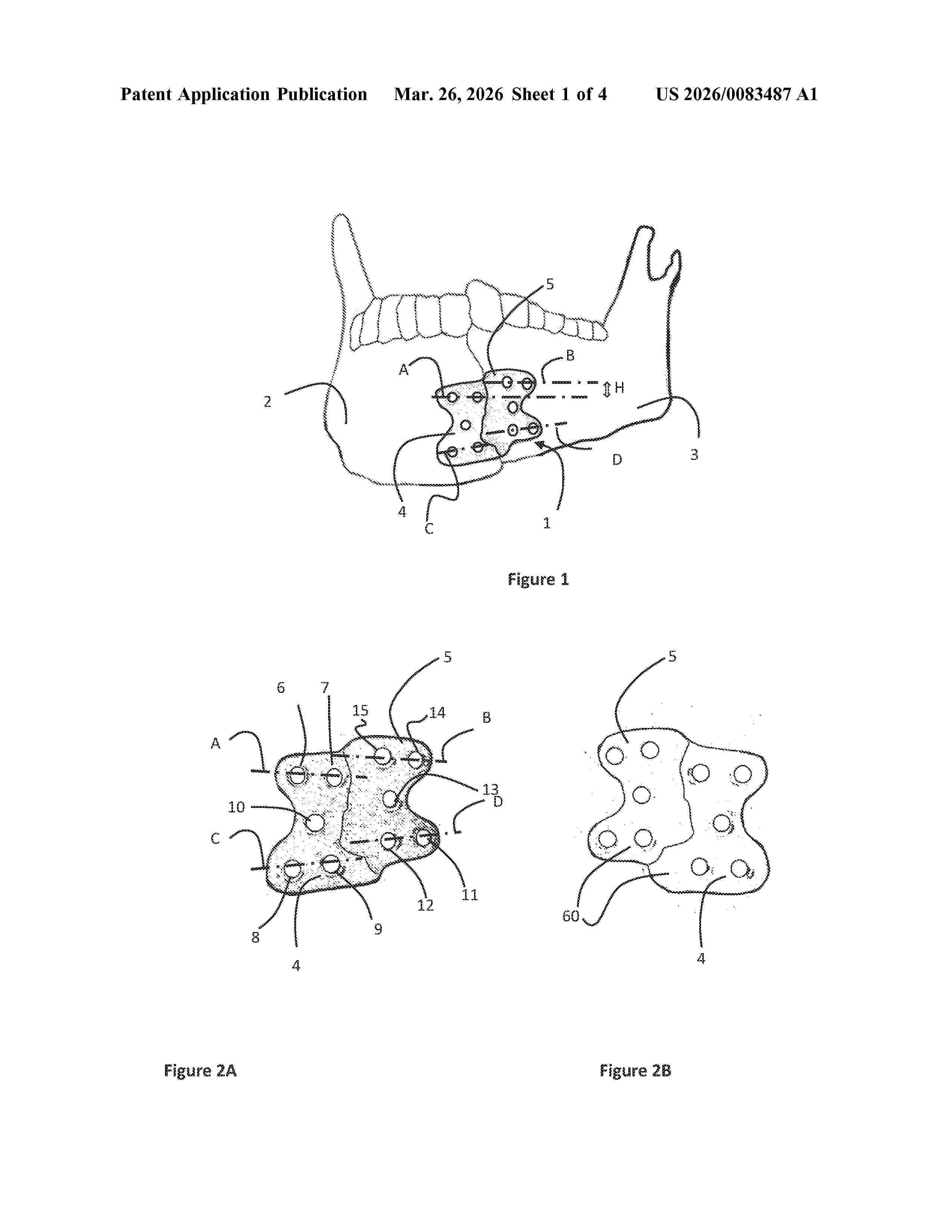
Resumen de: US20260083487A1
Guide plate for the anatomical realignment of a plurality of fractured bone portions, comprising a back surface configured such as it can be distinctively coupled to the surface anatomy of the area of said plurality of fractured bone portions and characterized by a first plurality of holes placed on a first portion of the guide plate and a second plurality of holes placed on a second portion of the guide plate, wherein the first plurality of holes is misaligned with respect to the second plurality of holes.
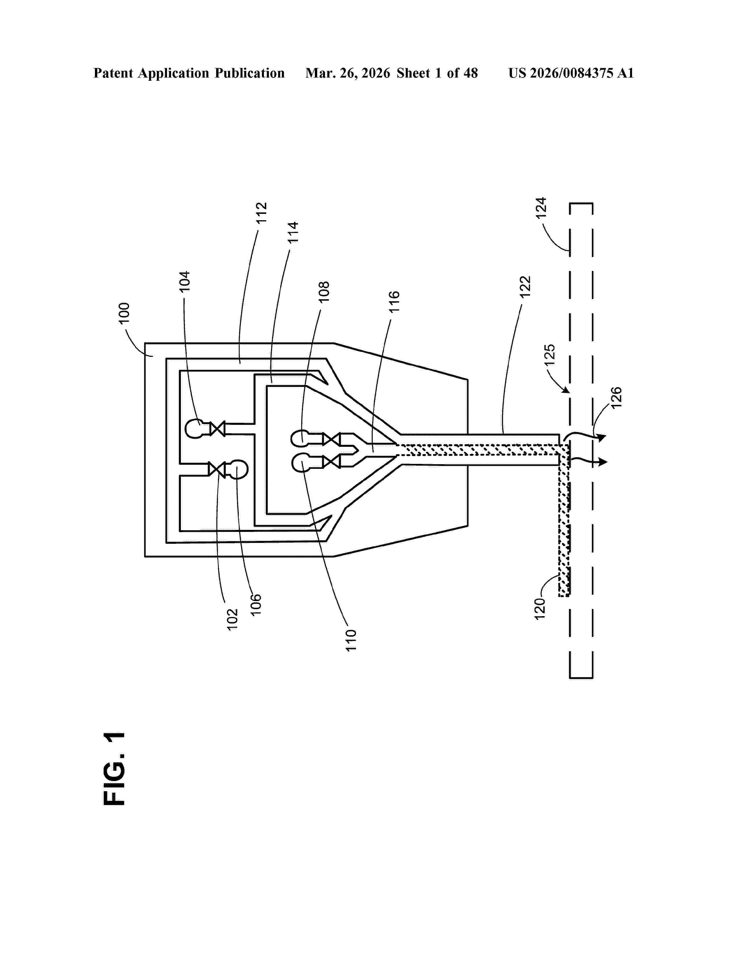
Resumen de: US20260084375A1
Aspects of the disclosure include a fabrication platform for supporting a bioprinted fiber structures during printing, patterning, and/or processing, comprising a frame with a plurality of posts for securing a cross-linkable fiber during printing thereof, and where a continuous length of the cross-linkable fiber is printed around a plurality of posts during the 3D bioprinting process. The fabrication platform enables the cross-linkable fiber to be suspended during one or more of printing, patterning, and/or processing. In this way, the bioprinted fiber structure comprises a uniform outer surface, and can be easily modified and/or further processed after printing and patterning are completed.
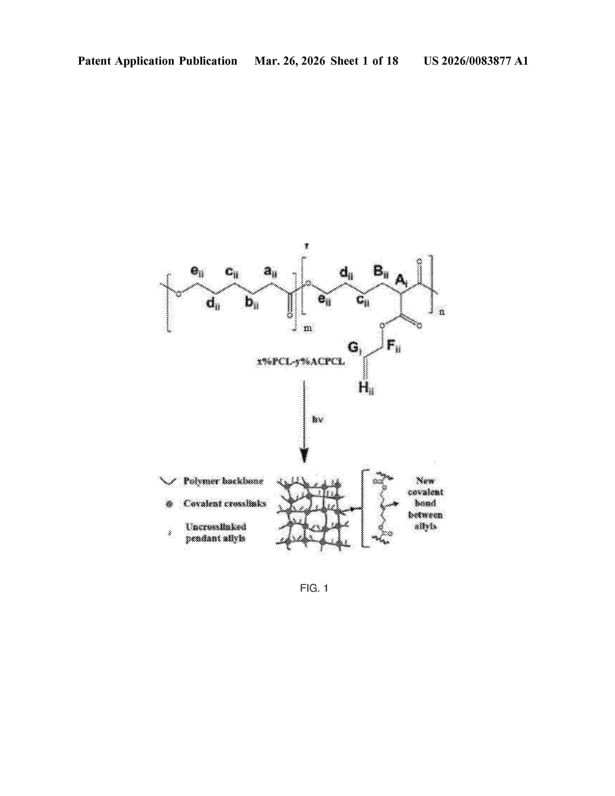
Resumen de: US20260083877A1
Various precursor solutions and methods of 3D printing and other additive manufacturing approaches are provided for the manufacture of articles using photocrosslinkable vinyl shape memory polymers. In various aspects, articles are manufactured by a process comprising (i) exposing a precursor solution to an intensity and frequency of light to initiate photo-polymerization of the precursor solution to form a layer of the article; and (ii) repeating step (i) a number of times to form the article in a layer-by-layer approach; wherein the precursor solution comprises a first polymeric precursor comprising a plurality of vinyl terminated side-chains attached thereto. A vinyl-functionalized, photocrosslinkable SMP used as an example herein is a novel variant of a previously disclosed SMP composition, with higher amounts of vinyl functionalization but similar thermomechanical properties to the SMP library previously disclosed. Moreover, a unique combination of chemistries is disclosed that incorporates the aforementioned vinyl-functionalized SMPs along with acrylate-based crosslinkers. Additional novel compositions are disclosed containing the vinyl-functionalized SMPs with additional dithiol functionalizations, vinyl-functionalized SMPs with dithiol crosslinkers, as well as vinyl-functionalized SMPs with both acrylate-based and dithiol crosslinkers, as another means to tune degradation rates and other material properties. The articles can include a variety of articles such as stents
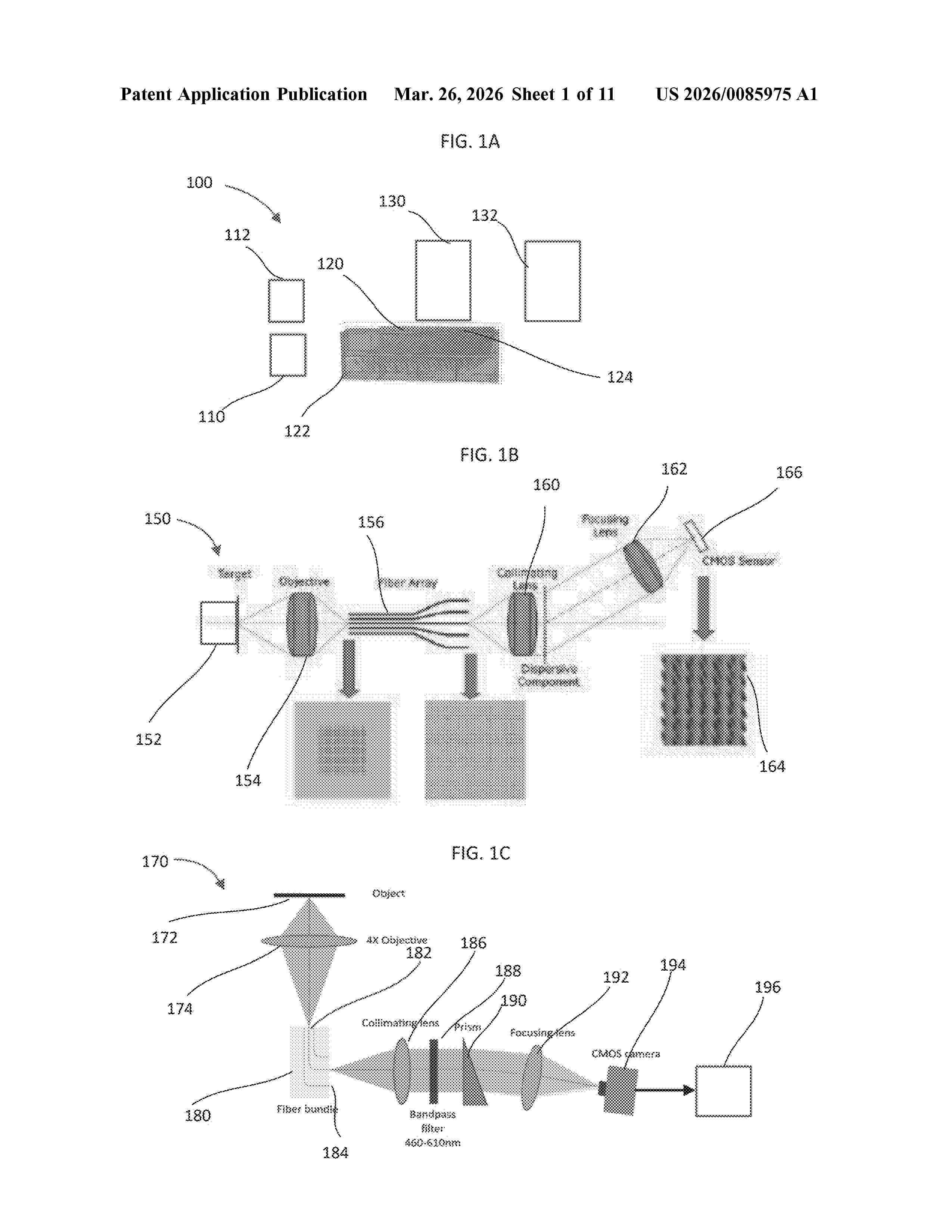
Resumen de: US20260085975A1
The present disclosure relates to a custom waveguide array to encode 3-dimensional data for snapshot imaging techniques like imaging spectrometry or volumetric spectral domain OCT. The custom waveguide array has a series of waveguides such as optical fibers having input ends and output ends. The input ends are grouped in a dense array input area. An array output area creates void spaces for the output ends. The output area thus may used to provide spectral information for an object imaged by the input area. The fiber arrays may be manufactured with an entirely automatic development process based on 3-D printing techniques such as 2-Photon Polymerization (2PP) additive manufacturing.
Resumen de: DE102024127337A1
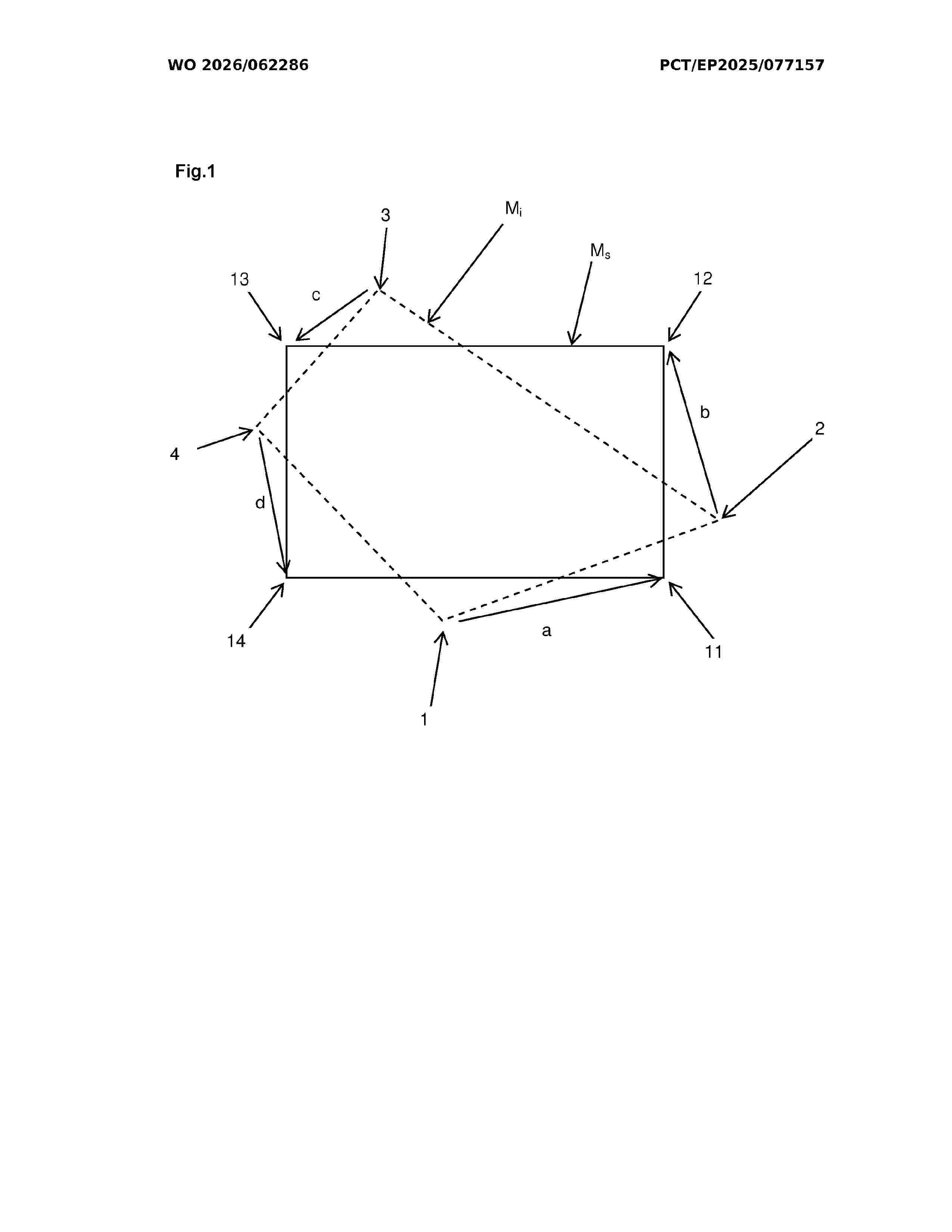
Die Erfindung betrifft eine Diagnoseverfahrens-Software zur Bereitstellung von Daten zur Herstellung einer Kiefer-Gebiss-Schiene für eine Person, wobei ein Ist-Zahnraum-Zustand und/oder Zahnstellung (1) und eine Ist-Kieferfunktion (2) erfasst werden, wobei- eine Ist-Körperhaltung (3) erfasst wird, und- ein Ist-Fußflächendruck (4) erfasst wird, und- der Ist-Zahnraum-Zustand und/oder Zahnstellung (1) mit einem Soll-Zahnraum-Zustand und/oder Zahnstellung (11) abgeglichen wird, um eine erste Abweichung (a) zwischen dem Ist-Zahnraum-Zustand (1) und dem Soll-Zahnraum-Zustand (11) zu ermitteln,- die Ist-Kieferfunktion (2) mit einer Soll- Kieferfunktion (12) abgeglichen wird, um eine zweite Abweichung (b) zwischen der Ist-Kieferfunktion (2) und der Soll-Kieferfunktion (12) zu ermitteln,- die Ist-Körperhaltung (3) mit einer Soll-Körperhaltung (13) abgeglichen wird, um eine dritte Abweichung (c) zwischen der Ist-Körperhaltung (3) und der Soll-Körperhaltung (13) zu ermitteln,- der Ist-Fußflächendruck (4) mit einem Soll-Fußflächendruck (14) abgeglichen wird, um eine vierte Abweichung (d) zwischen dem Ist-Fußflächendruck (4) und dem Soll-Fußflächendruck (14) zu ermitteln, wobei- die erfassten Daten zur Herstellung der Kiefer-Gebiss-Schiene genutzt werden, geeignet um die Abweichungen (a, b, c, d) zu verringern.
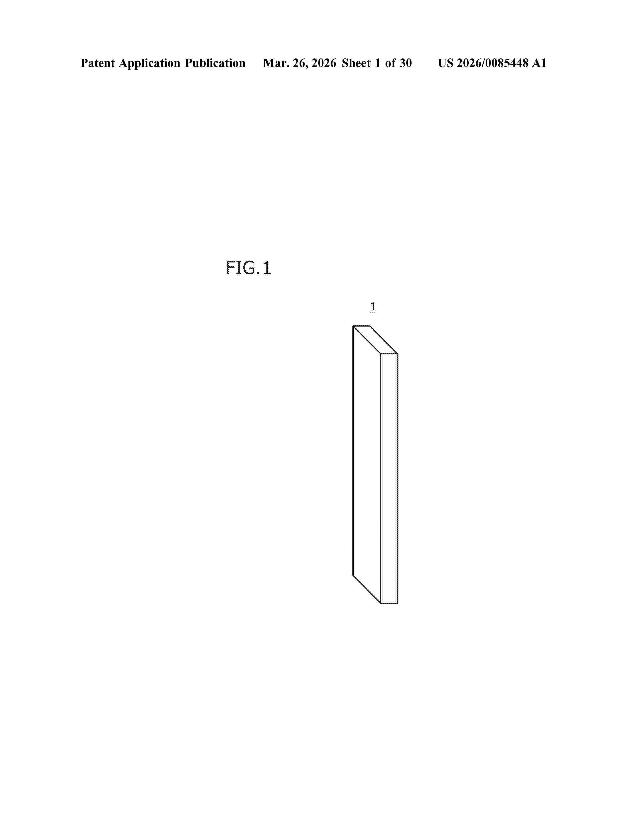
Resumen de: US20260085448A1
An inorganic structure having mechanical properties that differ depending on the region in the inorganic structure, and a method for manufacturing the inorganic structure are provided. An inorganic structure (1) of the present embodiment includes a plurality of solidified portions (SA) composed of an inorganic material. The plurality of solidified portions (SA) include a first solidified portion (SA1) having a first crystallographic direction (CO1) preferentially oriented in a predetermined direction, and a second solidified portion (SA2) having a second crystallographic direction (CO2) that is a different orientation from the first crystallographic direction (CO1).
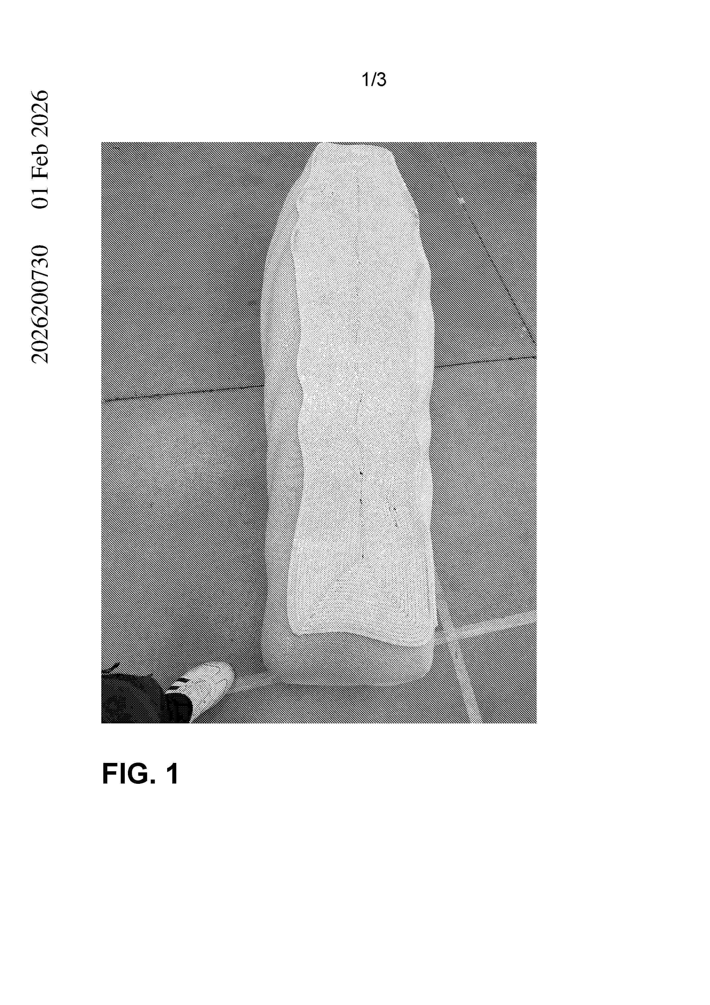
Resumen de: AU2026200730B1
Abstract Disclosed is a method for manufacturing a coffin that is configured to be burned via a cremation purpose. The method includes selecting a biocomposite printing material having the following properties: a primary material in the form of a plant-derived polylactic acid (PLA); a cellulose fibre content of between 5% and 20%; and less than 1% petroleum-based materials. The material is utilized to manufacture the coffin via a 3D printing process, wherein, upon assembly, the coffin has a shape having an irregular cross sectional area.
Resumen de: AU2025260012A1
The present invention relates to a 3D scaffold implant system for mandibular reconstruction, featuring a scaffold structure that includes at least four scaffold segments strategically positioned within the implant, the segments ensuring precise placement within a subject's jawbone and facilitate optimal integration with a surrounding bone tissue; a fluid delivery tube system integrated into each of the scaffold segments to administer a growth factor(s) and an antibiotic directly into the scaffold implant; a pump that facilitates controlled delivery of the growth factors into the scaffold segments; at least three fixation that provide stability to the implant and micro-motors that enable precise alignment of the scaffold implant to conform seamlessly to the natural curvature of the jawbone.
Resumen de: AU2024326857A1
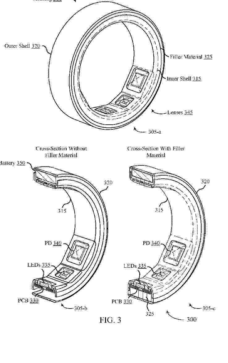
Methods, systems, and devices for a wearable ring device are described. A wearable ring device may include a ring-shaped housing and a printed circuit (PCB) that at least partially contacts an inner shell of the ring-shaped housing. Light-emitting components and light-receiving components may be disposed on a first surface of the PCB and may extend through an aperture(s) in the inner shell such that the lightemitting and light-receiving components are substantially flush with an inner ringshaped surface of the inner shell. Optical lenses may cover the light-emitting and lightreceiving components within the aperture(s). In some cases, the optical lenses may be molded over the light-emitting and light-receiving components before the PCB is inserted into the ring-shaped housing. In other cases, the optical lenses may be molded over the light-emitting and light-receiving components (and the aperture(s)) after the PCB is inserted into the ring-shaped housing.
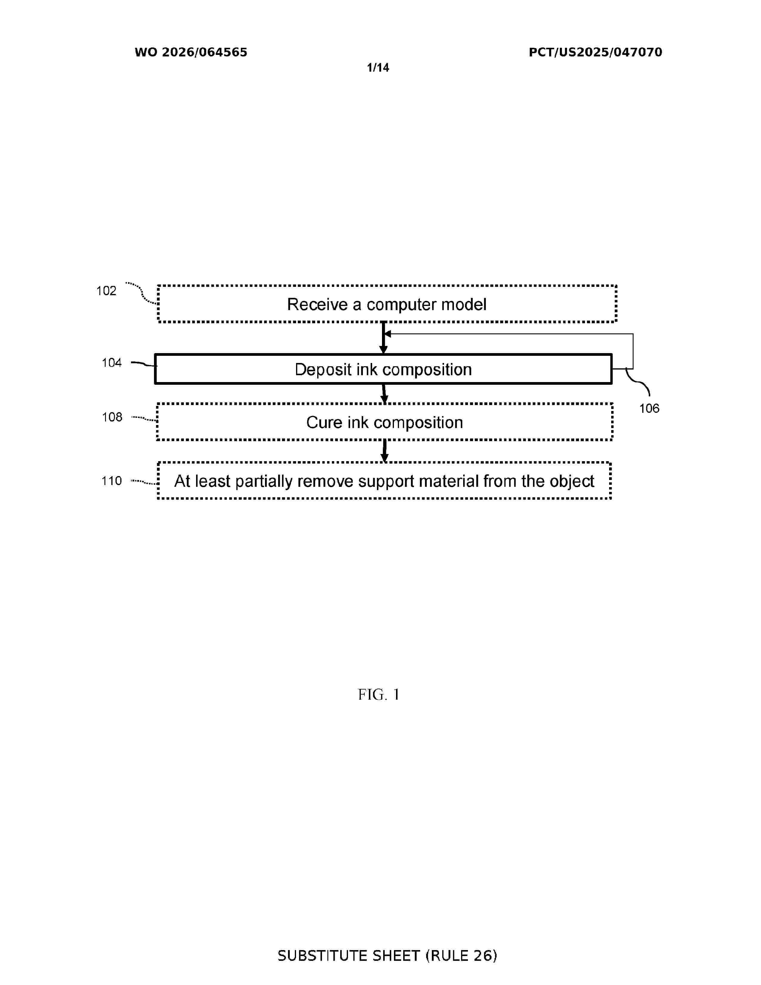
Resumen de: WO2026064565A1
Methods for additive manufacturing, biological structures, cultivated meats, and additive manufacturing systems are provided. The method comprises dispensing an ink composition from a nozzle of an additive manufacturing system. The ink composition comprise adipocytes and a hydrogel. The deposition occurs at a suitable shear stress such that the ink composition can flow through the needle and an integrity of the adipocytes is substantially maintained. The method comprises repeating, as necessary, repositioning of the nozzle and dispensing ink composition from the first nozzle, thereby forming the structure.
Resumen de: WO2026062286A1
The invention relates to diagnostic method software for providing data for producing an occlusive mouthguard for a person, wherein an actual tooth space state and/or tooth position (1) and an actual jaw function (2) are acquired, wherein - an actual body posture (3) is acquired, and - an actual plantar pressure (4) is acquired, and - the actual tooth space state and/or tooth position (1) is compared with a target tooth space state and/or tooth position (11) in order to determine a first deviation (a) between the actual tooth space state (1) and the target tooth space state (11), - the actual jaw function (2) is compared with a target jaw function (12) in order to determine a second deviation (b) between the actual jaw function (2) and the target jaw function (12), - the actual body posture (3) is compared with a target body posture (13) in order to determine a third deviation (c) between the actual body posture (3) and the target body posture (13), - the actual plantar pressure (4) is compared with a target plantar pressure (14) in order to determine a fourth deviation (d) between the actual plantar pressure (4) and the target plantar pressure (14), wherein - the acquired data are used to produce the occlusive mouthguard, in a manner suitable for reducing the deviations (a, b, c, d).
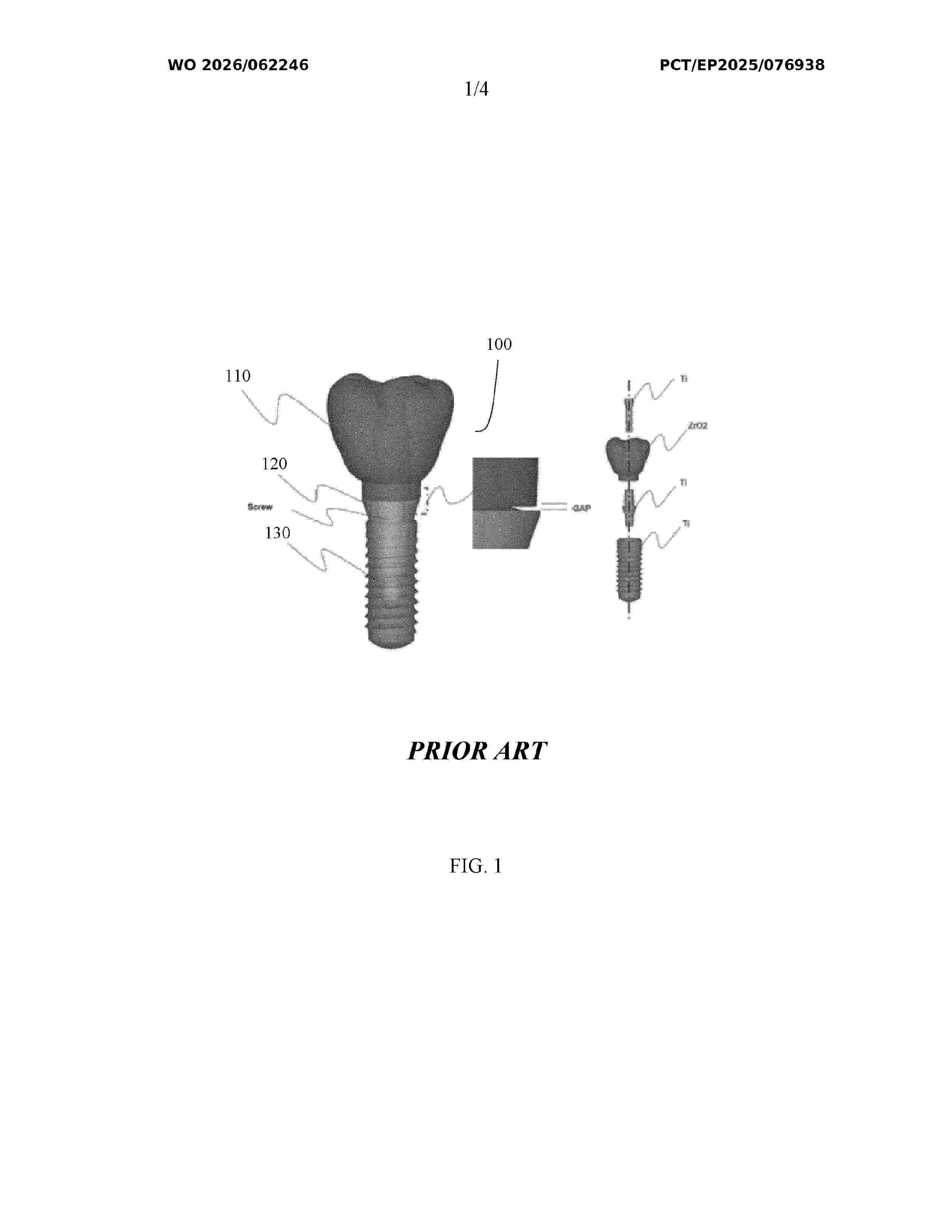
Resumen de: WO2026062246A1
The present disclosure provides methods for manufacturing of gap free, cement free, abrasion free dental implant devices. Dental implant devices manufactured according to the disclosed methods are also provided.
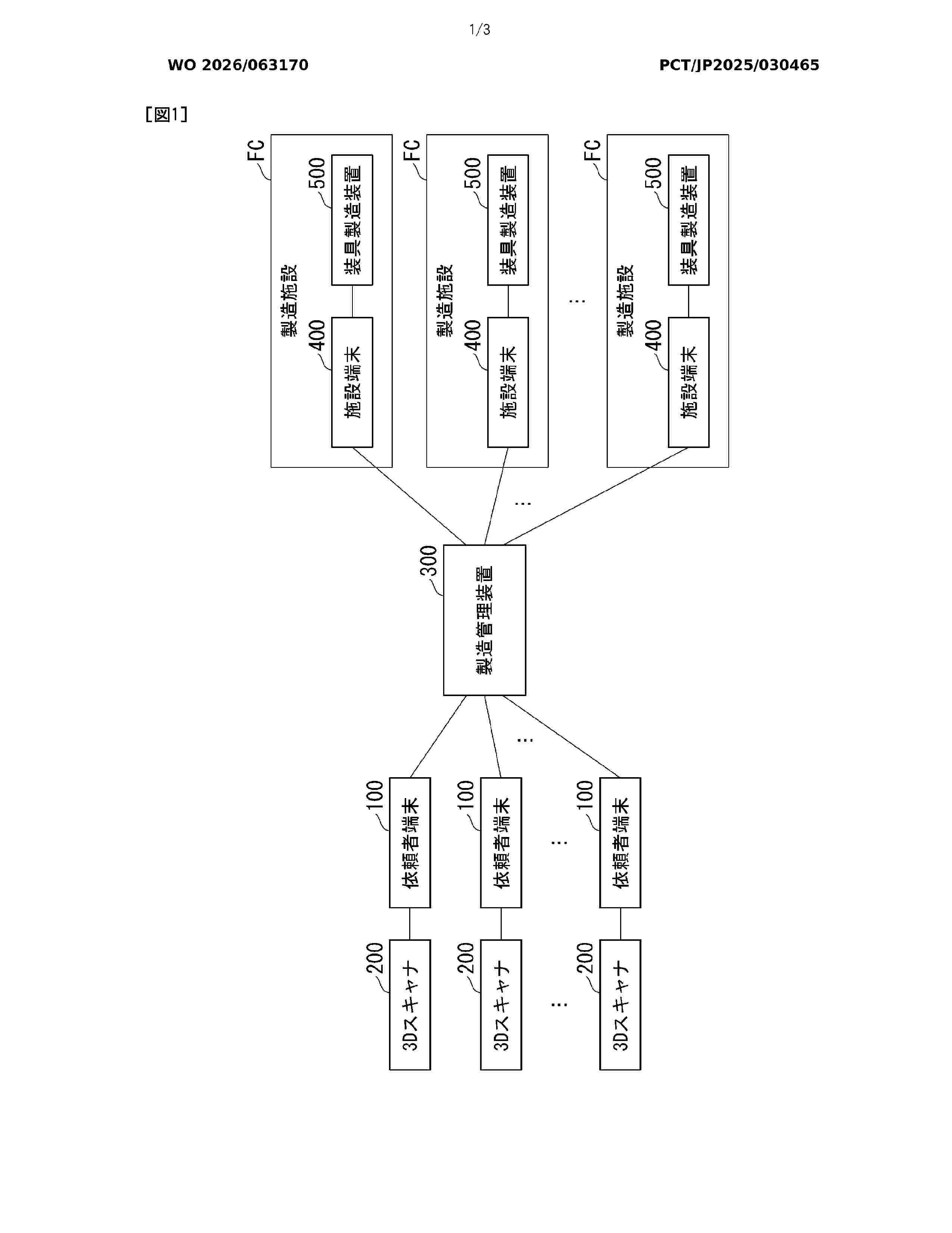
Resumen de: WO2026063170A1
Provided is an orthosis production system comprising: an information acquisition unit that acquires individual shape information indicating the shape of a body site of an orthosis user to which an orthosis is attached; an information generation unit (321) that generates, on the basis of the individual shape information, individual orthosis information indicating the shape of the orthosis to be used by the orthosis user; and a production instruction unit (322) that causes an orthosis production device, employing a pellet-based 3D printer which produces an orthosis according to an input of orthosis shape information indicating the shape of the orthosis, to produce the orthosis using the individual orthosis information.
Resumen de: WO2026063889A1
The present invention relates to a nerve conduit obtained by plant decellularization, which does not require 3D printing, is multi-channel, low-cost and capable of drug release. This nerve conduit is functionalized with GelMA and has properties that promote nerve healing.
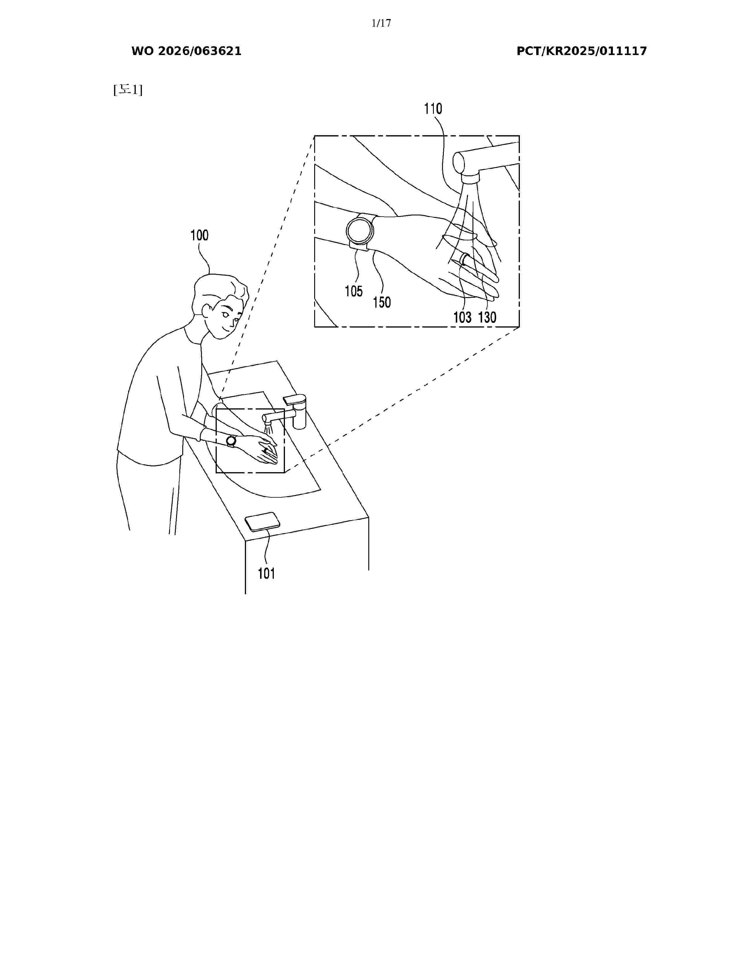
Resumen de: WO2026063621A1
This finger-wearable electronic device may comprise: a ring-shaped housing; a printed circuit board (PCB) disposed in the ring-shaped housing; and a sensor package mounted on the PCB. The sensor package can include an enclosure including a first light-transmitting portion, a second light-transmitting portion and a partition wall between the first light-transmitting portion and the second light-transmitting portion. The partition wall can include a surface that defines a portion of the exterior of the sensor package. The sensor package can include a first sensor. The first sensor can include: a light-emitting unit disposed under the first light-transmitting portion; and a reception unit disposed under the second light-transmitting portion. The sensor package can include a second sensor including electrodes. The electrodes of the second sensor can be disposed on the surface of the partition wall.
Resumen de: WO2026062644A1
Photoblocker usable in additive manufacturing a three-dimensional object are disclosed. Photocurable formulations comprising the photoblockers are also disclosed. The photoblocker can be beneficially included in formulations usable in additive manufacturing of 3D objects featuring a biological material in at least a portion thereof.
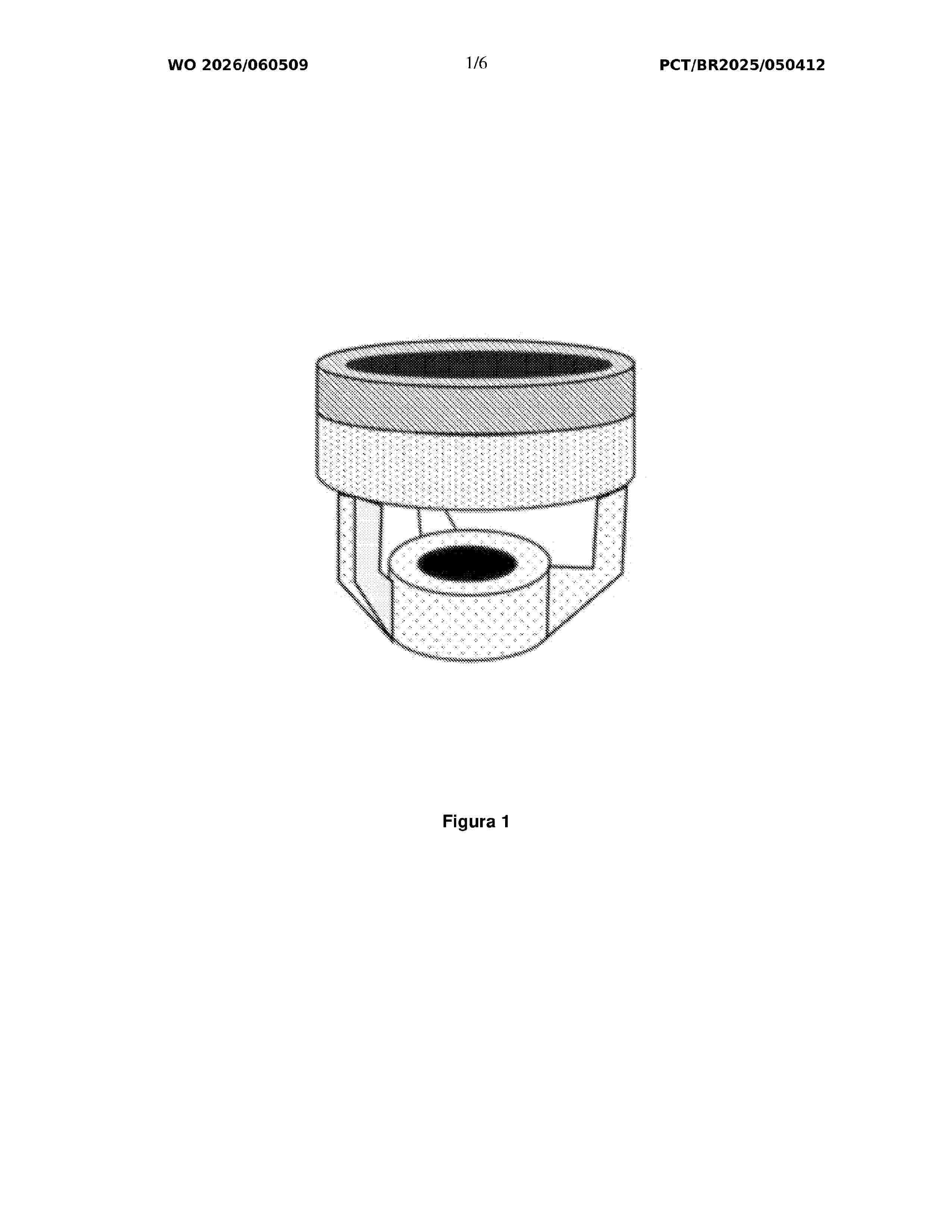
Resumen de: WO2026060509A1
The present patent of invention relates to the field of medicine and medical devices, and more specifically to an innovative fluorescent cervical cup for use in uterine manipulators during colpotomy surgical procedures. This improved device aims to increase the safety and efficacy of conventional or minimally invasive gynaecological procedures, including laparoscopic, robotic, and endoscopic surgery, particularly under conditions of limited visibility.
Nº publicación: ES3060116T3 25/03/2026
Solicitante:
AKIRA SCIENCE AB
Akira Science AB
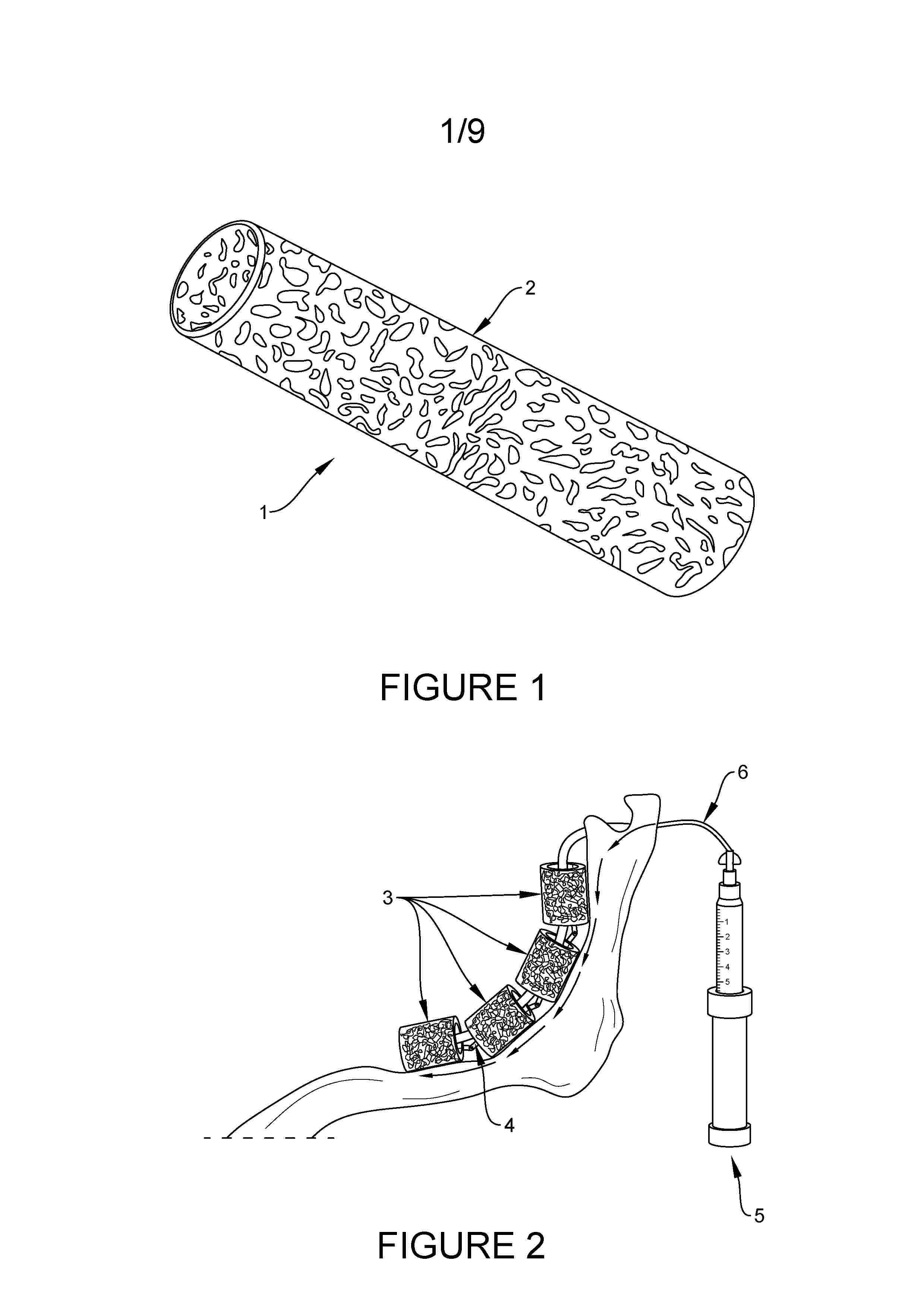
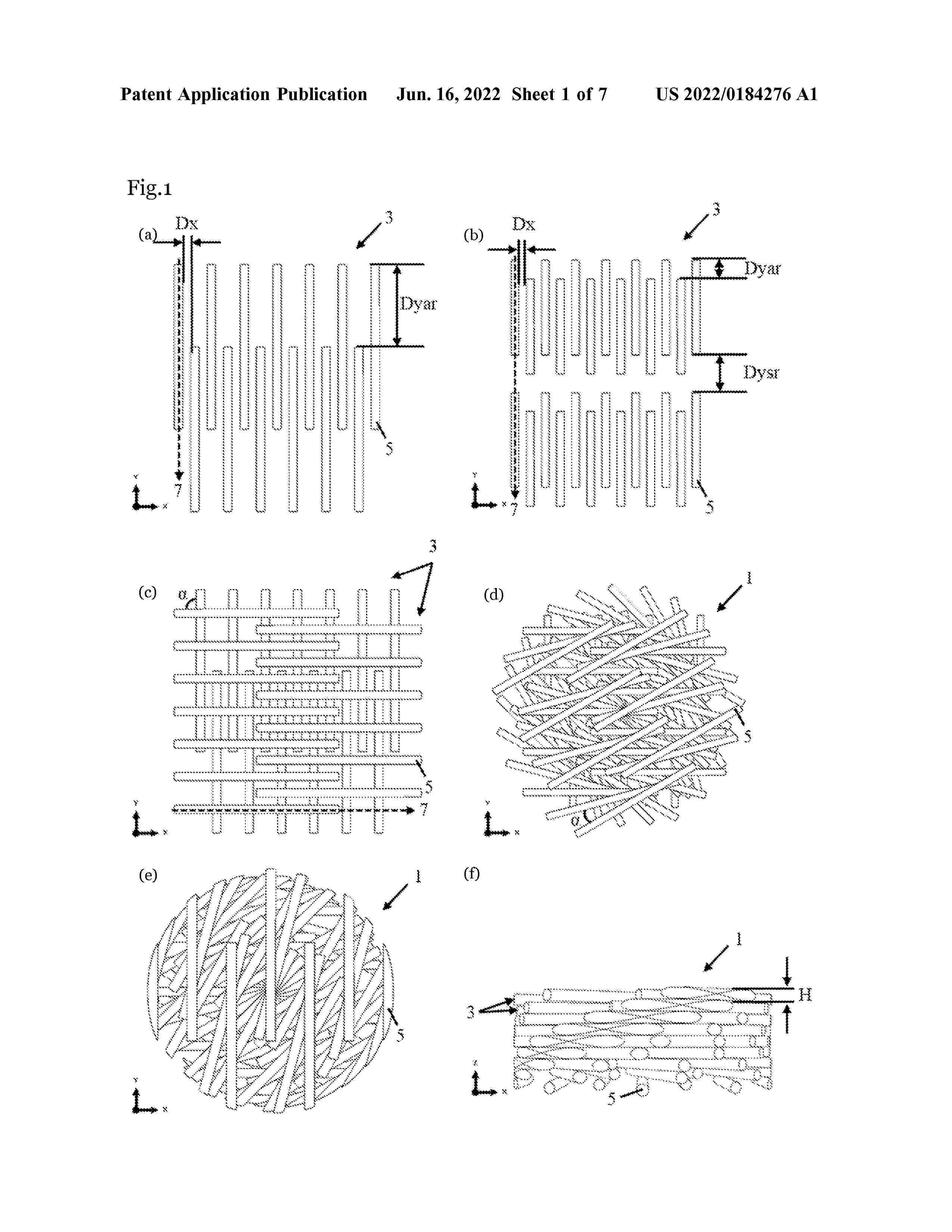
Resumen de: US2022184276A1
The present disclosure is directed to a degradable 3D-printable scaffold for use in tissue engineering, which scaffold has a combined gradient and staggered structure. Further provided is a medical device for use in tissue engineering, comprising such a scaffold. The present disclosure also provides a method for preparing a scaffold by additive manufacturing, e.g. 3D-printing, a method for in vivo tissue engineering, use of the scaffold in an in vitro cell culture system, in an in vitro method for culturing of cells and/or in an in vitro method for regenerating tissue. Also provided is a scaffold and a medical device for use in a method for in vivo tissue engineering. Further disclosed is a novel degradable copolymer of ε-caprolactone and p-dioxanone, which can be printed without degradation and which is particularly suitable for use as scaffold material in the scaffold and method according to the present disclosure.
 BOPI
BOPI
 Sede Electrónica