Si deseas distinguir tus productos, servicios o ambos de los de otra empresa, es posible que necesites una marca o nombre comercial. Descubre qué son, en qué consiste su procedimiento de registro y qué implica.

Información sobre los plazos de presentación de solicitudes de transformación de marcas de la Unión Europea en marca nacional española. Más información


Si tienes un nuevo dispositivo, producto o procedimiento que resuelva un problema técnico o tenga una ventaja práctica, existen distintas formas de protegerlo en España y en otros países. Descubre cómo hacerlo.

¿Tu innovación reside en la estética, la ornamentación o la apariencia de tu producto? Protégela mediante un diseño industrial. Descubre qué derechos confiere el registro y cómo realizar la tramitación.

Las indicaciones geográficas protegen el nombre de un producto originario de una zona geográfica, a la cual le debe una determinada calidad, reputación u otra característica. Descubre qué son, en qué consiste su procedimiento de registro y qué beneficios conceden.

Las patentes publicadas en todo el mundo son una valiosa fuente de información científica, técnica y comercial.

Si eres emprendedor/a o una empresa y quieres potenciar y mejorar la rentabilidad de tu negocio protegiendo de forma adecuada los activos intangibles de tu organización, en este espacio encontrarás lo necesario.


 484
resultados
484
resultados
 Última actualización
30/07/2026 [08:11:00]
Última actualización
30/07/2026 [08:11:00]


 Resultados 150 a 175 de 484

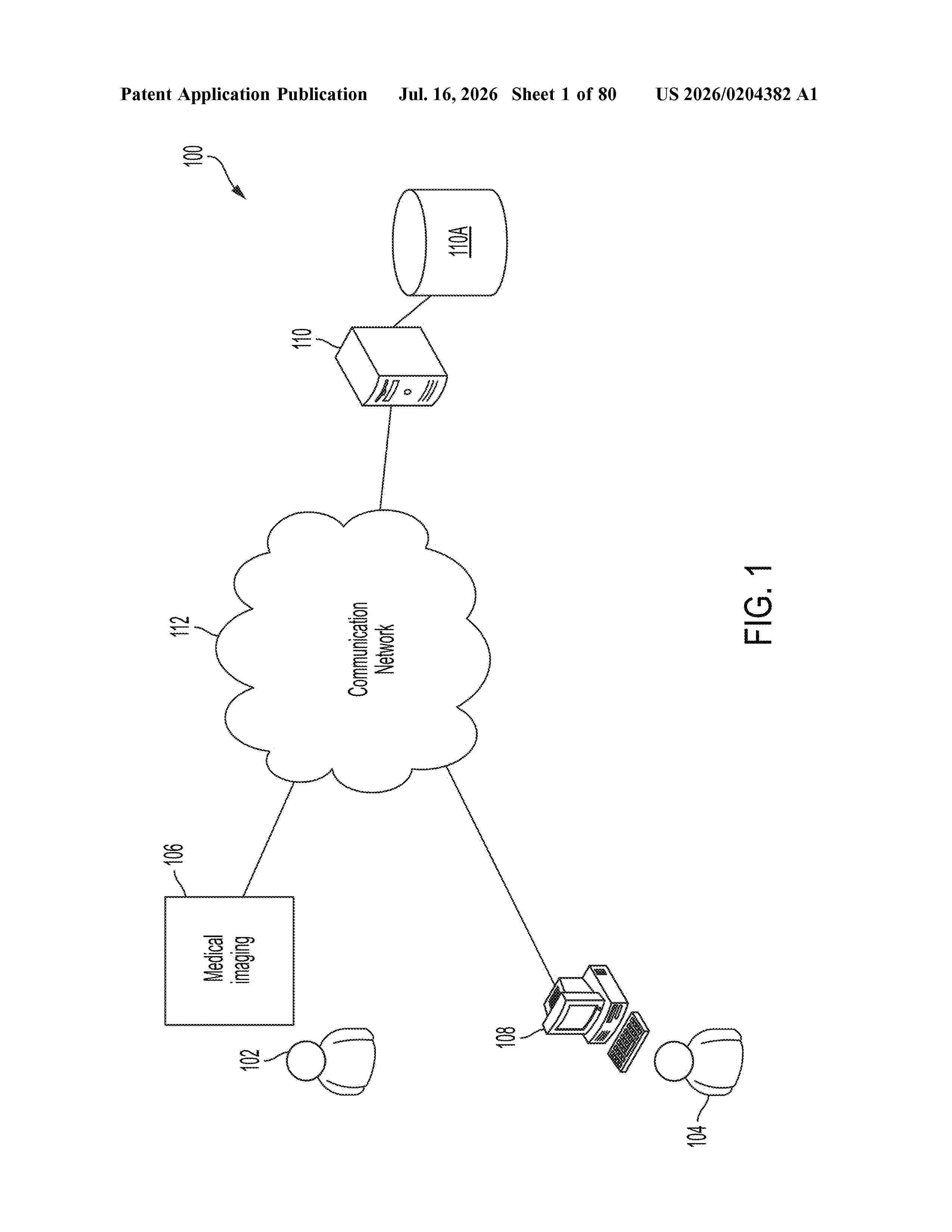
Resumen de: US20260204382A1
0000 Described herein are example techniques for evaluating morphology and/or operation of a joint of an animal. In embodiments, a computer model of the joint is prepared and analyzed. An amount of translation movement and an amount of rotation movement to be simulated are determined and used in simulated movement of the bone(s) of the joint across many different positions. The positions that are simulated with the computer model may correspond to different positions of the bone during operation of the joint, such as (in the case of a human hip joint) positions a bone may take during walking, running, or other movements. These positions may be automatically simulated based on the amounts of translation and rotation. In some embodiments, determined information regarding the morphology and/or operation of the joint from the simulation may be used to determine a recommendation of whether and/or how to treat improper operation of the joint.

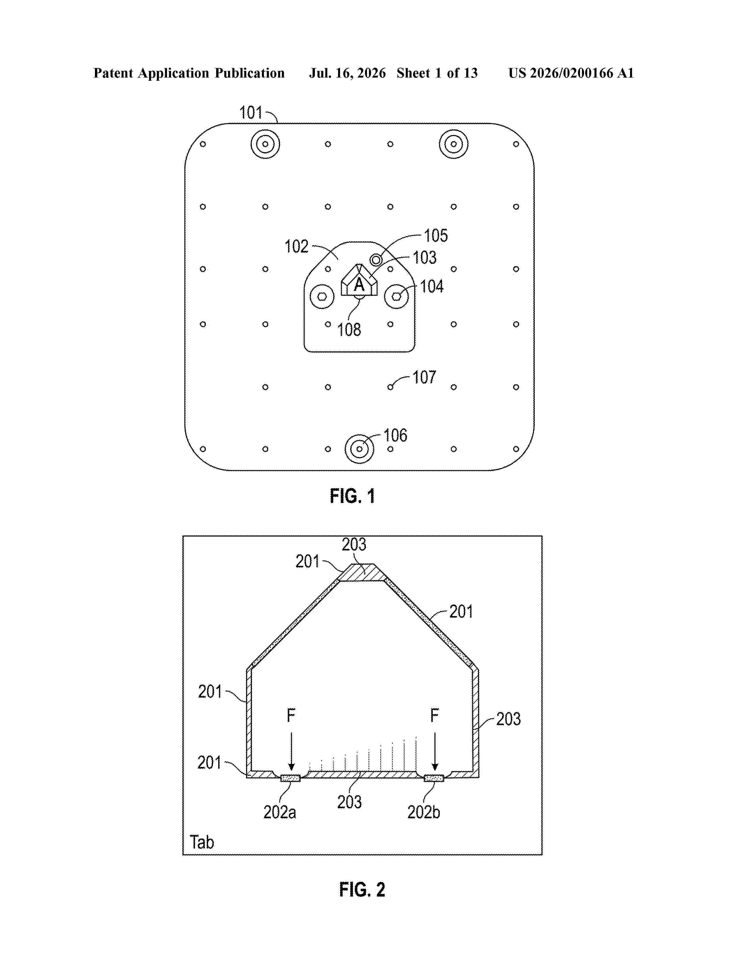
Resumen de: US20260200166A1
0000 The present disclosure discusses techniques for securing thermoform models, including a locator plate for receiving and securing individually unique dental models having a polygon cutout, wherein the locator plate has at least one ball plunger positioned within a raised polygon, the at least one ball plunger having a cylindrical body, a spring, and a ball extending partially outside the cylindrical body. The present disclosure also includes a system for thermoforming an orthodontic aligner and methods thereof.
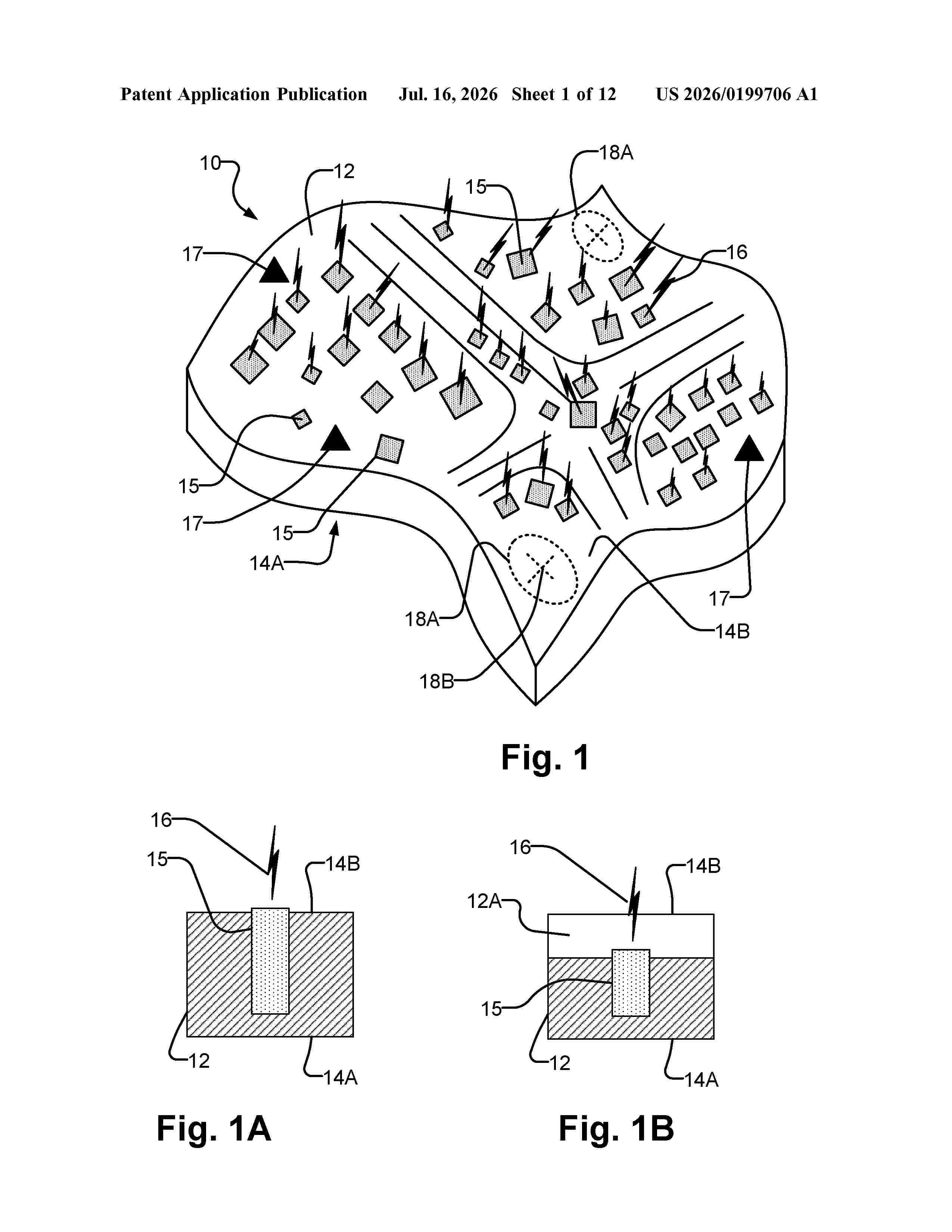
Resumen de: US20260199706A1
A radiation dosimetry apparatus comprises a body that supports scintillator elements that emit light when exposed to radiation. The body may be shaped to conform to contours of a portion of a patient's anatomy. Radiation intensity and dose may be determined by processing images obtained by one or more cameras to measure intensity of light emitted by different ones of the scintillator elements. The scintillator elements may be distributed non-uniformly based on a patient-specific radiation treatment plan. Scintillator elements may be arranged to provide increased spatial resolution and/or dose resolution in higher dose regions and/or regions corresponding to organs at risk. Radiation dosimeter apparatuses may be fabricated by additive manufacturing. Design and fabrication of the radiation dosimetry apparatuses may be integrated into workflows in clinical radiation treatment settings. Methods and systems for fabricating, calibrating, and using the radiation dosimetry apparatuses are described.
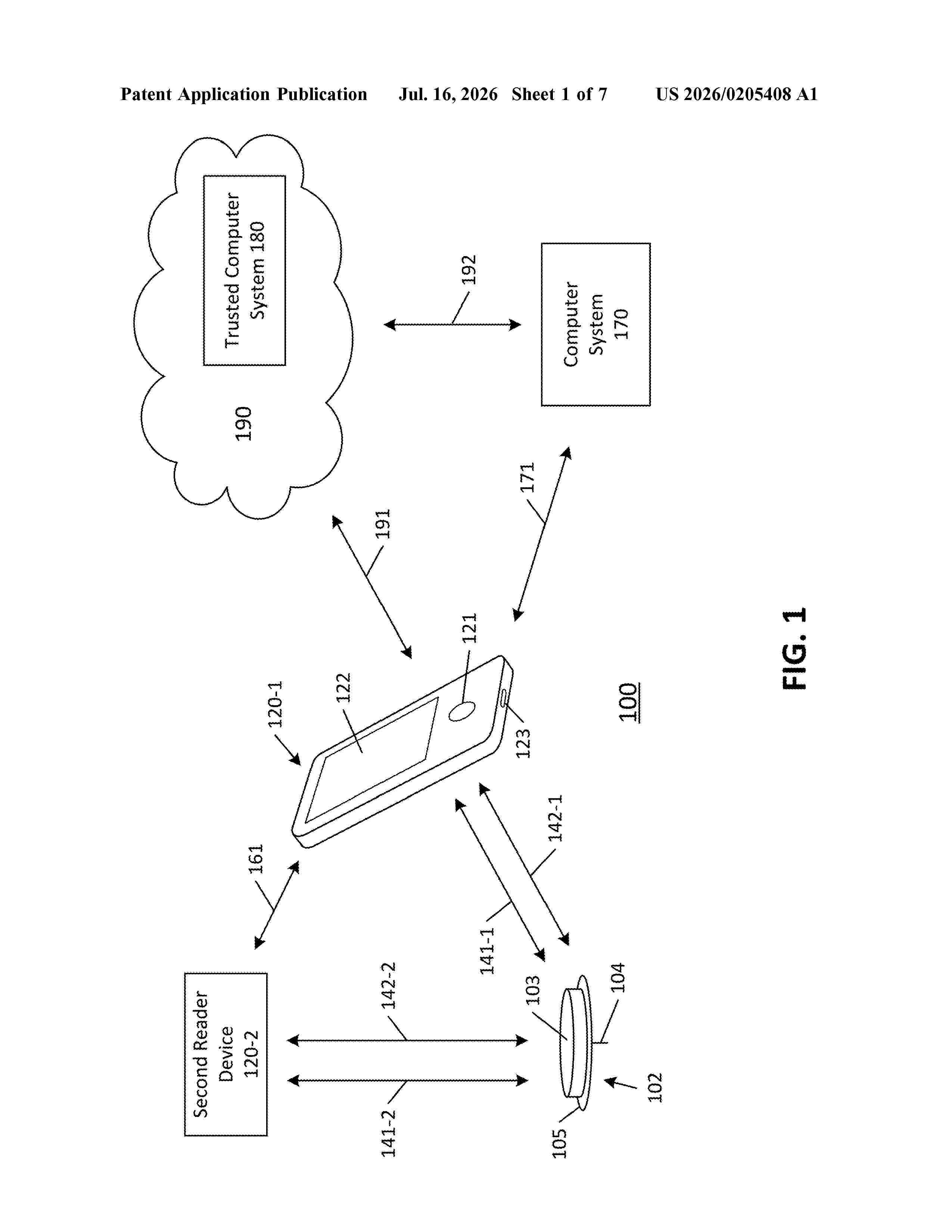
Resumen de: US20260205408A1
Example embodiments of systems, devices, and methods are described for communication in an analyte monitoring system in accordance with an applicable communication protocol. A first device of the system may transmit a command to a second device of the system and the second device may encounter a processing delay in preparing data responsive to the command. The second device may transmit dummy data to the first device in order to maintain compliance with the communication protocol until such time that the second device is ready to transmit data responsive to the command. Numerous different embodiments for incorporating and/or accommodating the presence of dummy data in a communication hierarchy are provided.
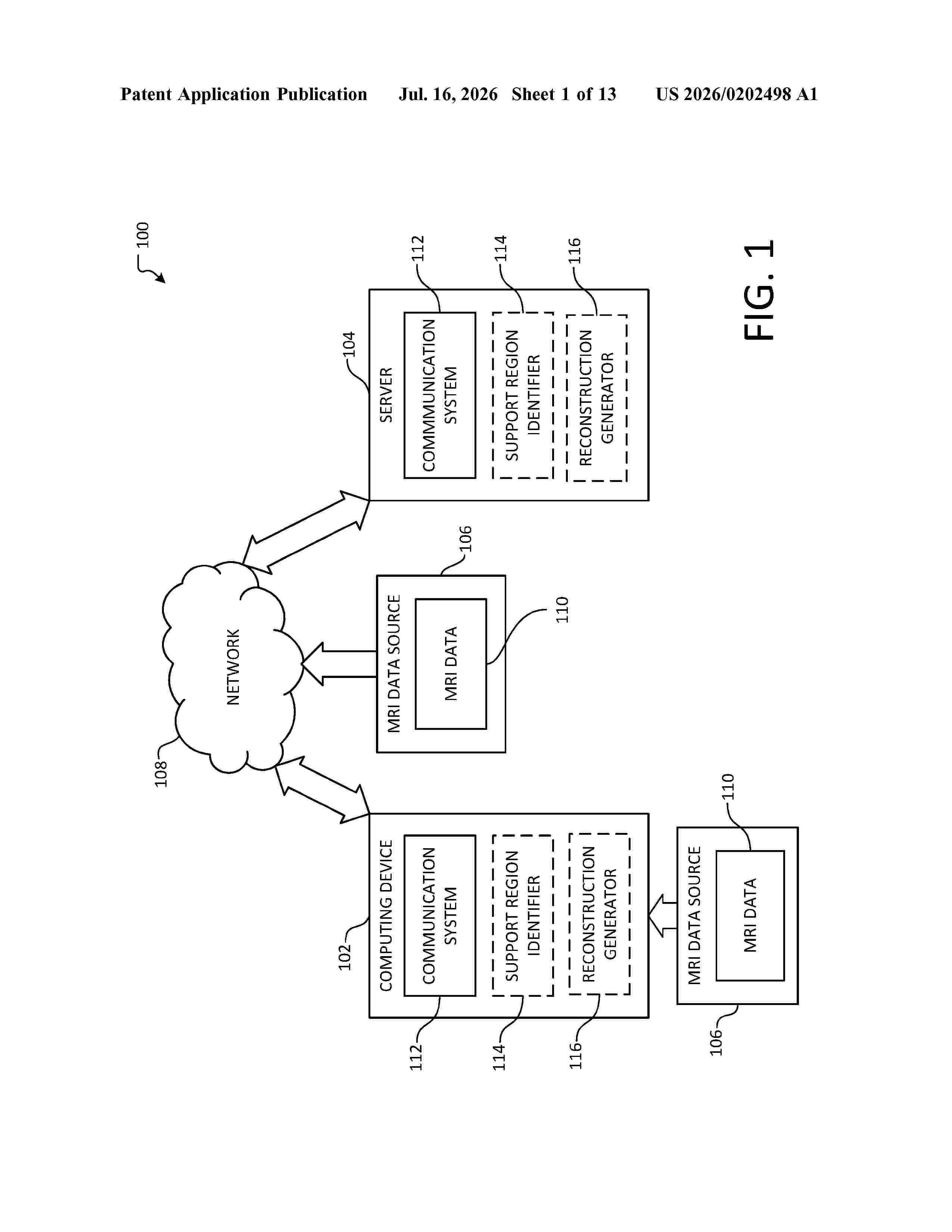
Resumen de: US20260202498A1
0000 A method to reconstruct an image, from a magnetic resonance imaging (MRI) machine, with reduced sampling is provided. The method includes receiving a first indication corresponding to a support region, obtaining a set of data samples associated with the first indication, determining a subset of data samples, from the set of data samples, that does not overlap with the one or more regions having aliasing, generating a first reconstruction, based on the determined subset of data samples, generating a frequency representation corresponding to the first reconstruction, subtracting the frequency representation from the set of data samples, thereby generating a modified set of data samples, generating a second reconstruction, based on the modified set of data samples, generating a reduced sampling reconstruction corresponding to the support region, by combining the first and second reconstructions, and outputting the reduced sampling reconstruction.
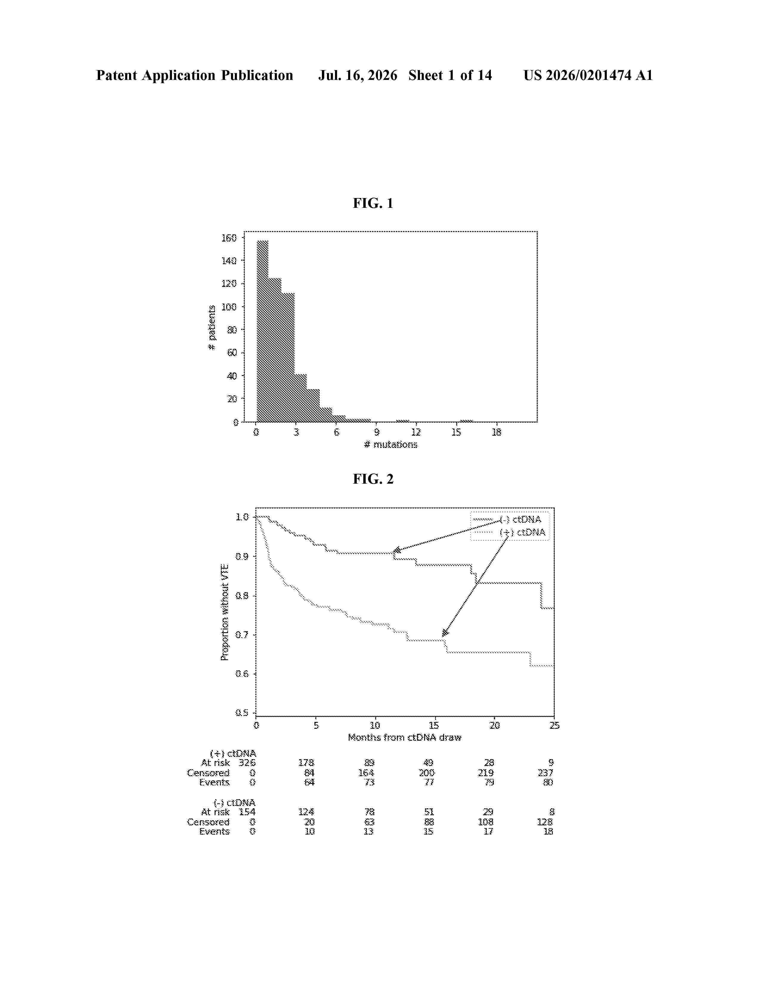
Resumen de: US20260201474A1
0000 The present disclosure relates generally to methods for accurately predicting the risk of cancer-associated venous thromboembolism (CAT) and/or preventing CAT in cancer patients using ctDNA as a biomarker.
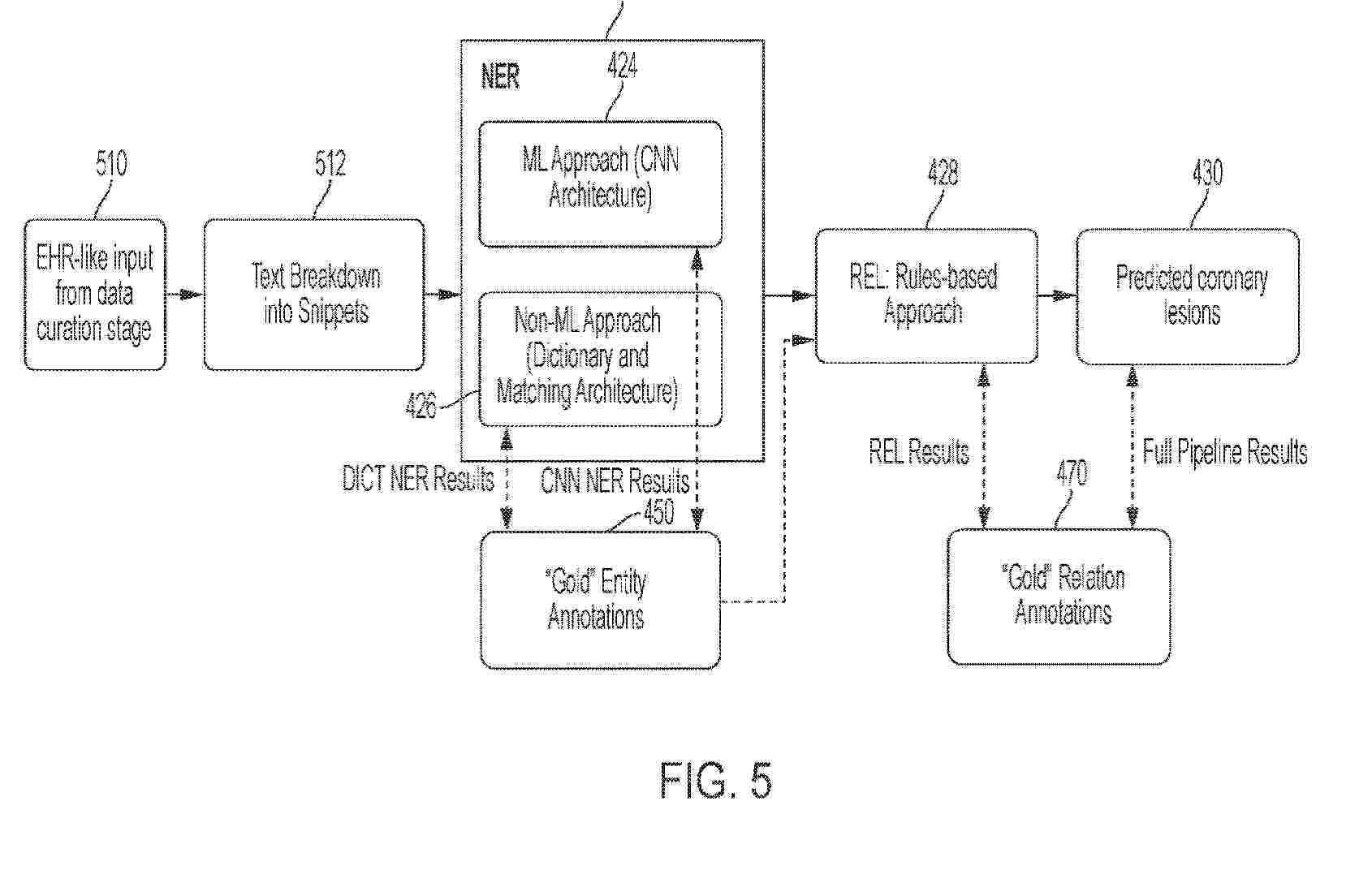
Resumen de: AU2024395361A1
Methods and apparatus for extracting coronary artery disease (CAD) information from unstructured medical text are provided. The method includes receiving unstructured medical text, processing the unstructured medical text using one or more trained natural language processing (NLP) models, wherein the one or more trained NLP models are trained to output CAD information, wherein the CAD information includes predicted coronary lesion information, and displaying an indication of the predicted coronary lesion information on a user interface associated with a computing device.
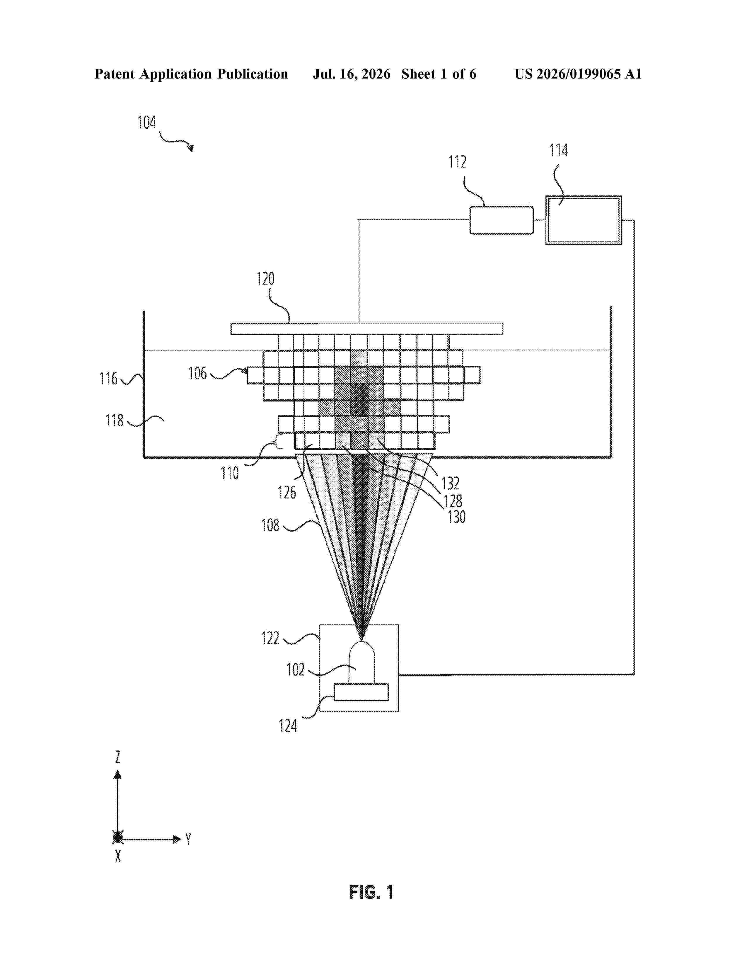
Resumen de: US20260199065A1
Manufacturing a dental part with volumetric units that vary in translucency, shading and/or color. A beam translation module of a 3D printing system is used to translate an input pattern into an output for controlling a spatial structuring of light for 3D printing an object from a resin material that is sensitive to light intensity, light color or light exposure time.
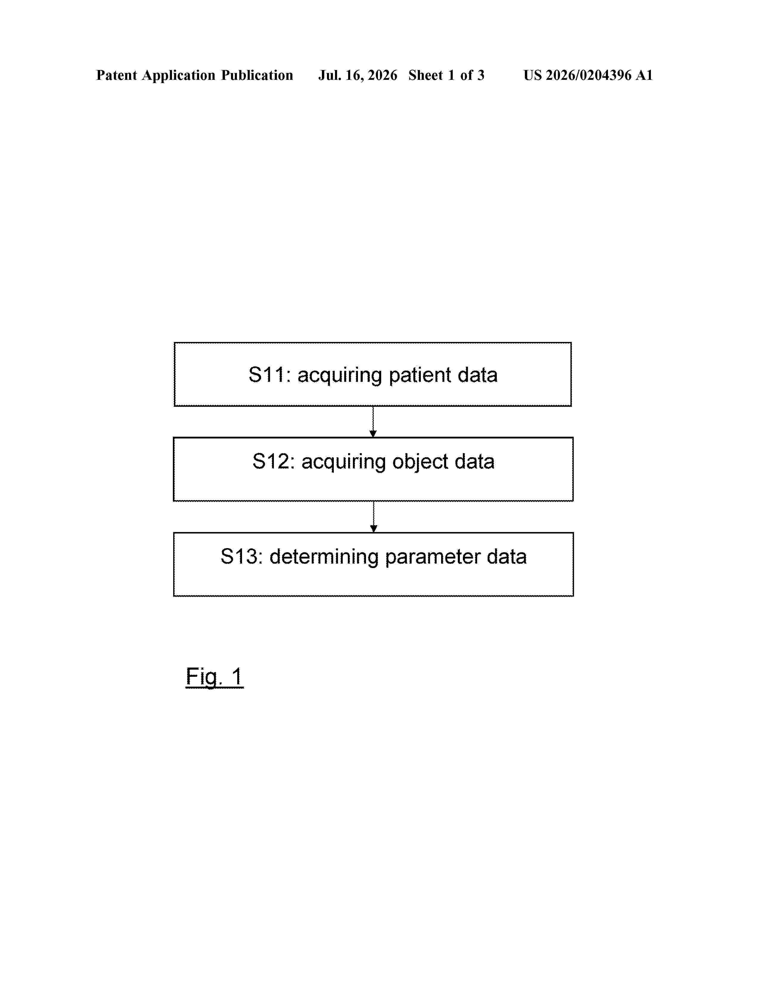
Resumen de: US20260204396A1
The present application relates to a computer-implemented medical method of determining a patient-specific setting of at least one imaging parameter of an imaging device. The method includes acquiring patient data describing information relating to a patient and having impact on the setting of at least one imaging parameter of the imaging device and acquiring object data describing at least one property of at least one object of interest associated with the anatomy of the patient. The imaging parameter data is determined based on the patient data, describing a patient-specific setting of the at least one imaging parameter of the imaging device.
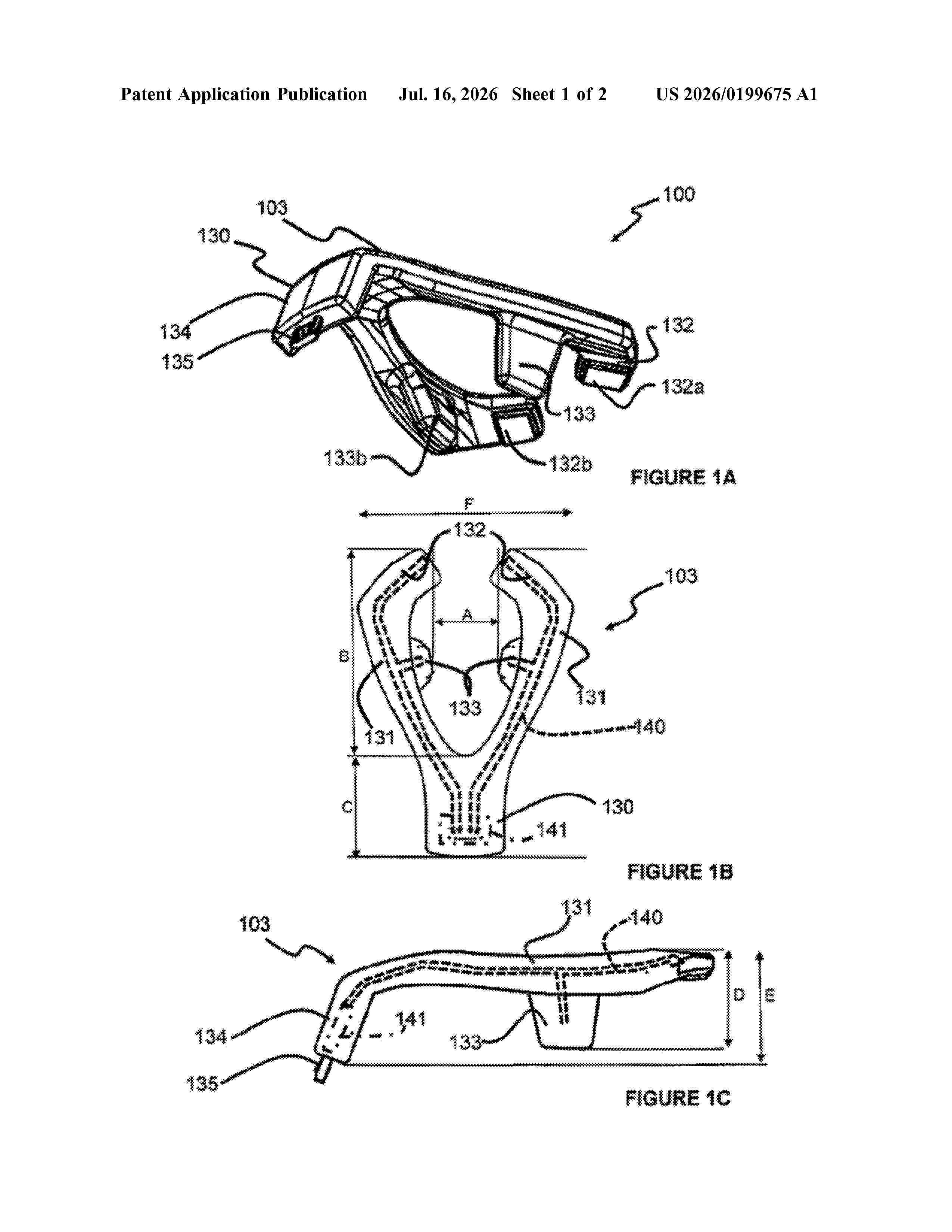
Resumen de: US20260199675A1
A device (100) for training oral muscle tone, the device (100) includes a mouthpiece (103) having a first and second arm (131) diverging away from one another and each carrying at least one electrode means (132a, 132b, 133a, 133b), at least one of the at least one electrode means protruding proud of adjacent portions of the arms (131), electrical circuitry operatively connected to the electrode means (132a, 132b, 133a, 133b), at least one of the at least one electrode means comprising an electrically conductive polymeric or plastics material.
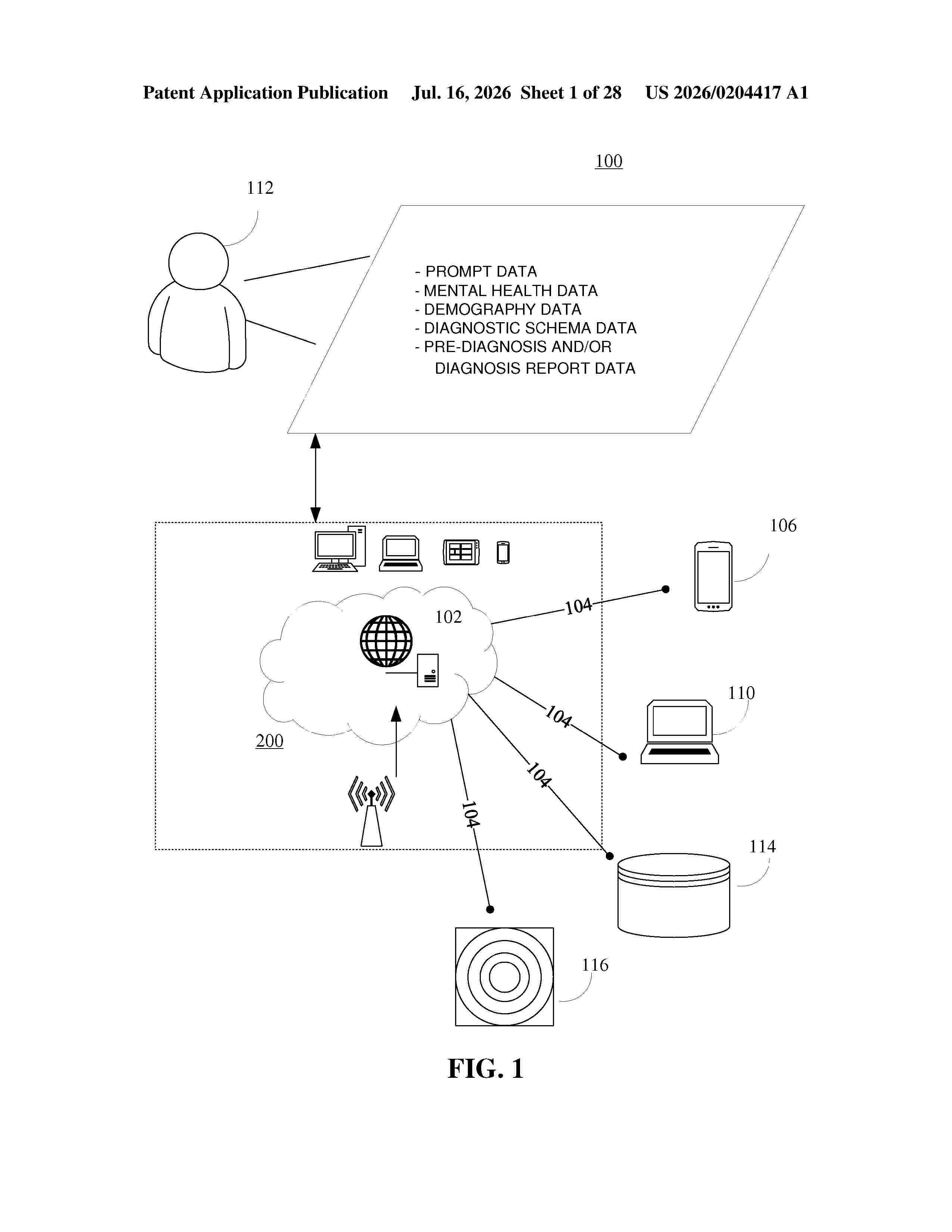
Resumen de: US20260204417A1
The present disclosure provides a method of facilitating AI-driven refinement of mental health diagnostics and personalized treatment. The method may include transmitting a prompt data to a user device. Further, the method may include receiving a mental health data from the user device based on the prompt data. Further, the mental health data may be associated with a user. Further, the method may include identifying a demography data based on the mental health data. Further, the method may include determining a diagnostic schema data to provide diagnostic insights based on the demography data. Further, the method may include generating a pre-diagnosis and/or diagnosis report data to provide personalized insights based on the diagnostic schema data and the mental health data. Further, the method may include transmitting the pre-diagnosis and/or diagnosis report data to the user device.
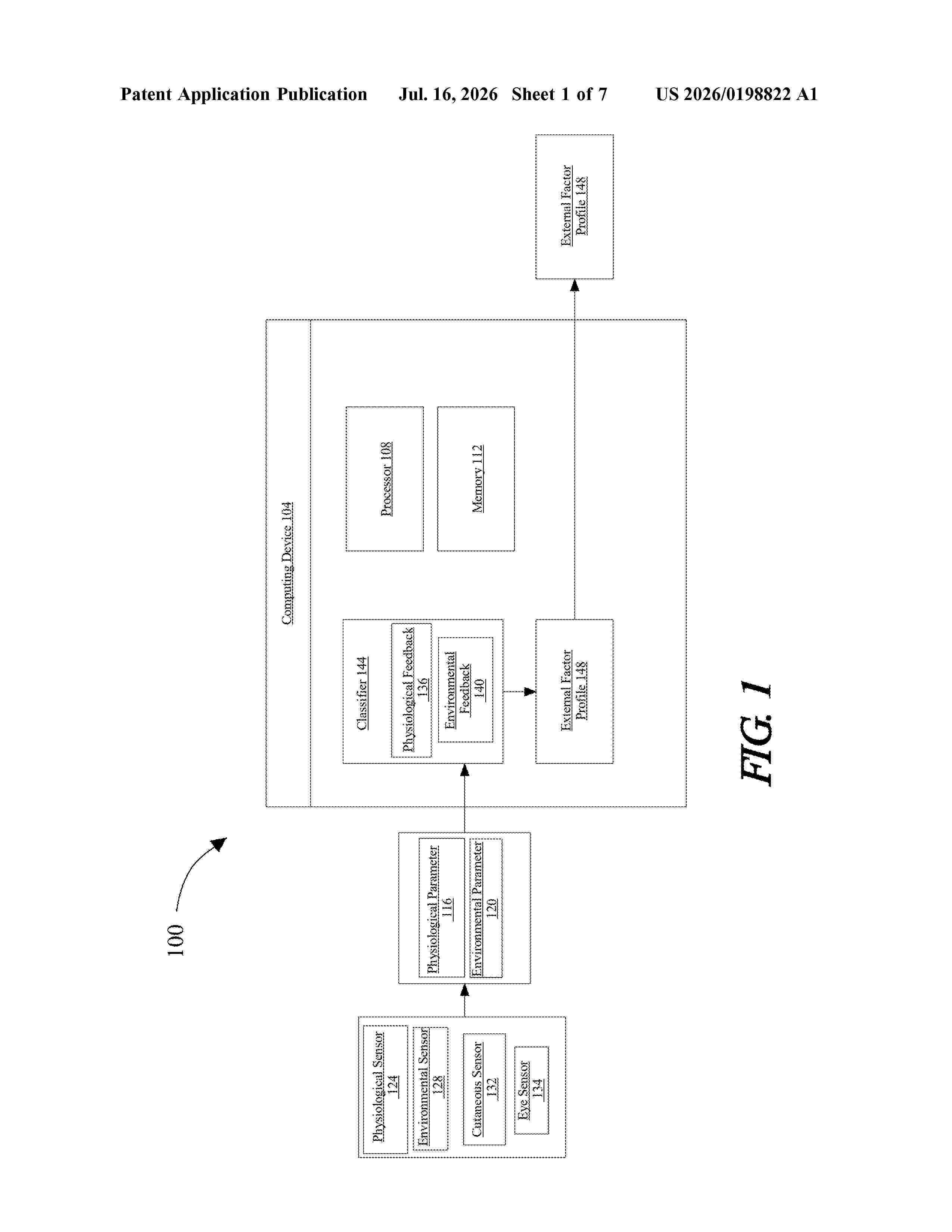
Resumen de: US20260198822A1
0000 An apparatus and method for gauging the effect of external factors relating to a user cognitive performance. The apparatus includes a plurality of sensors configured to detect at least a physiological parameter and an environmental parameter, at least a processor, and a memory communicatively connected to the at least a processor, the memory containing instructions configuring the at least a processor to receive physiological feedback from the plurality of sensors, receive environmental feedback from the plurality of sensors, generate an external factor profile containing a plurality of external factors selected by the processor configured to improve the user cognitive performance as a function of the received biological feedback and environmental feedback.
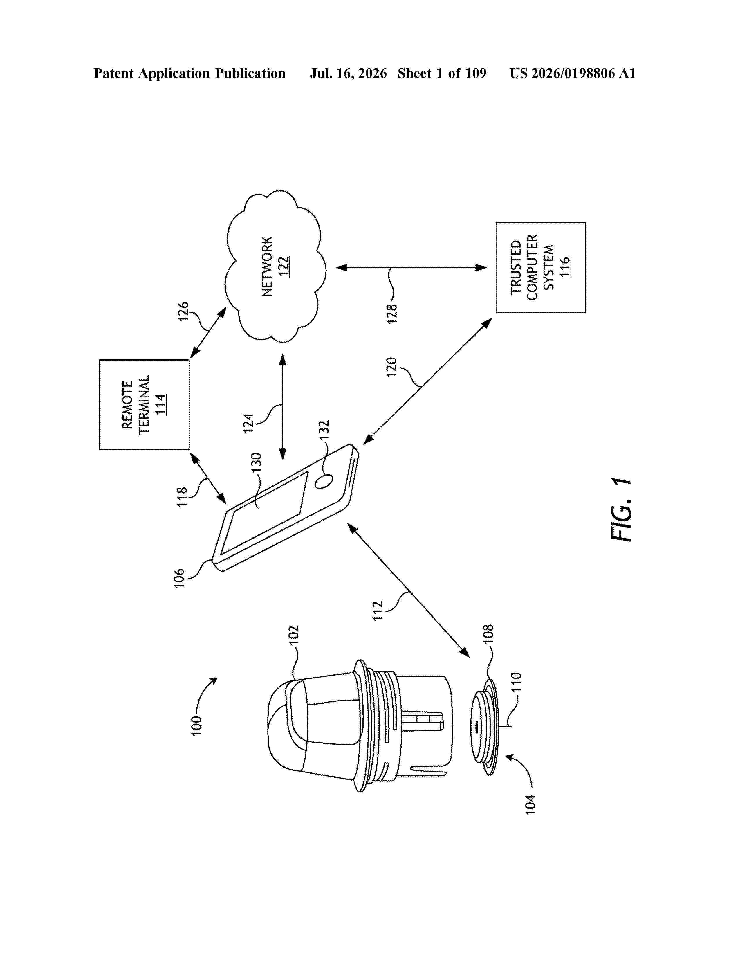
Resumen de: US20260198806A1
A system includes a sensor applicator, a sensor control device arranged within the sensor applicator and including an electronics housing and a sensor extending from a bottom of the electronics housing, and a cap coupled to one of the sensor applicator and the sensor control device, wherein the cap is removable prior to deploying the sensor control device from the sensor applicator.
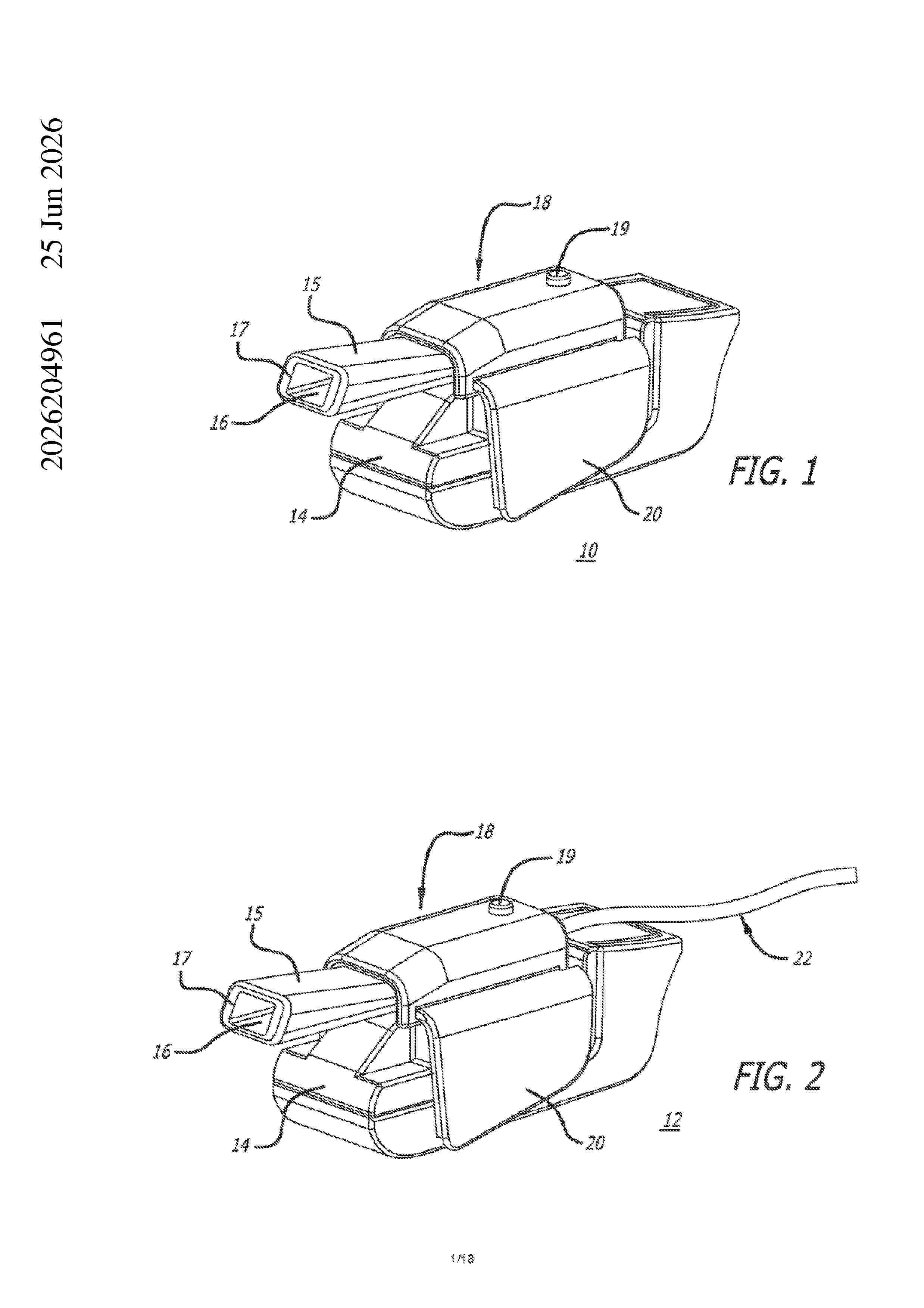
Resumen de: AU2026204961A1
The invention relates to an inhaler accessory apparatus comprising: a body configured for mounting onto or connection with an inhaler; a microprocessor, at least two pressure sensors, a first pressure sensor for detecting information on environmental conditions around said inhaler; and a second pressure sensor for detecting signals generated from said inhaler when in use, and a pressure equalization channel between said first pressure sensor and said second pressure sensor, wherein said first pressure sensor and said second pressure sensor generate at least one signal each which is processed in said microprocessor resulting in customized microprocessor output and wherein said microprocessor output generates a pressure differential versus time curve on a display concurrently with or immediately after a user’s inhalation. user's inhalation. un u n u s e r ' s i n h a l a t i o n
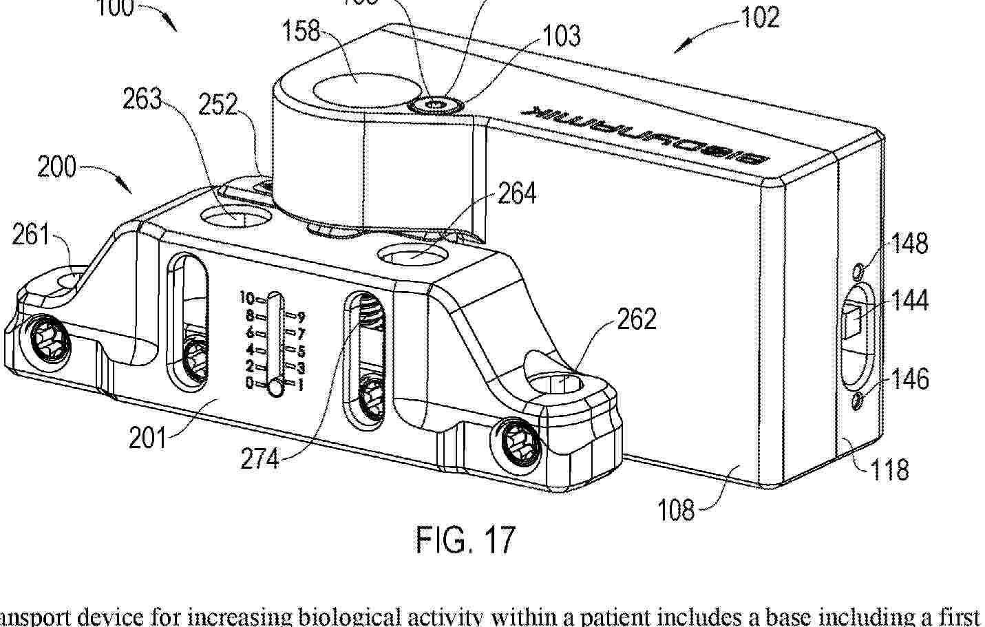
Resumen de: AU2024394477A1
A transport device for increasing biological activity within a patient includes a base including a first end, a second end, an upper surface located between the first end and the second end, and a lower surface located between the first end and the second end, a base anchor configured to statically couple the base to a first portion of a bone of a subject, a translatable anchor configured to engage a growth stimulator, a leadscrew dynamically coupling the base to the translatable anchor, wherein the leadscrew is configured to rotate about a leadscrew axis while substantially maintaining its longitudinal position along the leadscrew axis in relation to the base, such that the growth stimulator, when engaged with the translatable anchor, is capable of independent movement in relation to the base along a stimulation axis that includes at least some transverse displacement with respect to the bone when the base is coupled to the bone via the base anchor, and a screw drive rotatably coupled to the leadscrew and configured to couple to a rotatable mating tool that is configured to rotate the leadscrew via the screw drive.
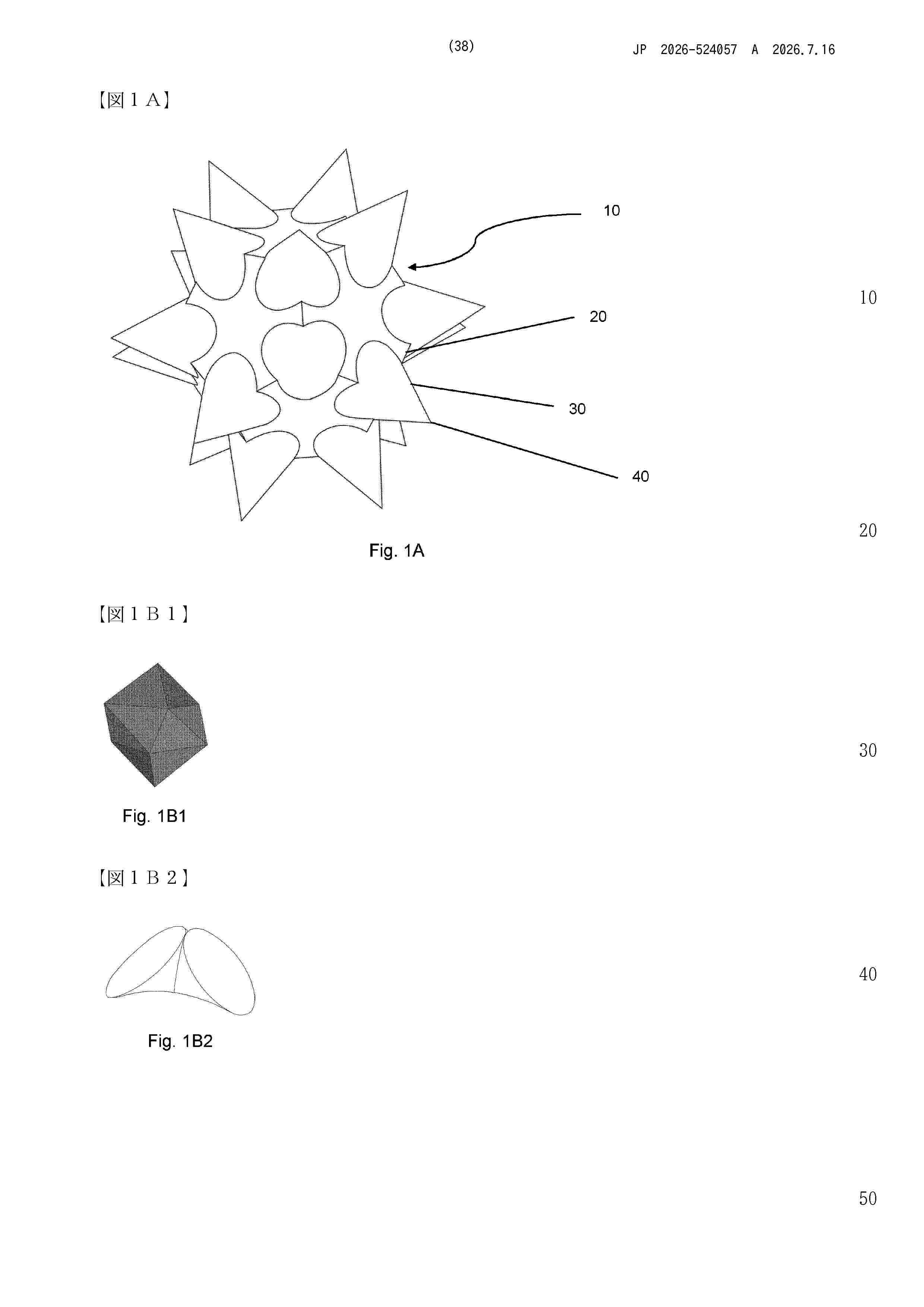
Resumen de: WO2024235974A1
A microneedle particle arranged to be rolled and massaged on a subject's skin (12) to penetrate the stratum corneum (14) is disclosed. The microparticle comprises a support body (20), and a plurality of microneedles (30) protruding from the support 5 body (20), each microneedle comprising a tip (40). The support body (20) maintains the microneedles (30) and tips (40) in a predetermined conformation allowing the microneedle particle to be rolled on the skin. The microneedles have an average tip sharpness radius of less than 20 µm, an average microneedle length of less than 100 µm, and an average tip sharpness to microneedle length ratio of 1:5 to 1:200. 10 Formulations and kits for said use are provided. A microneedle particle manufacturing method using a non-linear two-photon absorption process, a non- therapeutic cosmetic method of skin treatment, and a method of drug delivery through the skin for said microneedle particle are disclosed.
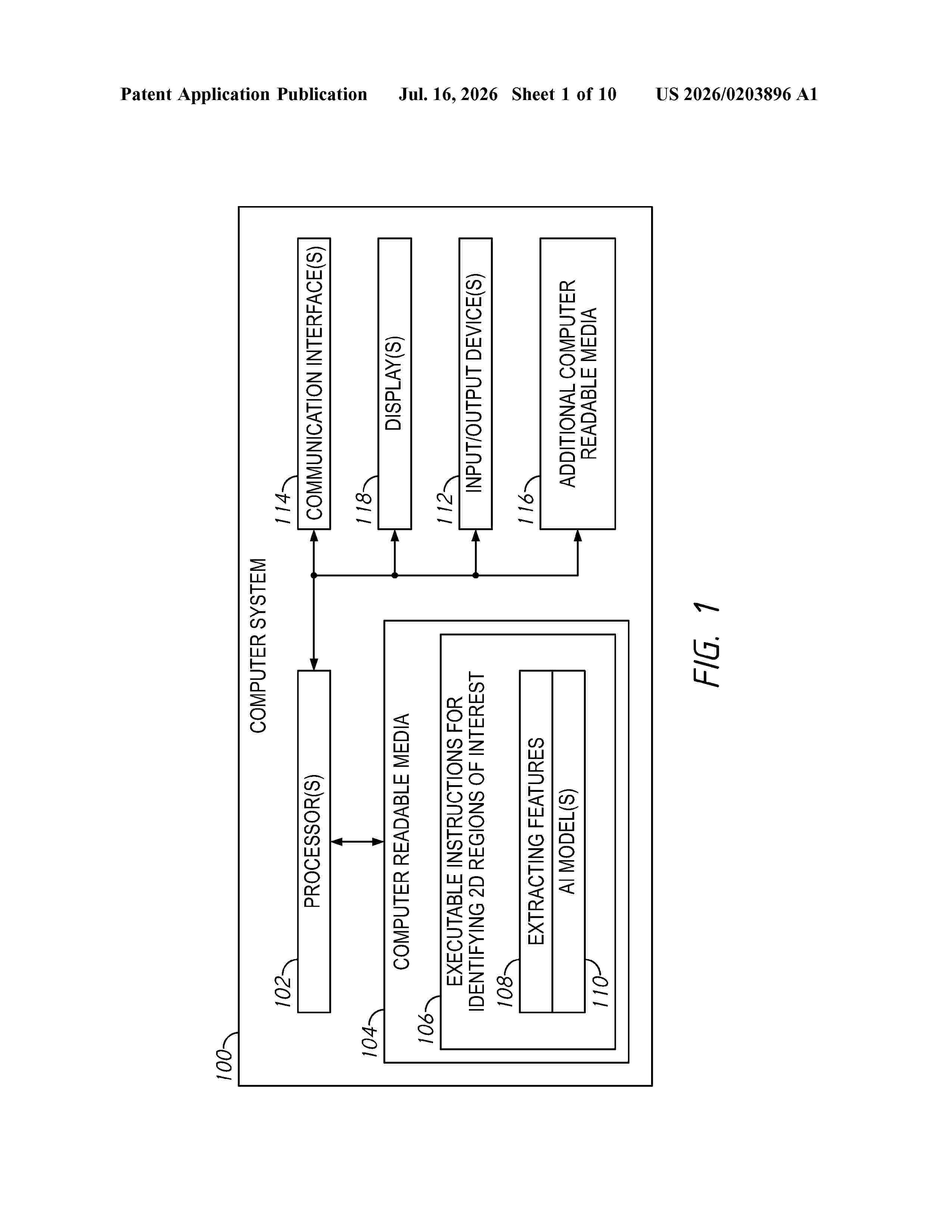
Resumen de: US20260203896A1
0000 Embodiments of the present disclosure provide methods and systems for identifying regions of interest from three-dimensional (3D) anatomical image data. An example method includes analyzing a three-dimensional (3D) data set of one or more medical images using an artificial intelligence (Al) model to identify two-dimensional (2D) regions of interest in the 3D data set having an increased probability of a diagnostic state, and providing the regions of interest to another process for additional analysis. In some embodiments, said analyzing the 3D data set includes generating a 3D heat map, a 3D segmentation, or both, of the 3D data set using the Al model, and identifying the regions of interest using another Al model, wherein each region of the regions of interest includes a respective 2D data set.
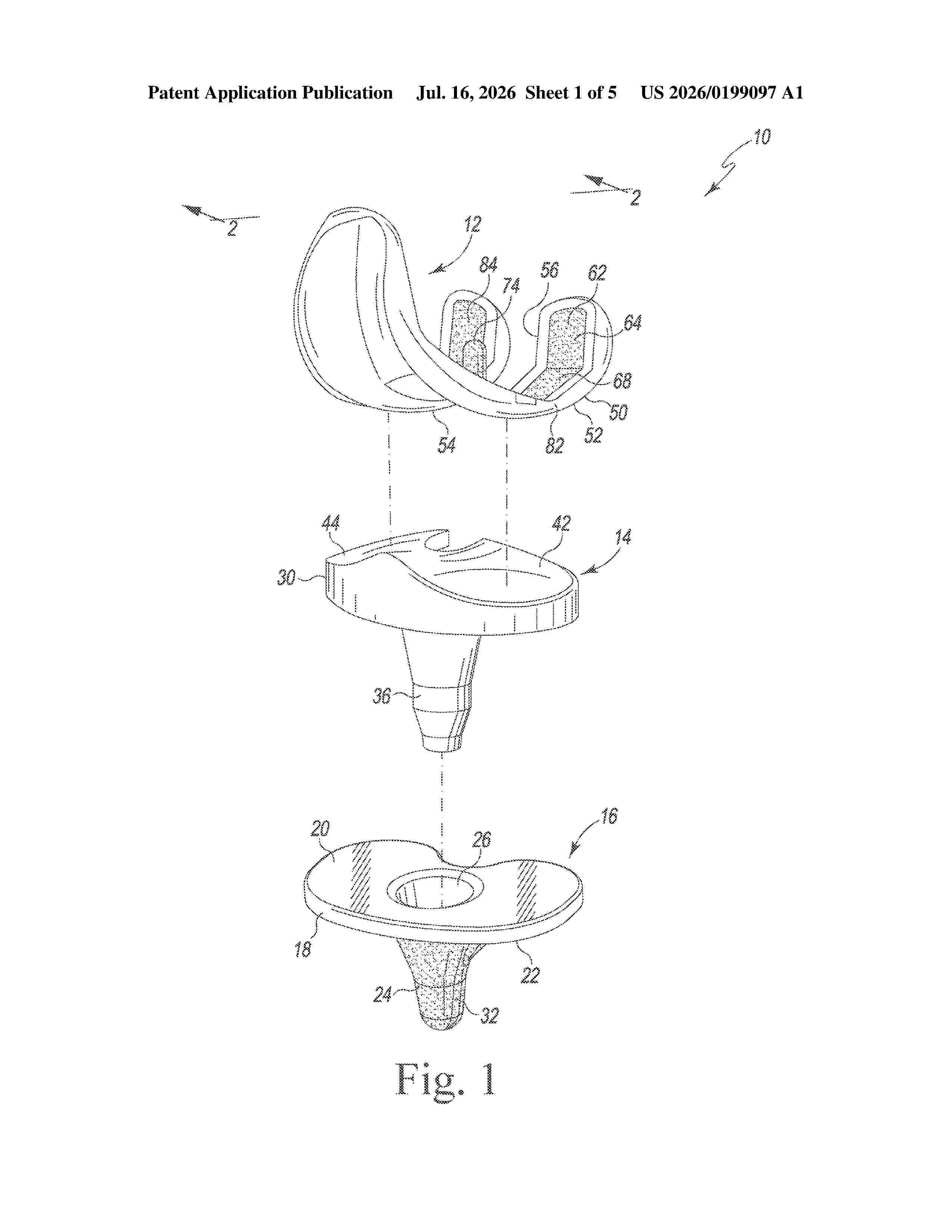
Resumen de: US20260199097A1
0000 An orthopaedic knee prosthesis includes a femoral component having a metal base with a polymer articular layer molded thereto. A method for making a metal-reinforced femoral component of an orthopaedic knee prosthesis is also disclosed.
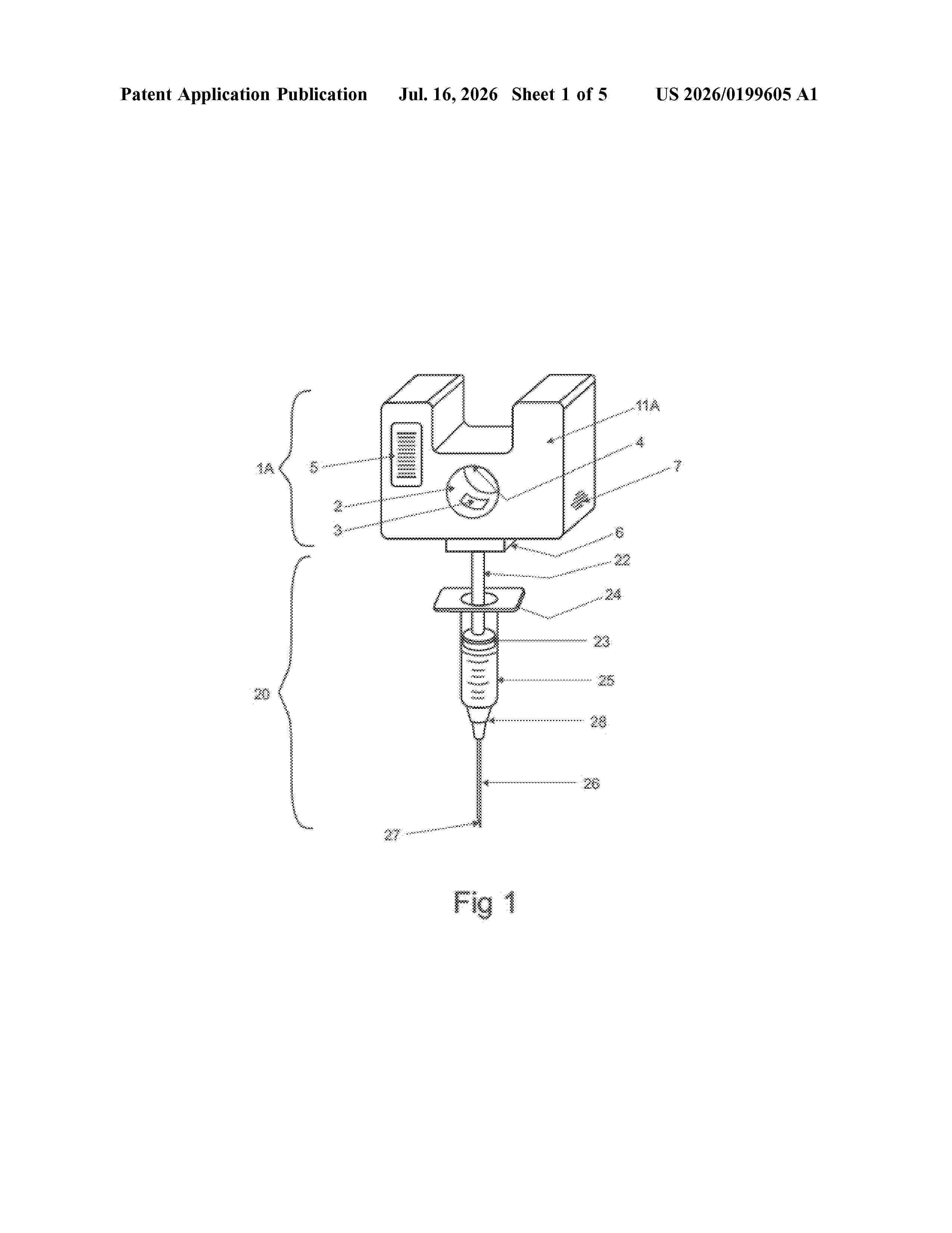
Resumen de: US20260199605A1
A syringe use monitoring device that can attach to a syringe and detect the type of medication being loaded and measure the force applied to the plunger and thus monitor subsequent delivery of medication and delivery pressure as a substance is being injected, and at least one display and/or alarm can indicate normal and/or abnormal conditions so that feedback can be employed during the injection process is provided.
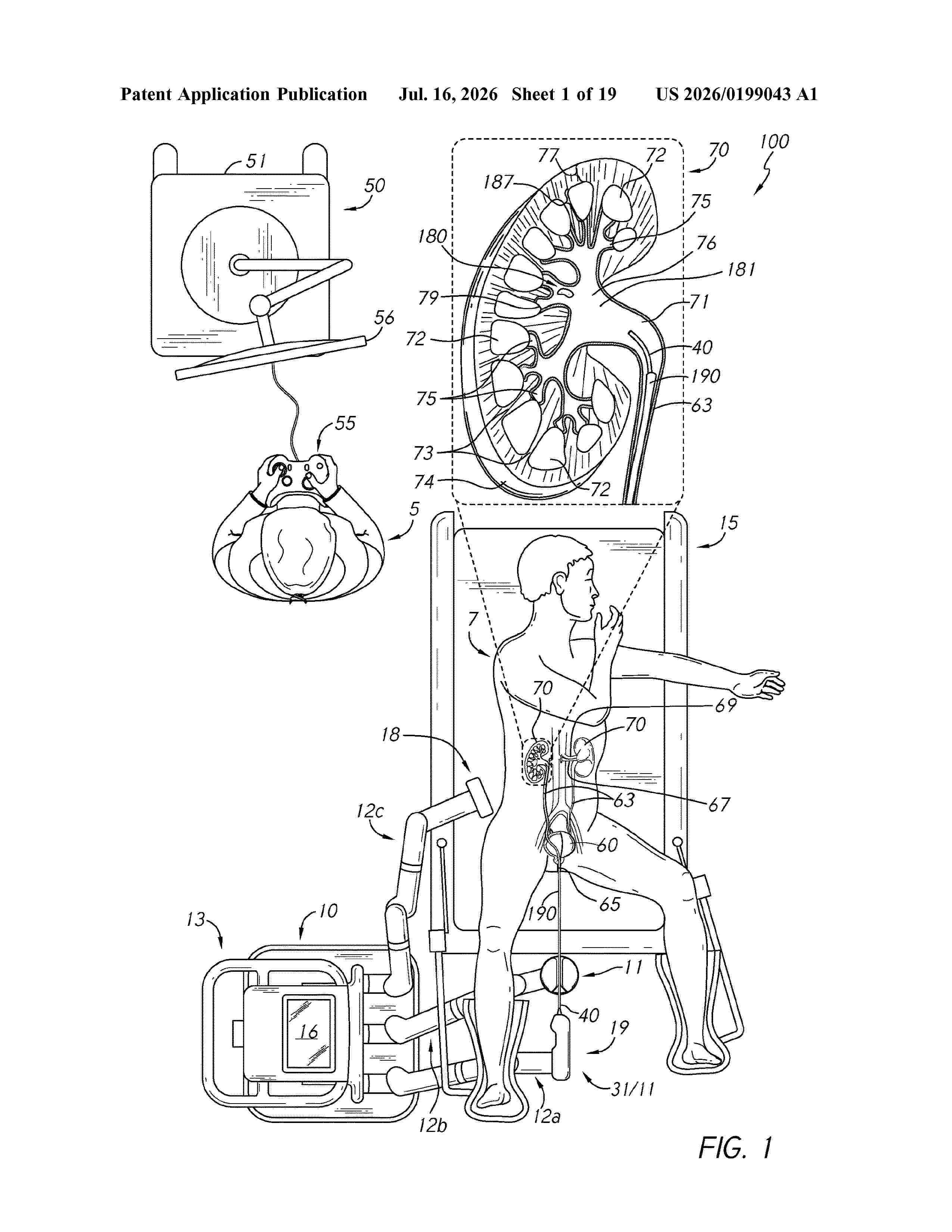
Resumen de: US20260199043A1
0000 A method for robotically controlling an endoscope comprises: navigating an elongate shaft in a patient body, the elongate shaft including a tip at a distal end, articulating the tip in a first direction with a first pull wire coupled to a dual-wire pulley, reversing articulation of the tip to a second direction with a second pull wire coupled to the dual-wire pulley based on a nonlinear response region of a kinematic model, determining an end point of the nonlinear response region, and, when articulation of the tip in the second direction reaches the end point, articulating the tip based on a linear response region.
Resumen de: WO2023159230A1
Systems and methods for distribution, storage, transport, administration and/or disposal of one or more therapeutic or diagnostic agents are disclosed. A storage device configured to connect to a delivery system for delivering the therapeutic or diagnostic agent has a housing with a chamber, a vessel having an access port positioned within the chamber. The door is movable relative to the housing between a closed position and an open position. In the closed position, the door covers an opening in the housing to enclose the chamber, and, in the open position, the door reveals the opening for accessing the access port of the vessel. The door is moveable between the closed position and the open position in response to actuation by an access mechanism of the delivery system.
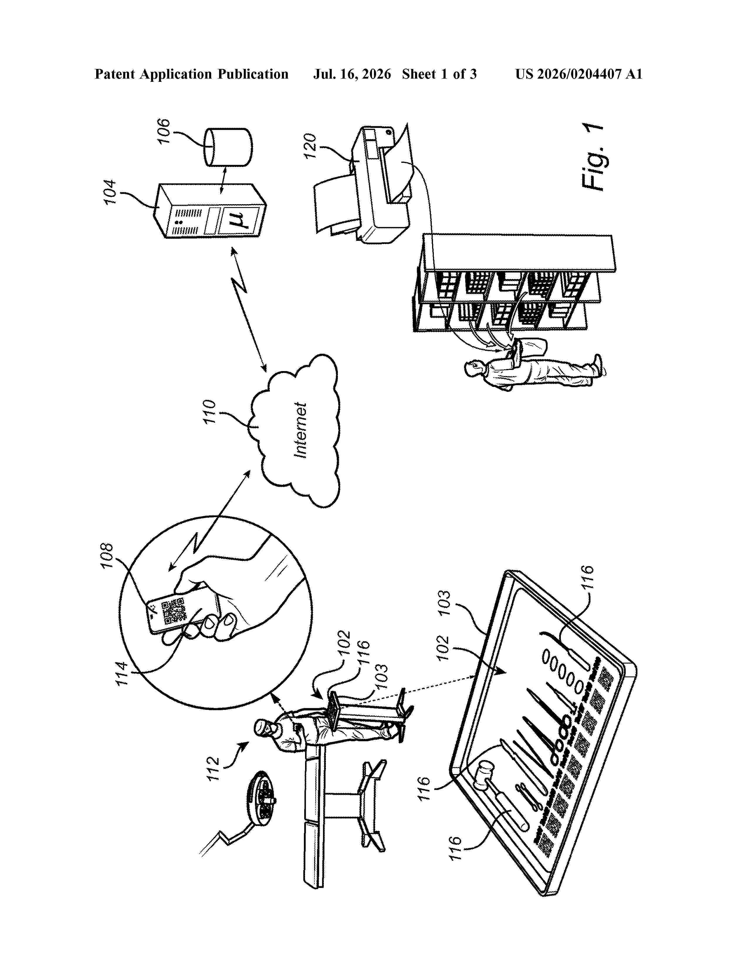
Resumen de: US20260204407A1
The present disclosure generally relates to a method for generating a tray instrument layout, where the tray instrument layout is adapted taking into account a type of surgical procedure to be performed, available surgical instruments and a procedural order for performing the surgical procedure. The present disclosure further relates to a corresponding computer system and computer program product.
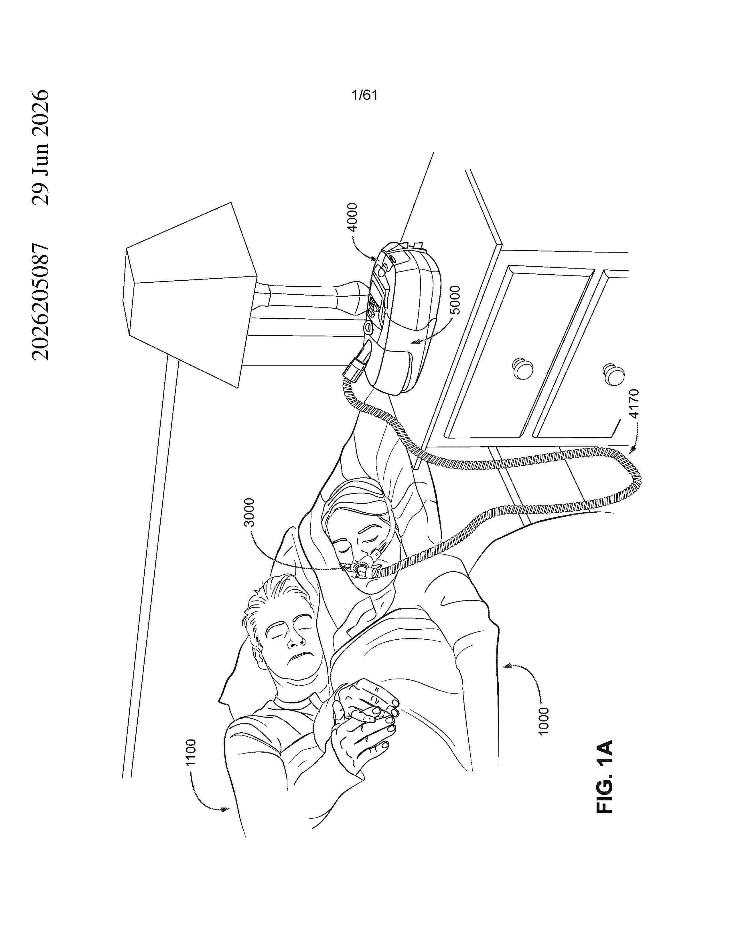
Resumen de: AU2026205087A1
A patient interface includes a plenum chamber pressurizable to a therapeutic pressure, a seal-forming structure configured to form a seal against the patient’s face, and a positioning and stabilising structure configured to provide a force for maintaining the seal-forming structure in a therapeutically effective position. The patient interface also includes an RPT device connected directly to the plenum chamber. The PRT device includes an electric blower for providing airflow at the therapeutic pressure. The positioning and stabilising structure supports at least part of the weight of the RPT device. The patient interface may also include an electrical power source electrically connected to the RPT device. connected to the RPT device.20 un u n c o n n e c t e d t o t h e d e v i c e
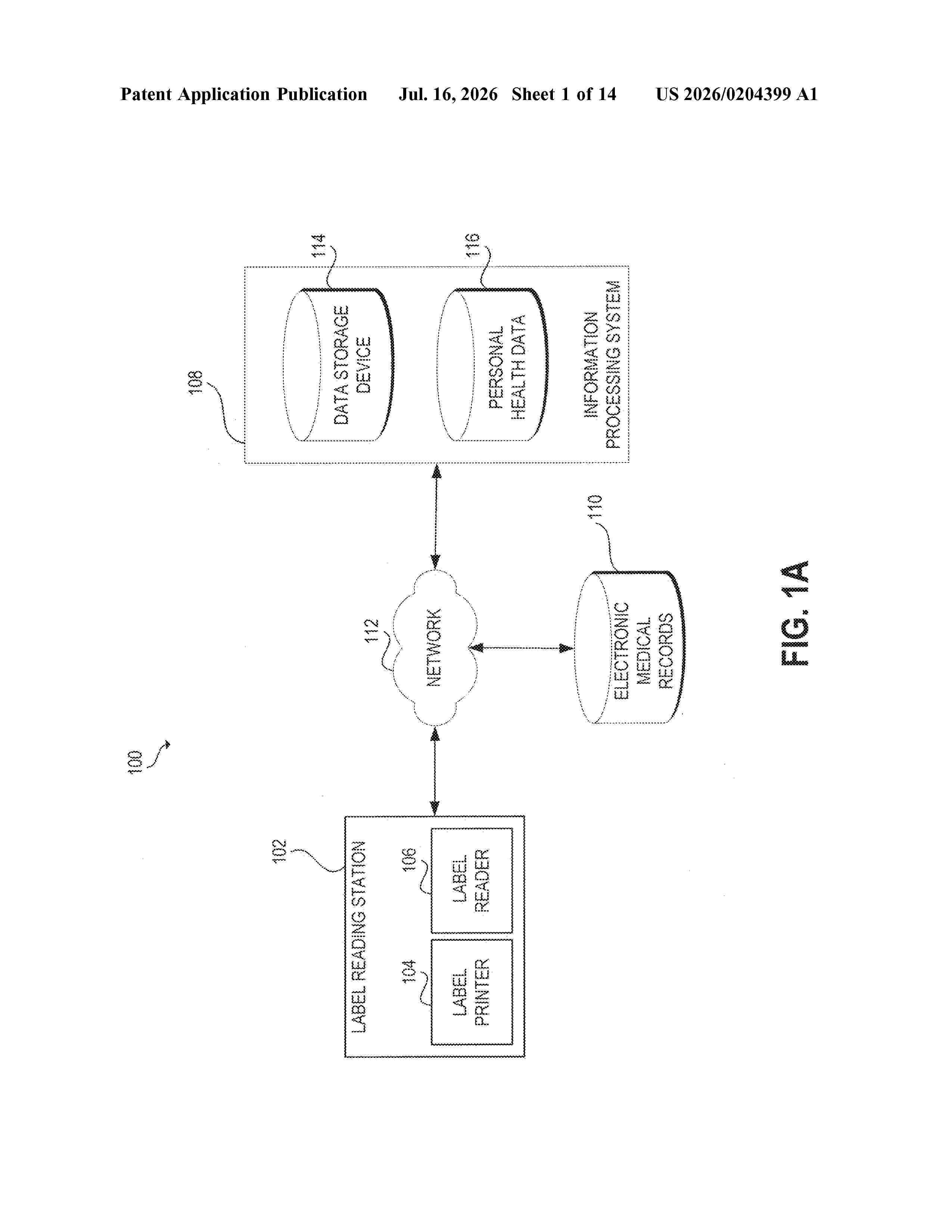
Resumen de: US20260204399A1
0000 A system and method is described for printing a label with an RFID tag. The system includes an RFID reader that queries a first RFID tag coupled to a first medicinal container that includes a medication. In response, the system receives a first unique identifier and uses the first unique identifier to determine a status of the medication, associate the first medicinal container with a medical provider and print a second label that includes a second RFID tag for a second medicinal container.
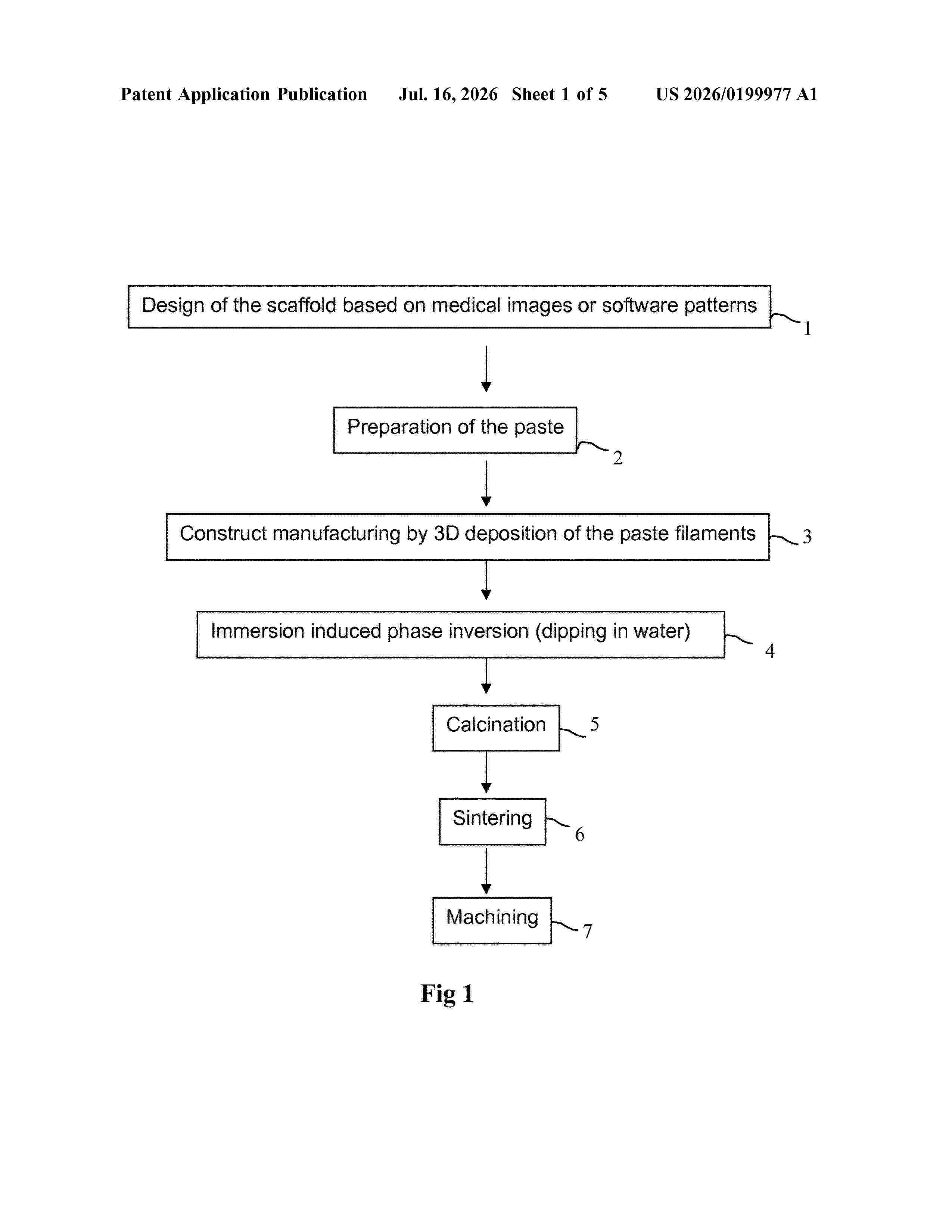
Nº publicación: US20260199977A1 16/07/2026
Solicitante:
VITO NV [BE]
VITO NV
Resumen de: US20260199977A1
A method for producing a three-dimensional macro-porous filament construct having interconnected microporous filaments may include preparing a mixture of particles of metal(s), metal alloy(s), binder(s), a first liquid solvent for the binder(s), and optionally dispersant(s). Particles containing a removable additive are dispersed in the mixture. The mixture is deposited in a three-dimensional pattern of interconnected filaments, creating a three-dimensional filament-based porous green structure that is then contacted with a second solvent (a non-solvent for the binder(s)) to induce phase inversion. At least part of the filaments are transformed to a solid state. The removable additive is removed to create additional micro-porosity in the phase inverted porous structure, and the structure is subjected to a thermal treatment. The removal agent includes the second liquid solvent or is a third liquid solvent distinct from the second.
 BOPI
BOPI
 Sede Electrónica